Int Adv Otol 2015; 11(1): 77-80 • DOI: 10.5152/iao.2015.288
Original Article
OBJECTIVE: To define clinical and audiological findings in patients with temporal bone posterior wall defects (TBPWD) and to investigate possible relationships between these findings and the characteristics of the defect.
MATERIALS and METHODS: The computed tomography (CT) views of 1198 patients with vestibulocochlear symptoms between 2007 and 2012 were retrospectively evaluated, and TBPWD and associated anomalies were investigated. Patients who had TBPWD were called back, and clinical and audio-logical examinations (tympanometry, pure tone audiometry, acoustic reflexes, and otoacoustic emission) were performed.
RESULTS: Twenty-eight (2.34%) patients had TBPWD. Twenty-three of them were eligible for the study. Size of the defect was significantly correlated with the presence of tinnitus and/or vertigo (p<0.005). The cut-off values for the largest size of TBPWD were 1.65 mm [sensitivity: 0.67 and specific-ity: 0.77 (95% CI: 0.58–0.97); p=0.04] in case of the presence of tinnitus and 1.85 for vertigo (sensitivspecific-ity: 0.78 and specificspecific-ity: 0.86 (95% CI: 0.67–0.99); p=0.006). In pure tone audiometry tests, mixed-type hearing loss was present in four (17%) patients, sensorineural hearing loss was present in three (13%) patients, and conductive-type hearing loss was present only in one (4%) patient. Otoacoustic emission tests revealed significant differences in signal/noise ratios at frequencies of 500, 750, 1000, and 6000 Hz.
CONCLUSION: For the first time in the literature, we defined clinical and audiological findings in patients with TBPWDs. These defects seem to cause more prevalent symptoms of vertigo and tinnitus and disturb the audiological characteristics of patients.
KEYWORDS: Temporal bone, vertigo, tinnitus, audiometry INTRODUCTION
The posterior wall of the temporal bone is thick, and it is rid of natural dehiscences when compared with the petrous bone, which is located in the middle cranial fossa. In the literature, arachnoid granulations originating from the posterior wall of the temporal bone, which result in temporal bone defects because of pulsations of the arachnoid villi, have been defined [1]. These bony defects may be overlooked by the clinicians, and rare but important complications may occur in the presence of this anomaly.
In two studies investigating temporal bone posterior wall defects (TBPWD), only histological and radiological data were available [2, 3]. For the first time in our study, we aimed to define clinical and audiological findings in patients with TBPWD and to investigate the possible relationship between these findings and the characteristics of the defect.
MATERIALS and METHODS
Computed tomography (CT) views of 1198 patients admitted to our outpatient clinics with vestibulocochlear symptoms between 2007 and 2012 were retrospectively evaluated. TBPWD was found in 32 patients. After the exclusion of patients who had a history of ear surgery or cholesteatoma causing bone destruction, 28 (2.34%) patients were enrolled.
The local ethics committee approved the study protocol and informed consent was obtained from all participants.
Computed Tomography Imaging
A 16-detector CT machine (GE Lightspeed; GE Healthcare, Wisconsin, USA) was used for CT imaging. Imaging protocol parameters were as follows: scan time of 1 s, 0.6-mm section thickness, 140-kVp peak kilovolt, 260-mAs miliampere-second value, matrix of 512×512, 26-cm field-of-view (FOV), mean number of slices was 140, and mean volume computed tomography dose index (CT-DIvol). All CT images were produced in an axial plane without gantry angle. CT views of patients who had TBPWD were examined in a dedicated double-monitored workstation with 3-megapixel resolution (Advanced Workstation 4.2 GE Healthcare, USA). All images were retrieved from hospital-based digital radiological image archive (Centricity, GE Healthcare, USA). Two radiology res-idents examined CT images in high-resolution algorithm and at a window width of 4000 HU and a window level of 400 HU. They
This study was presented at the 11th International Otolaryngology and Head and Neck Surgery Congress, 17-19 April 2014, Ankara, Turkey.
Corresponding Address:
Hande Ezerarslan, Department of Ear Nose Throat, Ufuk University, Ankara, Turkey Phone: +90 312 204 43 83; E-mail: [email protected]
Submitted: 22.05.2014 Revision received: 27.10.2014 Accepted: 09.04.2015
Copyright 2015 © The Mediterranean Society of Otology and Audiology
77
Temporal Bone Posterior Wall Defect, a New Clinic
Entity?
Hande Ezerarslan, Gökçe Kaan Ataç, Safinur Keskin, Güçlü Kaan Beriat, Sinan Kocatürk
Department of Ear Nose Throat, Ufuk University, Ankara, Turkey (HE, GKB, SiK) Department of Radiology, Ufuk University, Ankara, Turkey (GKA, SaK)
performed measurements together, and a final decision was reached regarding the existence of a bony defect and its size. Location of the defect in the posterior fossa on an axial plane was not mentioned in the evaluation. Bony defect measurements were taken in two planes perpendicular to each other. One experienced radiologist reviewed abnormal cases and made corrections. Other temporal bone anoma-lies, such as high jugular bulb, jugular dehiscence, and superior semi-circular channel dehiscence, were also investigated.
Clinical and Audiological Evaluation of Patients
The hospital records of 28 patients were retrospectively evaluated, and patients were phoned and called back for physical and audiolog-ical examination. Five (17.8%) of the patients were unavailable; thus, they were excluded. Rest of the patients were invited to our clinics for detailed examination.
The presence or absence of tinnitus and vertigo was reported. Af-ter the physical examination of the ear, patients underwent audio-logical tests, including tympanometry (AZ 26 Clinical Audiometer; Interacoustics Assens, Denmark), pure tone audiometry (AC 33 Clin-ical Audiometer; Interacoustics Assens, Denmark), and otoacoustic emission [distortion product otoacoustic emission, DPOAE; (Integrity V500; Vivosonic, Toronto, Canada)].
In a completely isolated cabin using hearing thresholds that were ob-tained between the frequencies of 250–8000 Hz pure tone averages were determined. Audiogram results were classified into the three following groups: conductive-type hearing loss (CHL), mixed-type hearing loss (MHL), and sensorineural-type hearing loss (SNHL). Acoustic stapes reflex (ASR) and tympanograms of patients were ob-tained. ASR was separately interpreted as present or absent at 500, 1000, and 2000 Hz. Tympanogram curves were interpreted as type A, B, C, and As [4].
DPOAE measurements for both ears were made in an acoustically iso-lated room. DPOAEs (2f1-f2) were evoked by the system using an f2/ f1 ratio of 1.2. For L1, the amplitude of the stimulus was 65 dB SPL, and for L2, it was 55 dB SPL. At a constant stimulus amplitude and time, distortion-dependent otoacoustic emission data were saved at different frequency zones from low-to-high frequency range of f2=0.5–8.0 kHz, and distortion dependent audiogram was attained. In a patient with CHL who had sclerosis at the bilateral mastoid bone, no acoustic reflexes, and a type B curve on the tympanogram, a mid-dle ear disease was also thought to be accompanied. Except this pa-tient, all patients underwent the DPOAE test. In patients with unilat-eral TBPWD, signal/noise ratio (SNR) values were also calculated.
Statistical Analysis
Data analysis was performed using SPSS for Windows, version 15.0 (SPSS Inc.; Chicago, IL, USA). Whether continuous variables were distributed normally or not was determined using the Kolmogor-ov–Smirnov test. Data were shown as mean±standard deviation and median range (minimum–maximum). Normally distributed variables were compared with t-test and non-normally distributed variables with Mann–Whitney U-test. For categorical variables, chi-square test was used. Degrees of associations between continuous vari-ables were calculated by the Spearman’s correlation coefficient. Re-ceiver-operating characteristics analysis was used to determine the cut-off values. A p value of <0.05 was considered to be statistically significant.
RESULTS
After the exclusion of patients who are ineligible for the study, 23 patients [12 (52.2%) women; mean age: 52.0±16.6)] were enrolled. TBPWD was present at the right side in eight (34.8%) patients, at the left side in 13 (56.5%) patients, and bilaterally in two (8.7%) patients. All TBPWDs were non-lytic circular defects with irregular borders, and they did not have calcification or bony particles inside (Figure 1). The mean largest diameter of defects was calculated as 1.51±1.15 mm. Two (8%) patients had high jugular bulb at the same side with the defect. Other two (8%) patients have jugular dehiscence, and one (4%) patient had carotid dehiscence ipsilaterally located. In all, four (17%) patients had superior semicircular channel dehiscence (SSCD) accompanying TBPWD, with three patients having ipsilaterally locat-ed and one having bilaterally locatlocat-ed SSCD.
A significant positive correlation was found between age and size of the TBPWD (r=0.581; p=0.004; Figure 2).
The symptomatology of patients was as follows: three (13.0%) of the patients had vertigo, six (26.1%) had tinnitus, and three (13.0%) had both symptoms. Out of 23 patients, 11 (47.8%) were asymptomat-ic by means of vestibulocochlear symptoms. The size of the TBPWD was higher in patients who had tinnitus (2.33±1.21 vs. 0.98±0.74 mm; p=0.006) and in patients who had vertigo (2.12±1.15 vs. 1.29±1.10 mm; p=0.048). As other anomalies in the temporal bone may be responsible for the symptoms, we also separately investigated 16 patients without additional anomalies and found similar results in patients who had tinnitus (2.40±1.28 vs. 1.05±0.83 mm; p=0.022). However, size of the defect did not differ between patients with and without vertigo (1.75±0.95 vs. 1.43±0.31 mm; p=0.38).
The cut-off values of the maximum size of TBPWD were 1.65 mm [sen-sitivity: 0.67 and specificity: 0.77 (95% CI: 0.58-0.97); p=0.04] for the presence of tinnitus and 1.85 for vertigo [sensitivity: 0.78 and specific-ity: 0.86 (95% CI: 0.67-0.99); p=0.006; Figures 3A and B]. When patients with additional anomalies were excluded, cut-off values of the
max-78
Int Adv Otol 2015; 11(1): 77-80
Figure 1. CT view of an 81-year-old woman with a defect of size 39 mm in the posterior wall of the temporal bone (arrow) located at the left side
imum size of TBPWD for tinnitus were 1.65 mm [sensitivity: 0.80 and specificity: 0.82 (95% CI: 0.68-0.99); p=0.023; Figure 3c]. However, no relationship between the size of TBPWD and the presence of vertigo was found after the exclusion of patients with additional anomalies. One patient had a type B curve in the tympanogram, and ASR was not found. All the other patients had type A curves and ASRs were present. In pure tone audiometry tests, MHL was present in four (17%) pa-tients, SNHL was present in three (13%) papa-tients, and CHL was pres-ent only in one (4%) patipres-ent.
In 21 patients with unilateral DPTWB, we compared our study param-eters between defective and non-defective sides. In pure tone audi-ometry tests, hearing thresholds did not differ between patients with and without TBPWD. However, at 1000 Hz and 1500 Hz DPOAE mea-surements were significantly lower at the defective side (17.48±5.9 dB vs. 19.41±6.5 dB; p=0.006 and 18.63±3.9 dB vs. 20.81±4.8 dB; p=0.003, respectively).
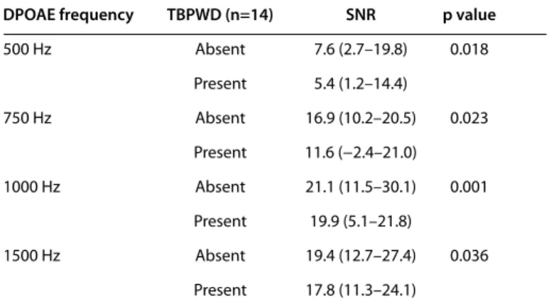
In 14 patients with unilateral TBPWD without additional anomalies, hearing thresholds at 2000 Hz were significantly higher for bone conductions at the defective side (25.71±16.4 dB vs. 21.07±12.43
dB; p=0.046). In these patients, we also investigated SNR at different DPOAE frequencies and found significant differences between the defective side and anatomically normal side (Table 1).
DISCUSSION
Defects are rarely seen in the thick, posterior portion of the tempo-ral bone. Ear infections, cholesteatoma, surgical procedures, trauma, tumors, radiotherapy, and congenital defects can be counted in etiol-ogy. Besides, arachnoid granulations, which may cause spontaneous otorrhea, can also play a role in the formation of bony defects [5]. Arachnoid granulations are formed by the overgrowth of the arachnoid membrane to the dural sinuses [6]. In addition to spontaneous otorrhea, these lesions may also cause meningeal tumors [7]. The defects formed by arachnoid granulations are usually lobulated with irregular borders, and the radiologic appearance of the defect is almost always sufficient to diagnose arachnoid granulations [8]. In our study, the radiologic ap-pearances of all defects were compatible with arachnoid granulations. Although unlikely, they could not be differentiated from the emissary veins of the posterior fossa because magnetic resonance and/or CT an-giography views were not available. Endolymphatic sac tumors may be another cause of TBPWD. However, in our study, none of the patients had calcification at the lesion borders, bony particles inside the lesion, and abnormal localization of the endolymphatic sac. As a result, the di-agnosis of endolymphatic sac tumor was ruled out.
Scarce data in the literature have shown that TBPWDs are usually associated with serious life-threatening complications such as spon-taneous otorrhea and meningitis [3]. In our study, we disclosed that these lesions may also cause common otological symptoms, i.e., tinnitus and vertigo. It is important because an underestimation of these common symptoms may cause the clinician to miss out this potentially life-threatening disease.
SSCD was present in four (17%) patients in our study. The incidence of dehiscence of the superior semicircular channel, which completes its ossification first, is between 3.6%–4.9% [9, 10]. The higher incidence of this anomaly in patients with TBPWD may support the hypothesis, implying that multiple defects may occur in case of discontinuation of ossification in the temporal bone at several steps in the postpar-tum period [11]. However, if we consider that TBPWD may occur be-cause of arachnoid granulations or idiopathic increases of the intra-cranial pressure at older ages, it may be said that erosion of the thin
79
Ezerarslan et al. Temporal Bone Posterior Wall Defect
Figure 2. Correlation between age and size of the TBPWD
r: 0.581; p: 0.004 Siz e of the TBPWD (mm) Age 20 4 3 2 1 0 40 60 80
Figure 3. a-c. Receiver-operating characteristics (ROC) curve analysis of the size of the defect and symptoms tinnitus (a), vertigo (b), and tinnitus (c) in patients without additional anomalies) Sensitivit y Sensitivit y Sensitivit y 1 - Specificity 0.0 0.0 0.0 1.0 0.8 0.6 0.4 0.2 0.0 1.0 0.8 0.6 0.4 0.2 0.0 1.0 0.8 0.6 0.4 0.2 0.0 0.2 0.4 0.6 0.8 1.0 0.2 0.4 0.6 0.8 1.0 0.2 0.4 0.6 0.8 1.0 Sensitivity: 0.67, Specificity: 0.77 (95% CI: 0.58-0.97) p=0.04 Sensitivity: 0.78, Specificity: 0.86 (95% CI: 0.67-0.99) p=0.006 Sensitivity: 0.80, Specificity: 0.82 (95% CI: 0.68-0.99) p=0.023 a b c 1 - Specificity 1 - Specificity
tegmen tympani with time has an increased possibility in causing TBPWD [12-15]. In our study, older patients had larger TBPWDs in size, which was consistent with this theory.
The phenomenon of conductive hyperacusis has been defined by Stenfelt et al. [16] in subjects with SSCDs. Patients with this disorder may hear their eyeball movements, their heart beat at night, the movement of their joints while jogging, or their own voice too loudly in the ear with superior semicircular canal dehiscence. This symptom can be very disturbing as well as a hindrance to the patients’ life. The cause of conductive hyperacusis is the negative bone conduction in certain subjects with SSCDs, which creates a higher sensitivity to bone-conducted sound at frequencies below 2 kHz. Interestingly, in our study, patients with TBPWD did not have conductive hyperacusis. A second defect in the bony conduction may prevent the develop-ment of conductive hyperacusis.
In our study, high jugular bulb was defined when the jugular bulb was more superior than the inferior annulus of the tympanic mem-brane. The incidence of high jugular bulbs varies among authors and studies because of the variation in its definition. It was found to be approximately 3% when the inferior annulus had been selected as the reference structure [17]. Again, we have found an increased inci-dence of high jugular bulbs (8%) in patients with TBPWD, which sup-ports the general knowledge implying that temporal bone anoma-lies commonly accompany each other.
In our study, the pattern of DPOAE decrease was similar to the pat-tern of patients who had tinnitus after exposure to noise [18, 19]. We thought that this phenomenon may be due to the lack of a barrier at the posterior temporal bone, which may cause an increased effect of the inner voice on the external hairy cells in the cochlea. However, further studies should be conducted to explain this finding.
For the first time in the literature, we defined clinical and audiological findings in patients with TBPWDs. These defects seem to cause more prevalent symptoms of vertigo and tinnitus and disturb the audio-logical characteristics of the patients.
Ethics Committee Approval: The approval of local Institutional Review board has been obtained (04.09.2013/2).
Informed Consent: Written informed consent has been obtained from all par-ticipants.
Peer-review: Externally peer-reviewed.
Author Contributions: Concept - H.E., S.K. Design - G.K.A., H.E.; Supervision - S.K., G.K.B.; Funding - H.E., S.K.; Materials - H.E., G.K.A.; Data Collection and/or Processing - H.E., S.K.; Analysis and/or Interpretation - H.E., G.K.A.; Literature Review - H.E., S.K.; Writing - H.E., G.K.A.; Critical Review - G.K.B., S.K.
Conflict of Interest: No conflict of interest was declared by the authors. Financial Disclosure: The authors declared that this study has received no financial support.
REFERENCES
1. Gacek R. Arachnoid granulation cerebrospinal fluid otorrhea. Ann Otol Rhinol Laryngol 1990; 99: 854-62. [CrossRef]
2. Yew M, Dubbs B, Tong O, Nager GT, Niparko JK, Tatlipinar A, et al. Arach-noid granulations of the temporal bone: a histologic study of dural and osseous penetration. Otol Neurotol 2011; 32: 602-9. [CrossRef]
3. Lee MH, Kim HJ, Lee IH, Kim ST, Jeon P, Kim KH. Prevalence and appear-ance of the posterior wall defects of the temporal bone caused by pre-sumed arachnoid granulations and their clinical significance: CT find-ings. AJNR Am J Neuroradiol 2008; 29: 1704-7. [CrossRef]
4. Jerger J. Clinical experience with impedance audiometry. Arch Otolaryn-gol 1970; 92: 311-24. [CrossRef]
5. Perry BP, Rubinstein JT. Meningitis due to acute otitis media and arach-noid granulations. Ann Otol Rhinol Laryngol 2000; 109: 877-9. [CrossRef]
6. Schuknecht HF, Gulya AJ. Anatomy of the temporal bone with surgical implications. Philadelphia: Lea & Febiger; 1986: 125-6.
7. al-Rodhan NR, Laws ER Jr. Meningioma: a historical study of the tumor and its surgical management. Neurosurgery 1990; 26: 832-46. [CrossRef]
8. Mukherji SK, Albernaz VS, Lo WW, Gaffey MJ, Megerian CA, Feghali JG, et al. Papillary endolymphatic sac tumors: CT, MR imaging, and angio-graphic findings in 20 patients. Radiology 1997; 202: 801-8. [CrossRef]
9. Crovetto M, Whyte J, Rodriguez OM, Lecumberri I, Martinez C, Eléxpuru J. Anatomo-radiological study of the superior semicircular canal dehis-cence radiological considerations of superior and posterior semicircular canals. Eur J Radiol 2010; 76: 167-72. [CrossRef]
10. Russo JE, Crowson MG, DeAngelo EJ, Belden CJ, Saunders JE. Posterior semicircular canal dehiscence: CT prevalence and clinical symptoms. Otol Neurotol 2014; 35: 310-4. [CrossRef]
11. Yu KK, Mukherji S, Carrasco V, Pillsbury HC, Shores CG. Molecular genetic advances in semicircular canal abnormalities and sensorineural hearing loss: a report of 16 cases. Otolaryngol Head Neck Surg 2003; 129: 637-46.
[CrossRef]
12. Cox KM, Lee DJ, Carey JP, Minor LB. Dehiscence of bone overlying the superior semicircular canal as a cause of an air-bone gap on audiometry: a case study. Am J Audiol 2003; 12: 11-6. [CrossRef]
13. Hirvonen TP, Carey JP, Liang CJ, Minor LB. Superior canal dehiscence: mechanisms of pressure sensitivity in a chinchilla model. Arch Otolaryn-gol Head Neck Surg 2001; 127: 1331-6. [CrossRef]
14. Lee MH, Kim HJ, Lee IH, Kim ST, Jeon P, Kim KH. Prevalence and appear-ance of the posterior wall defects of the temporal bone caused by pre-sumed arachnoid granulations and their clinical significance: CT find-ings. AJNR Am J Neuroradiol 2008; 29: 1704-7. [CrossRef]
15. Harvey RS, Hertzano R, Kelman SE, Eisenman DJ. Pulse-synchronous tin-nitus and sigmoid sinus wall anomalies: descriptive epidemiology and the idiopathic intracranial hypertension patient population. Otol Neuro-tol 2014; 35: 7-15. [CrossRef]
16. Stenfelt S, Goode RL. Bone-conducted sound: physiological and clinical aspects. Otol Neurotol 2005; 26: 1245-61. [CrossRef]
17. Wadin K, Thomander L, Wilbrand H. Effects of high jugular fossa and jug-ular bulb diverticulum on the inner ear. A clinical and radiologic investi-gation. Acta Radiol Diagn 1986; 27: 629-36. [CrossRef]
18. Satar B, Kapkin O, Ozkaptan Y. Evaluation of cochlear function in patients with normal hearing and tinnitus: a distortion product otoacoustic emis-sion study. Kulak Burun Bogaz Ihtis Derg 2003; 10: 177-82.
19. Ami M, Abdullah A, Awang MA, Liyab B, Saim L. Relation of distortion prod-uct otoacoustic emission with tinnitus. Laryngoscope 2008; 118: 712-7.
[CrossRef]
80
Int Adv Otol 2015; 11(1): 77-80
DPOAE frequency TBPWD (n=14) SNR p value
500 Hz Absent 7.6 (2.7–19.8) 0.018 Present 5.4 (1.2–14.4) 750 Hz Absent 16.9 (10.2–20.5) 0.023 Present 11.6 (−2.4–21.0) 1000 Hz Absent 21.1 (11.5–30.1) 0.001 Present 19.9 (5.1–21.8) 1500 Hz Absent 19.4 (12.7–27.4) 0.036 Present 17.8 (11.3–24.1)
Median (min–max) values are given for SNR values.
DPOAE: distortion product otoacoustic emission; TBPWD: temporal bone posterior wall defect; SNR: signal/noise ratio
Table 1. Signal noise ratio (SNR) comparisons between defective and non-defective sides at different DPOAE frequencies in patients with defect in the posterior wall of the temporal bone