Original Article
Circulating levels of Cyclophilin A in women with PCOS:
correlation with clinical and biochemical parameters
Akin Usta1, Mine Taskin1, Ozgur Baykan2, Ertan Adali1
1Department of Obstetrics and Gynecology, School of Medicine, Balikesir University, Balikesir, Turkey; 2Department
of Biochemistry, Balikesir Ataturk State Hospital, Balikesir, Turkey
Received October 16, 2017; Accepted May 16, 2018; Epub October 15, 2018; Published October 30, 2018 Abstract: Women with polycystic ovary syndrome (PCOS) are more likely to suffer from obesity, insulin resistance, and chronic low-grade inflammation than other women. Cyclophilin A (CyPA) is known as an inflammatory media-tor that is secreted by various types of cells in response to inflammamedia-tory stimuli. Previous studies have shown that immunohistochemical expressions and/or circulating levels of CyPA are high in many diseases that cause inflam-matory conditions in the body. This study aimed to evaluate serum levels of CyPA and their correlation with clinical and biochemical parameters of women with PCOS. In the study population, we analyzed 49 consecutive patients with PCOS and 30 age and body mass index (BMI)-matched non-PCOS healthy volunteers (Control group). PCOS was diagnosed using Rotterdam criteria. Serum CypA levels were measured using a CyPA ELISA Kit. The relationship between serum CyPA levels and the clinicopathological variables of PCOS were also evaluated. Average levels of CyPA were lower in PCOS subjects than the non-PCOS subjects (21.5±3.1 and 38.5±4.2, respectively, P=0.0015). Serum CyPA levels were significantly correlated with homeostasis model assessment-insulin resistance (HOMA-IR) and high sensitivity C-reactive protein (hsCRP) levels in the PCOS group (r=0.230, P=0.04 and r=0.302, P=0.006, respectively). There was no correlation between serum CyPA levels and other clinical and biochemical parameters. Our study demonstrates that patients with PCOS have lower circulating levels of CyPA than women with normal ovaries. Decreased CyPA levels may be related to increased insulin resistance in PCOS patients. Further research is needed to evaluate the association between CyPA and PCOS.
Keywords: Cyclophilin A, low-grade inflammation, hyperandrogenemia, insulin resistance
Introduction
Polycystic ovary syndrome (PCOS) is one of the most common complex endocrine pathologies. It is characterized by oligo/anovulation, hor-monal and/or clinical hyperandrogenism, and the appearance of polycystic ovaries on ultra-sound [1]. It affects 6-14% of women in repro-ductive age, and long-term complications include type 2 diabetes mellitus, cardiovascu-lar diseases (CVD), and infertility [2, 3]. Therefore, it is important to understand the molecular basis of the pathophysiology of this syndrome.
Previous studies have reported that various biomarker alterations are associated with low-grade inflammation, endothelial dysfunction, hyperandrogenemia, dyslipidemia, obesity, and insulin resistance in women with PCOS [4-6].
Cyclophilin A (CyPA), an 18 kDa protein, is a member of the immunophilin family. It is a well-known cellular protein that is present in various human tissue cells [7, 8]. Previous studies have shown that immunohistochemical expressions and/or circulating levels of CyPA are high in many disorders including diabetes mellitus, asthma, rheumatoid arthritis, abdominal aortic aneurism, CVD, and sepsis which couse inflam-matory conditions in the body [9-14].
A recent study conducted by Satoh reported that plasma CyPA levels were significantly high-er in patients with significant coronary stenosis than those without it. Moreover, they found that CyPA levels were correlated with a number of atherosclerotic risk factors associated with oxi-dative stress (OS) [11]. OS is generated by reac-tive oxygen species (ROS), which activate a pathway that induces CyPA secretion [15].
Secreted CyPA mediates cell proliferation and the migration of inflammatory cells in vascular smooth muscle cells (VSMCs) and endothelial cells [16, 17]. Furthermore, CyPA activates DNA synthesis, and it inhibits nitric oxide-induced apoptosis in VSMCs [18].
Women with PCOS are more likely than other women to have increased cardiovascular risk factors, such as hyperinsulinemia, abnormal plasma lipids, hypertension and endothelial dysfunction (ED), as well as increased levels of C-reactive protein (CRP), endothelin-1, and homocysteine [19]. Of the mentioned risk fac-tors in PCOS, ED is crucial in the development of CVD. ED is caused by imbalance between the production and bioavailability of endothelium-dependent relaxing factors and endothelium constricting factors, characterized as OS, which is associated with increased ROS generation and decreased antioxidant concentration. ED contributes to the increased risk of
atheroscle-A cross-sectional study was designed and con-ducted at Balikesir University, School of Medicine, Education and Research Hospital between January 2016 and December 2016 to evaluate the serum CyPA levels in women with PCOS. Eighty women (including 50 with PCOS and 30 age- and body mass index (BMI)-matched healthy volunteers) were included in the study population. All the women with PCOS were selected consecutively. After giving a blood sample, one of the participants in the PCOS group declared that she wanted to with-draw from the study. Thus, data of 79 partici-pants (49 PCOS subjects and 30 with non-PCOS subject [control group]) were evaluated in this study.
Patients who had other disorders with clinical features similar to PCOS, such as Cushing’s syndrome, congenital adrenal hyperplasia, and androgen-secreting tumors, were excluded from the study. Patients who had taken medi-Table 1. Clinical, hormonal, and biochemical characteristics of PCOS
and non-PCOS groups
Variables PCOS (n=49) Non-PCOS (n=30) P-Value
Age (years) 25.2±0.61 26.4±0.91 0.247a BMI (kg/m2) 26.4 (17.5-43.8) 24.2 (18.5-36.5) 0.091b WHR 0.79±0.01 0.74±0.01 0.001a Hirsutism score 5 (1-14) 1 (0-4) <0.001b Parental history of DM (%) 23/49 (46%) 8/30 (26.6%) 0.120c Parental history of CVD (%) 10/49 (20.4%) 10/30 (33.3%) 0.309c Smoking (%) 11/49 (22.4%) 2/30 (6.6%) 0.127c
Menstrual Cycle length (day) 39 (27-65) 28 (24-33) <0.001b
Systolic blood pressure (mmHg) 111 (89-160) 100 (92-121) 0.010b
Diastolic blood pressure (mmHg) 71 (61-114) 62 (52-85) <0.001b
CyPA (ng/mL) 21.5±3.1 38.5±4.2 0.0015a FSH (mIU/mL) 6.2±0.2 6.8±0.3 0.086a LH (mIU/mL) 8.1±0.7 5.4±0.4 0.008a E2 (pg/mL) 34.1±2.3 39.2±3.9 0.237b Total Testosteron (ng/dL) 1.2 (0.04-4.7) 0.1 (0.07-0.3) <0.001b Triglyceride (mg/dL) 97 (46-538) 66.5 (44-109) <0.001b Total Cholesterol (mg/dL) 176 (116-263) 173 (102-226) 0.302b LDL (mg/dL) 101 (45-185) 95 (39-138) 0.317b HDL (mg/dL) 52 (27-86) 63 (36-82) <0.001b HOMA-IR 3.4 (0.6-39.7) 1.4 (0.5-5.4) <0.001b hsCRP 3.5 (0.2-33.0) 1.2 (0.2-9.8) <0.001b
aIndependent samples t-test, bMann-Whitney test, cChi-squared test. Values: mean ± SEM or median (min-max). Note: Clinical, hormonal, and biochemical characteristics of our PCOS and non-PCOS groups. In PCOS patient significantly higher WHR, hirsutism score, menstruel cycle lenght, sistolic and diastolic blood pressure, HOMA-IR and hsCRP levels than non-PCOS patients.
rosis and CVD in insulin-resistant subjects with PCOS [20].
Therefore, in this study, we addressed whether CyPA is a potential bio-marker for determining cardiovascular risks in patients with PCOS. This study aimed to evaluate serum CyPA levels and their correlation with cl- inical and biochemical parameters of women with PCOS.
Materials and methods The investigation proto-col was approved by the Ethics Committee of Ba- likesir University, and all subjects gave their in- formed consent before participating in the stu- dy. The study protocols were in accordance with Helsinki Committee re- quirements.
cation for the previous 3 months, such as oral contraceptives, antilipidemic and/or antihyper-tensive medication, steroids, anti-diabetic medication, anticoagulants, or antiplatelet drugs, were also excluded from the study. PCOS was diagnosed using Rotterdam criteria [1]. The presence of hirsutism was evaluated using the Ferriman-Gallwey scoring system [21]. A score equal to or greater than 8 was defined as hirsutism. Physical and gynecologi-cal examinations, ultrasonography monitoriza-tion, and peripheral venous blood sampling were performed during day 2 or day 3 of the study participants’ menstrual cycles. After overnight fasting, blood samples were collect-ed from an antecubital vein. Serum was col-lected after centrifugation at 2500×g for 10 min, and it was stored at -80°C until biochemi-cal and hormonal assessment was underta- ken.
Indices
Waist-to-hip ratio (WHR) was defined as the ratio of the waist measurement to the hip mea-surement. BMI was defined as the mass in kilo-grams divided by the square of the body height in meters (kg/m2).
Biochemical evaluation
Serum CyPA levels were evaluated using com-mercially avaliable enzyme-linked
immunosor-was measured with chemiluminescent immu-noassay using an ADVIA Centaur XP (Siem- ens Healthcare Diagnostics, NY, USA). Levels of fasting insulin were determined using commer-cial kits and an automatic hormone analyzer (Beckman Coulter; Unicel DXI 600; Access Immunoassay System). Homeostasis model assessment for insulin resistance (HOMA-IR) was defined as (insulin x glucose)/405.
Statistical analysis
MedCalc Statistical Software Program version 17.2 (MedCalc, Belgium) was used for the sta-tistical analysis. The distribution of all variables in both the PCOS and non-PCOS groups was studied by describing the mean, median, range, and standard error of mean. The CyPA levels in the two groups were compared using the inde-pendent samples t-test. Receiver operator characteristic (ROC) curves were plotted to detect a cut-off value for the serum levels of CyPA levels to predict the levels for patients in both the PCOS and non-PCOS groups. A P-value of <0.05 was considered to be statistically significant.
Results
A total of 49 women with PCOS and 30 women without PCOS were included in the study. The clinical, hormonal, and biochemical character-istics of the women in the PCOS and non-PCOS groups are summarized in Table 1.
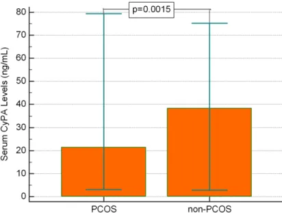
Figure 1. Average levels of serum CyPA of the two groups. Data represent-ed lower average levels of CyPA in PCOS group than in non-PCOS group (P=0.0015).
bent assay kits (Sunredbio, Shangai, China). Serum levels of follicle-stimulating hormo- ne (FSH), luteinizing hormo- ne (LH), estradiol (E2), thyroid-stimulating hormone (TSH), and total testosterone were determined using commercial-ly avaliable enzyme-linked im- munosorbent assay (ELISA) kits (eBioscience, Austria) on a diagnostic instrument (BioTek, ELx 800, USA). Levels of glu-cose, total cholesterol, low-density lipoprotein (LDL) cho-lesterol, and high-density lipo-protein (HDL) cholesterol were measured using commercially available kits on a chemistry AutoAnalyzer (Cobas Integra 800; Roche Diagnostics Gm- bH; Mannheim, Germany). Ser- um C-reactive protein (CRP)
blood pressure, the HOMA-IR and hsCRP levels in the PCOS group (P=0.0105, P=0.0001 and P=0.0005, respectively). There was no correla-tion between the serum CyPA levels and the other clinical and biochemical parameters in PCOS group. Also, there was no correlation between serum CyPA levels and clinical, bio-chemical and hormonal variables in non-PCOS group (Table 2).
The cut-off value of CyPA was 16.02 ng/mL for the prediction of PCOS in our study population
ROC curves were plotted to detect a cut-off value for the serum CyPA levels to the predict levels in the PCOS and non-PCOS groups. The ROC analysis demonstrated that the cut-off value of the serum CyPA levels was 16.02 ng/ mL with a sensitivity of 63.3% and a specificity of 80% (Figure 2). The majority of women with PCOS had serum CyPA levels below this cut-off
Serum levels of CyPA were higher in PCOS patients
As shown in Table 1, the serum CyPA levels were significantly lower in the PCOS group than the non-PCOS group (21.5±3.1 and 38.5±4.2, respectively, P=0.0015) (Figure 1).
Biochemically, PCOS is characterized by increased insulin resistance, hyperandr-ogenemia, and low-grade inflammation
Regarding the other hormonal and bio-chemical parameters, median levels of total testosterone, triglyceride, HOMA-IR and high sensitivity C-reactive pro- tein (hsCRP) were significantly higher in the PCOS group than the non-PCOS group (P<0.001, P<0.001, P<0.001 and P<0.001, respectively). However, the median levels of HDL were significantly lower in the PCOS group (P<0.001). Serum levels of FSH, E2, total choles-terol, and LDL were similar between the two groups.
Serum levels of CyPA strongly corre-lated with presence of hypertension, insulin resistance and inflammation in PCOS patients
The information presented in Table 2 shows that the serum CyPA levels were significantly correlated with systolic Table 2. Correlation between serum levels of CypA and
clinical, biochemical and hormonal variables of patients with and without PCOS
Variables PCOSCyPA Non-PCOSCyPA
Age (years) 0.145 P=0.3202 P=0.58770.103 BMI (kg/m2) -0.0564 P=0.7001 P=0.38250.165 WHR -0.0276 P=0.8506 P=0.75000.0607 Hirsutism score 0.148 P=0.3094 P=0.63270.0909
Systolic blood pressure (mmHg) 0.362
P=0.0105 P=0.67130.0808
Diastolic blood pressure (mmHg) 0.173
P=0.2356 P=0.67340.0802 Total testosterone (ng/dL) 0.154 P=0.2921 P=0.17790.253 Triglyceride (mg/dL) -0.114 P=0.4337 P=0.27130.207 Total cholesterol (mg/dL) -0.189 P=0.1925 P=0.94600.0129 LDL (mg/dL) -0.176 P=0.2272 P=0.5863-0.103 HDL (mg/dL) 0.0605 P=0.6798 P=0.1547-0.266 HOMA-IR 0.520 P=0.0001 P=0.43370.148 hsCRP 0.479 P=0.0005 P=0.27080.208 Spearman’s rank correlation.
There was no difference in age and BMI (P=0.247 and P=0.091, respectively) between the two groups. Parental history of DM and CVD and smoking rates were similar between the women in the PCOS and non-PCOS groups (P=0.120, P=0.309, and P=0.127, respective- ly).
Clinically, PCOS is characterized by an in-crease in WHR, a prolonged duration of the menstrual cycle and high blood pressure
WHR and hirsutism scores were significantly higher in the PCOS group than the non-PCOS group (P=0.001, and P<0.001, respectively). Menstrual cycle length was longer in the PCOS group than the non-PCOS group (P<0.001). Median systolic and diastolic blood pressures were higher in the PCOS group than the non-PCOS group (P=0.010, and P≤0.001, respec- tively).
point. However, it was observed that the distri-bution range of the serum CyPA levels was wide. Some of the patients in the PCOS group had extremely high CyPA levels, but the only common common characteristics they shared were insulin resistance and high CRP levels. Discussion
In the present study, we evaluated clinical and biochemical variables in PCOS subjects and compared them with age- and BMI-matched non-PCOS subjects. Our clinical and biochemi-cal parameters confirmed that women with PCOS have higher triglyceride, HOMA-IR, total testosterone, and hsCRP levels, higher systolic and diastolic blood pressure readings, higher WHR and lower HDL values. We also found that serum CyPA levels were significantly lower in the PCOS group than the non-PCOS group. Also, correlation analysis showed that there was a positive correlation between serum CyPA levels and systolic blood pressure, HOMA-IR and hsCRP levels in patients with PCOS. To our knowledge, this is the first study to examine cir-culating levels of CyPA in women with PCOS. PCOS is a complex endocrine and metabolic disease associated with obesity, insulin resis-tance and compensatory hyperinsulinemia,
and chronic low-grade inflammation [22, 23]. Various biomarker alterations are associated with insulin resistance and low-grade inflam-mation in PCOS [4-6]. Insulin resistance and compensatory hyperinsulinemia in PCOS seem to have a stimulatory effect on chronic low-grade inflammation. Recent studies have also shown that obesity is associated with insulin rezistance and low-grade inflammation in PCOS [24].
CyPA, a multifunctional protein, is known to be an inflammatory mediator that is secreted from various types of cells in response to inflamma-tory stimuli, such as hypoxia, OS and infection, and many previous studies have shown that expression and/or circulating levels of CyPA are higher in disorders related to inflammatory con-ditions [12, 16, 18, 25]. Billich reported that CyPA levels increased in the synovial fluids of patients with rheumatoid arthritis (RA) [10]. Tegeder reported that CyPA activity was significantly higher in patients with severe sep-sis than in healthy subjects [9]. Stemmy showed higher concentrations of extracellular CyPA in the chronic phase of asthma using a murine model [13]. Ramachandra reported that in comparison to the non-diabetic population, patients with type 2 diabetes mellitus have higher circulating levels of CyPA [12]. Yan reported that serum CyPA concentrations in unstable angina and acute myocardial infarc-tion subjects were significantly higher than those in patients with stable angina and in the controls [26]. A recent study by Nigro found that CyPA expression was significantly higher in the atherosclerotic plaque of the arterial wall, and they concluded that CyPA is an inflammatory mediator that promotes atherosclerosis [14]. In the present study, contrary to the initial expectation, we found lower CyPA levels in the PCOS subject than non-PCOS subjects. This result may be due to differences in complex molecular mechanisms, as well as the severity of the inflammation in different inflammatory conditions. In most previous studies, increased levels of CyPA were found in severe inflamma-tory conditions, such as RA, sepsis, asthma, type 2 diabetes mellitus, unstable angina and acute myocardial infarction [9-14]. In contrast, the results of the study showed that women in the PCOS group had relatively low grade inflam-mation accompanied by insulin resistance and
Figure 2. ROC analysis of study population. ROC analysis demonstrated that the cut-off value of the serum CyPA levels was 16.02 ng/mL with the sensi-tivity of 63.3% and specificity of 80%.
mild hsCRP elevation and these results are compatible the findings reported in previous PCOS studies [22, 23]. Furthermore, we found that serum CyPA levels were positively corre-lated with systolic blood pressure, the HOMA-IR and hsCRP levels in patients with PCOS. These results indicate serum CyPA levels tend to ele-vate with increased severity of inflammatory conditions in patients with PCOS.
Additionally, differences between the sociode-mographic features, such as age, gender, and BMI, and different sample sizes of studied pop-ulations are potential confounding factors that can cause these conflicting results. For exam- ple, a study by Li reported that the expression of CyPA in skin tissue increased with aging [27]. Similarly, a study comparing young rats with older rats reported that, CyPA expression was significantly higher in older rats [28]. In our study, for the reduced possibility of errors, the women in the PCOS group and the non-PCOS group were matched according to age and BMI. Low-grade inflammation in PCOS is associated with an increase in plasma levels of hsCRP [29, 30]. It is known that hsCRP is a simple inflam-matory biomarker, and it reflects future CVD risk [31]. We found that hsCRP levels in the women in the PCOS group were significantly higher than the women in the non-PCOS group, and our current results correspond to the find-ings reported in previous studies [29, 30]. According to our results, the women in the PCOS group are more likely to have a future risk of CVD than the women in the non-PCOS group. Previous studies have reported that the pres-ence of dyslipidemia is the most common met-abolic co-morbidity in women with PCOS, and a lower HDL cholesterol level is the most frequent lipid abnormality in PCOS patients [32]. Lower HDL poses a cardiac risk even if other choles-terol levels are normal. In the present study, women in the PCOS group had lower HDL cho-lesterol levels than women in the non-PCOS group. Our present results are consistent with the findings reported in previous studies [33]. HDL cholesterol is referred to as good choles-terol because HDL particles can prevent ath-erosclerosis of the walls of blood vessels. Based on our results, the women in the PCOS group are at high risk for future atherosclerotic disease.
Regarding hyperandrogenism and insulin resis-tance, we found that women in the PCOS group had higher total testosterone and HOMA-IR lev-els than women in the non-PCOS group. Recent reports have confirmed these results [6]. The mechanism underlying hyperandrogenism and insulin resistance in PCOS patients is associat-ed with an abnormal activation of the ERK1/2 pathway [34]. Recent studies have reported an alteration in ERK1/2 pathway function in skel-etal muscle cells, in granulosa cells, and in theca cells for women with PCOS. These find-ings implicate abnormally lower ERK1/2 path-way activation in the pathogenesis of insulin resistance and excessive ovarian androgen production in PCOS patients [34, 35].
Interestingly, an animal model study conducted by Satoh found that, after complete ligation of arteria carotis, the intimal, medial, and adventi-tial thickening were significantly lower in CyPA knockout (CyPA-/-) mice than in wild-type (WT)
mice and mice overexpressing CyPA specifically in VSMC (VSMC-Tg). They also found that ERK1/2 activation and Ki67 cells were signifi-cantly decreased in the CyPA-/- mice [36]. These
studies indicate that lower CyPA levels have a protective effect against tissue damage in inflammatory conditions. Additionally, in our previous study, we found that there were no sig-nificant differences in carotis intima media thickness between the PCOS group and the control group as reported in other previous studies [37, 38]. These results might be associ-ated with lower levels of CyPA.
During the inflammatory process, CyPA medi-ates cell proliferation and migration in VSMCs and adhesion molecule expression in endothe-lial cells [11, 15, 18]. This proliferation is accrued by activation of the ERK1/2 pathway [16]. Thus, CyPA can stimulate DNA synthesis, and it inhibits nitric oxide induced apoptosis in VSMCs [18]. In addition, previous tumor cell line and culture medium studies have shown that secreted CyPA poses mitogenic activity on tumor cells via CD147. It is the only known sig-naling receptor for secreted CyPA, and blocking the secreted CyPA/CD147 interaction by mono-clonal antibody against CD147 significantly suppresses the effect of secreted CyPA on cell proliferation and ERK1/2 activation in cells. CyPA was found to have a positive correlation with the phosphorilation of ERK1/2, and
knock-down of CyPA inhibited phosphorylation of ERK1/2 in cells [39]. These results suggest that there may be a possible relationship between lower levels of CyPA and abnormal ERK1/2 activation. In light of these studies, we extended our research to investigate the inter-actions between CyPA/CD147 and ERK1/2 pathway in PCOS patients.
This present study has some limitations. It used a cross-sectional study design and it had a relatively small number of the participants. However, it is the first study to investigate the serum CyPA levels in patients with PCOS using a multivariable and age- and BMI-matched study design.
In conclusion, the results of the study have shown that women with PCOS have lower circu-lating levels of CyPA than women with normal functioning ovaries. The low serum CyPA levels in the women in the PCOS group suggest that there may be a possible defect in the produc-tion of CyPA within the patients’ cell or secreted out of their cells. Decreased CyPA levels in PCOS may be associated with abnormal activa-tion of ERK pathway. Clearly, further studies are necessary to test this hypothesis.
Acknowledgements
The study was supported by Scientific Invest- igations Foundation of Balikesir University (Project number: BAP. 2016. 0001).
Disclosure of conflict of interest None.
Address correspondence to: Dr. Akin Usta, Depart- ment of Obstetrics and Gynecology, University of Balikesir, Faculty of Medicine, Cagis Campusu Bigadic yolu 17. km, Altıeylul/Balikesir 10345, Tur- key. Tel: +905057956560; Fax: +902666121010; E-mail: [email protected]
References
[1] Rotterdam ESHRE/ASRM-Sponsored PCOS Consensus Workshop Group. Revised 2003 consensus on diagnostic criteria and long-term health risks related to polycystic ovary syn-drome. Fertil Steril 2004; 81: 19-25.
[2] Azziz R, Woods KS, Reyna R, Key TJ, Kno- chenhauer ES and Yildiz BO. The prevalence and features of the polycystic ovary syndrome
in an unselected population. J Clin Endocrinol Metab 2004; 89: 2745-2749.
[3] Sirmans SM and Pate KA. Epidemiology, diag-nosis, and management of polycystic ovary syndrome. Clin Epidemiol 2013; 6: 1-13. [4] Diamanti-Kandarakis E, Alexandraki K, Piperi
C, Protogerou A, Katsikis I, Paterakis T, Lekakis J and Panidis D. Inflammatory and endothelial markers in women with polycystic ovary syn-drome. Eur J Clin Invest 2006; 36: 691-697. [5] Karadeniz M, Erdogan M, Tamsel S, Zengi A,
Alper GE, Caglayan O, Saygili F and Yilmaz C. Oxidative stress markers in young patients with polycystic ovary syndrome, the relation-ship between insulin resistances. Exp Clin Endocrinol Diabetes 2008; 116: 231-235. [6] Bayram F, Kocer D, Ozsan M and Muhtaroglu
S. Evaluation of endothelial dysfunction, lipid metabolism in women with polycystic ovary syndrome: relationship of paraoxonase 1 activ-ity, malondialdehyde levels, low-density lipo-protein subfractions, and endothelial dysfunc-tion. Gynecol Endocrinol 2012; 28: 497-501. [7] Ryffel B, Woerly G, Murray M, Eugster HP and
Car B. Binding of active cyclosporins to cy-clophilin A and B, complex formation with calci-neurin A. Biochem Biophys Res Commun 1993; 194: 1074-1083.
[8] Tsai SF, Su CW, Wu MJ, Chen CH, Fu CP, Liu CS and Hsieh M. Urinary cyclophilin a as a new marker for diabetic nephropathy: A cross-sec-tional analysis of diabetes mellitus. Medicine (Baltimore) 2015; 94: e1802.
[9] Tegeder I, Schumacher A, John S, Geiger H, Geisslinger G, Bang H and Brune K. Elevated serum cyclophilin levels in patients with severe sepsis. J Clin Immunol 1997; 17: 380-386. [10] Billich A, Winkler G, Aschauer H, Rot A and
Peichl P. Presence of cyclophilin A in synovial fluids of patients with rheumatoid arthritis. J Exp Med 1997; 185: 975-980.
[11] Satoh K, Fukumoto Y, Sugimura K, Miura Y, Aoki T, Nochioka K, Tatebe S, Miyamichi-Yamamoto S, Shimizu T, Osaki S, Takagi Y, Tsuburaya R, Ito Y, Matsumoto Y, Nakayama M, Takeda M, Takahashi J, Ito K, Yasuda S and Shimokawa H. Plasma cyclophilin A is a novel biomarker for coronary artery disease. Circ J 2013; 77: 447-455.
[12] Ramachandran S, Venugopal A, Kutty VR, A V, G D, Chitrasree V, Mullassari A, Pratapchandran NS, Santosh KR, Pillai MR and Kartha CC. Plasma level of cyclophilin A is increased in pa-tients with type 2 diabetes mellitus and sug-gests presence of vascular disease. Cardiovasc Diabetol 2014; 13: 38.
[13] Stemmy EJ, Balsley MA, Jurjus RA, Damsker JM, Bukrinsky MI and Constant SL. Blocking cyclophilins in the chronic phase of asthma
re-duces the persistence of leukocytes and dis-ease reactivation. Am J Respir Cell Mol Biol 2011; 45: 991-998.
[14] Nigro P, Satoh K, O’Dell MR, Soe NN, Cui Z, Mohan A, Abe J, Alexis JD, Sparks JD and Berk BC. Cyclophilin A is an inflammatory mediator that promotes atherosclerosis in apolipopro-tein E-deficient mice. J Exp Med 2011; 208: 53-66.
[15] Suzuki J, Jin ZG, Meoli DF, Matoba T and Berk BC. Cyclophilin A is secreted by a vesicular pathway in vascular smooth muscle cells. Circ Res 2006; 98: 811-817.
[16] Jin ZG, Lungu AO, Xie L, Wang M, Wong C and Berk BC. Cyclophilin A is a proinflammatory cy-tokine that activates endothelial cells. Arterioscler Thromb Vasc Biol 2004; 24: 1186-1191.
[17] Damsker JM, Bukrinsky MI and Constant SL. Preferential chemotaxis of activated human CD4+ T cells by extracellular cyclophilin A. J Leukoc Biol 2007; 82: 613-618.
[18] Jin ZG, Melaragno MG, Liao DF, Yan C, Haendeler J, Suh YA, Lambeth JD and Berk BC. Cyclophilin A is a secreted growth factor in-duced by oxidative stress. Circ Res 2000; 87: 789-796.
[19] Fauser BC, Tarlatzis BC, Rebar RW, Legro RS, Balen AH, Lobo R, Carmina E, Chang J, Yildiz BO, Laven JS, Boivin J, Petraglia F, Wijeyeratne CN, Norman RJ, Dunaif A, Franks S, Wild RA, Dumesic D and Barnhart K. Consensus on women's health aspects of polycystic ovary syndrome (PCOS): the Amsterdam ESHRE/ ASRM-Sponsored 3rd PCOS Consensus Workshop Group. Fertil Steril 2012; 97: 28-38. e25.
[20] Paradisi G, Steinberg HO, Hempfling A, Cronin J, Hook G, Shepard MK and Baron AD. Polycystic ovary syndrome is associated with endothelial dysfunction. Circulation 2001; 103: 1410-1415.
[21] Ferriman D and Gallwey JD. Clinical assess-ment of body hair growth in women. J Clin Endocrinol Metab 1961; 21: 1440-1447. [22] Pandey V, Singh A, Krishna A, Pandey U and
Tripathi YB. Role of oxidative stress and low-grade inflammation in letrozole-induced poly-cystic ovary syndrome in the rat. Reprod Biol 2016; 16: 70-77.
[23] Puder JJ, Varga S, Kraenzlin M, De Geyter C, Keller U and Muller B. Central fat excess in polycystic ovary syndrome: relation to low-grade inflammation and insulin resistance. J Clin Endocrinol Metab 2005; 90: 6014-6021. [24] Repaci A, Gambineri A and Pasquali R. The role
of low-grade inflammation in the polycystic ovary syndrome. Mol Cell Endocrinol 2011; 335: 30-41.
[25] Kao HW, Lee KW, Chen WL, Kuo CL, Huang CS, Tseng WM, Liu CS and Lin CP. Cyclophilin A in ruptured intracranial aneurysm. A prognostic biomarker. Medicine (Baltimore) 2015; 94: e1683.
[26] Yan J, Zang X, Chen R, Yuan W, Gong J, Wang C and Li Y. The clinical implications of increased cyclophilin A levels in patients with acute coro-nary syndromes. Clin Chim Acta 2012; 413: 691-695.
[27] Li J, Xie H, Yi M, Peng L, Lei D, Chen X and Jian D. Expression of cyclophilin A and CD147 dur-ing skin agdur-ing. Zhong Nan Da Xue Xue Bao Yi Xue Ban 2011; 36: 203-211.
[28] Chen J, Rider DA and Ruan R. Identification of valid housekeeping genes and antioxidant en-zyme gene expression change in the aging rat liver. J Gerontol A Biol Sci Med Sci 2006; 61: 20-27.
[29] Turkcuoglu I, Kafkasli A, Meydanli MM, Ozyalin F and Taskapan C. Independent predictors of cardiovascular risk in polycystic ovarian syn-drome. Gynecol Endocrinol 2011; 27: 915-919.
[30] Ersoy AO, Tokmak A, Ozler S, Oztas E, Ersoy E, Celik HT, Erdamar H and Yilmaz N. Are progran-ulin levels associated with polycystic ovary syn-drome and its possible metabolic effects in adolescents and young women? Arch Gynecol Obstet 2016; 294: 403-409.
[31] Toulis KA, Goulis DG, Mintziori G, Kintiraki E, Eukarpidis E, Mouratoglou SA, Pavlaki A, Stergianos S, Poulasouchidou M, Tzellos TG, Makedos A, Chourdakis M and Tarlatzis BC. Meta-analysis of cardiovascular disease risk markers in women with polycystic ovary syn-drome. Hum Reprod Update 2011; 17: 741-760.
[32] Essah PA, Nestler JE and Carmina E. Differences in dyslipidemia between American and Italian women with polycystic ovary syn-drome. J Endocrinol Invest 2008; 31: 35-41. [33] Silfen ME, Denburg MR, Manibo AM, Lobo RA,
Jaffe R, Ferin M, Levine LS and Oberfield SE. Early endocrine, metabolic, and sonographic characteristics of polycystic ovary syndrome (PCOS): comparison between nonobese and obese adolescents. J Clin Endocrinol Metab 2003; 88: 4682-4688.
[34] Rajkhowa M, Brett S, Cuthbertson DJ, Lipina C, Ruiz-Alcaraz AJ, Thomas GE, Logie L, Petrie JR and Sutherland C. Insulin resistance in poly-cystic ovary syndrome is associated with de-fective regulation of ERK1/2 by insulin in skel-etal muscle in vivo. Biochem J 2009; 418: 665-671.
[35] Lan CW, Chen MJ, Tai KY, Yu DC, Yang YC, Jan PS, Yang YS, Chen HF and Ho HN. Functional microarray analysis of differentially expressed
genes in granulosa cells from women with polycystic ovary syndrome related to MAPK/ ERK signaling. Sci Rep 2015; 5: 14994. [36] Satoh K, Matoba T, Suzuki J, O’Dell MR, Nigro
P, Cui Z, Mohan A, Pan S, Li L, Jin ZG, Yan C, Abe J and Berk BC. Cyclophilin A mediates vas-cular remodeling by promoting inflammation and vascular smooth muscle cell proliferation. Circulation 2008; 117: 3088-3098.
[37] Taskin MI, Bulbul E, Adali E, Hismiogullari AA and Inceboz U. Circulating levels of obestatin and copeptin in obese and nonobese women with polycystic ovary syndrome. Eur J Obstet Gynecol Reprod Biol 2015; 189: 19-23.
[38] Buyukkaya R, Besir FH, Yazgan S, Karatas A, Kose SA, Aydin Y and Erdogmus B. The evalua-tion of carotid intima-media thickness and vis-ceral obesity as an atherosclerosis predictor in newly-diagnosed polycystic ovary syndrome. Clin Ter 2014; 165: e6-11.
[39] Obchoei S, Weakley SM, Wongkham S, Wongkham C, Sawanyawisuth K, Yao Q and Chen C. Cyclophilin A enhances cell prolifera-tion and tumor growth of liver fluke-associated cholangiocarcinoma. Mol Cancer 2011; 10: 102.