Akut İskemik İnmede Damar Okluzyonu, NIHSS Skoru (National Institues Of
Healt Stroke Scale Scores) İle Tutulan Hemisfer Arasındaki İlişki
Relationship Between NIHSS score (National Institues Of Healt Stroke Scale Scores), Vessel Occlusion And Affected Hemisphere In Acute Ischemic Stroke
Muhammed Nur Ögün1, Bahar Aksay Koyuncu3, Özlem Güngör Tuncer4, Gökhan Evcili1, Reha
Tolun3, Yakup Krespi2
1Derince Eğitim Ve Araştırma Hastanesi, Nöroloji Kliniği, Kocaeli 2İstanbul Memorial Hastanesi, Nöroloji Kliniği, İstanbul 3İstanbul Hizmet Hastanesi, Nöroloji Kliniği, İstanbul
4İstanbul Florence Nightingale Hastanesi, Nöroloji Kliniği, İstanbul
Özet Abstract
GİRİŞ ve AMAÇ: AMAÇ: Ulusal İnme Sağlık Ölçeği Skalası (NIHSS) akut iskemik inmenin şiddetini belirlemede kullanılır. Ancak dil fonksiyonu gibi sol hemisfer fonksiyonlarının, ihmal gibi sağ hemisfer fonksiyonlarına göre daha yüksek NIHSS skoru teşkil ettiği bilinmektedir. Bu durum sağ hemisfer inmeli düşük NIHSS skorlu hastaların tedaviden dışlanmasına sebep olabilir. Yüksek NIHSS skoru genellikle intrakranyal arter okluzyonu ile ilişkilidir, ancak tutulan hemisferin (Sağ ya da sol hemisfer inmesi) damar okluzyonunu ön gördüren NIHSS skoruna etkisi bilinmemektedir. Çalışmamızda inmenin ilk 12 saatinde başvuran hastaların NIHSS skoru ve arteriyel okluzyon varlığı değerlendirildi. Bu çalışmanın amacı akut intrakranyal MR anjiografi ile belirlenen damar okluzyonunun, NIHSS skoru ile ilişkisinin sağ ve sol taraflı inmeli hastalar arasında farklı olup olmadığının araştırmaktır. YÖNTEM ve GEREÇLER: Bu çalışma, 2006-2009 yılları arasında Florence Nigtingale İnme Merkezi’ne inme başlangıcından 12 saat içinde başvuran ve DWI MR ve İntrakranyal MR anjiografi yapılmış olan 139 hastanın verilerinden retrospektif olarak yapıldı. NIHSS skoru görüntülemenin hemen öncesinde bir Nöroloji hekimi tarafından kaydedildi. İntrakranyal MR anjiografi incelemeleri hastaların klinik verilerinden habersiz bir Nörolog ve Nöroradyolog
tarafından yapıldı.
BULGULAR: Mean (± SD) NIHSS skoru 9,7 ± 6,7 idi. ROC Eğrisi ile arter tıkanıklığını ön gördüren NIHSS Cut-Off değeri sağ ve sol taraflı inme arasında hastalarda benzerdi. Sol taraf inmeli hastalarda damar okluzyonunu ön gördüren Cut-off noktası 8 (duyarlılık % 83, özgüllük % 84) idi. Sağ taraf inmeli hastalarda damar okluzyonunu ön gördüren Cutoff noktası 9 (Duyarlılık % 72, özgüllük % 72) idi.
TARTIŞMA ve SONUÇ: SONUÇ: Arter tıkanıklığını ön gördürmede NIHSS skorunun Cut-Off değeri sağ ve sol taraflı (Sağ 9, Sol 8) inme arasında hastalarda benzerdi. Tüm grup göz önüne alındığında arteriyel okluzyonu ön gördüren NIHSS skoru 8 düzeyinde saptandı.
INTRODUCTION: The NIH Stroke Scale (NIHSS) is used to assess acute ischemic stroke severity and outcome, however, it can award more points for tests of presumed left-hemisphere function, such as language, than for tests of right-hemisphere function, such as neglect. This difference may be important if a low NIHSS score is used to exclude patients with right-hemisphere stroke from clinical trials or established treatments. High NIHSS scores are usually associated with arterial occlusion but it is unknown what the effect of side of stroke lesion to clinical evaluation in this association is. We tested the NIHSS scores as an instrument to determine arterial occlusion within 12 hours of stroke onset. The aim of this study was to investigate whether the relationship between acute NIHSS score and arteral occlusion as determined by acute MR angiography differs between right- and left-sided stroke. METHODS: This was a retrospective study of 139 patients with acute stroke seen at Florence Nightingale Stroke Center who underwent an MRI examination and NIHSS within 12 hours of stroke onset. NIHSS score was recorded prospectively by the admitting stroke fellow at the time of acute presentation, immediately preceding imaging. Computerized volumetric analysis of the MRI lesions, intracranial MR anjiography evaluation was performed by investigators blinded to clinical data.
RESULTS: Mean (±SD) NIHSS score was 9,7 ± 6,7 Receiver operating characteristic curves showed that the validity of NIHSS in predicting arterial occlusion was smilar in patients between left and right-sided stroke. The best cut-off point in patients evaluated with left-sided stroke was an NIHSS of 8 (83 % sensitivity, specificity 84 %), while in patients evaluated after with right-sided stroke best cut-off point was 9 (sensitivity 72 %, specificity 72 %).
DISCUSSION AND CONCLUSION: The validity of NIHSS in predicting arterial occlusion was smilar in patients between left and right-sided (Right 9, Left 8) stroke. S in predicting arterial occlusion was smilar in patients between left and right-sided (Right 9, Left 8) stroke. An NIHSS score of 8 seems to represent the cut-off for discriminating between patients with arterial occlusion and patients without.
Anahtar Kelimeler: Ulusal İnme Sağlık Ölçeği Skalası
(NIHSS), Akut İskemik İnme KeywordsScale Scores (NIHSS), Acute Ischemic Stroke: National Institues of Health Stroke INTRODUCTION
The NIHSS (The National Institute of Health Stroke Scale) is one of the most widely used and approved measurement tool for assessing the clinical severity of acute ischemic stroke. The NIHSS is strongly related to both functional
outcome and determining suitable candidates for thrombolytic therapy in patients with acute ischemic stroke. (1) The NIHSS has a potential weakness that it has a greater emphasis on deficits associated with left-hemisphere lesions
hemisphere stroke. (2)
The NIHSS may be less sensitive to right-hemisphere than left-right-hemisphere stroke. Presumably, it is based on the testing of cognitive deficits associated with right-hemisphere stroke is more difficult than testing aphasia. The NIHSS score is not more favorable in determining the ultimate functional outcome of patients with right-hemisphere stroke than for left-hemisphere stroke. (3)
The NIHSS score may cause a bias against patients with a right-hemisphere stroke when it is used to determine the enrolment in clinical trials or in clinical decision-making protocols. The volume of right-hemisphere strokes as determined by a chronic CT scan has been shown to be larger than the volume of left-hemisphere strokes for a given NIHSS score; however, the clinical implications of these findings remain uncertain.
Differences between hemispheres in relation to an NIHSS score and imaging parameters during the hyperacute phase of a stroke might play a greater role in clinical decision making. (4)
The aim of this study is to determine whether the relationship between an acute NIHSS score and the artery occlusion found through an acute MR angiography could indicate the differences between right- and left-sided strokes.
SUBJECTS AND METHODS
In this study, we made use of the data on the patients that were recorded prospectively and consecutively in the acute stroke database, between 2006 and 2009, at the Florence Nightingale Stroke Center. Among those registered in this database, the patients who were eligible to be included in this study is based using the following criteria: The patients with middle cerebral artery stroke; the patients who underwent an MRI (DWI and MRA) examination and NIHSS within 12 hours of stroke onset
infarction (simultaneously anterior and posterior circulation infarcts; hemispheric infarcts, chronic infarcts, the ultimate diagnose was transient ischemic attack (TIA); hemorrhagic transformation in initial MRI imaging) were excluded from the study. The data, demographics, stroke risk factors, time of stroke onset (determined as the time of onset of symptoms or in patients with wake-up stroke (the time of last seen healthy), the NIHSS score recorded by a neurologist, the type and the time of imaging, the anatomic localization of the stroke and also including the predicted mechanism of stroke (TOAST classification was used to classify the etiology of the stroke) were recorded prospectively for each patient. The study was performed in accordance with the local ethics committee, informed consent for data collection was obtained from all participants and we adhered to the Declaration of Helsinki.
Imaging Protocol
Images of patients were acquired using Siemens Sonata 1.5 Tesla (T) and GE-HD 3-T MRI device echo-planar system from the Radiology Department at the Florence Nightingale Stroke Center. All lesions were classified by including an arterial territory; anterior circulation: anterior cerebral artery, middle cerebra artery (proximal, distal and lentriculostriate), single penetrant artery in profound white matter, anterior choroidal artery territory and border zone territories; posterior circulation: posterior cerebral artery, basilar artery, cerebellar arteries (superior, anterior inferior and posterior inferior) and cerebellar border zone territories. In addition, patients with multiple infarction in cortical and/or internal border zone were verified.
Determining the Artery Occlusion and Artery Stenosis
Intracranial MR angiography studies were performed on a 3D TOF sequence. Reformatted images were interpreted visually and the percentage of vascular stenosis was determined. (Figure-1) A presence of >50% artery stenosis or arterial occlusion in the right and left anterior circulation on the intracranial MR angiography were recorded, respectively. The anterior circulation artery segments were determined as ICA distal, ICA supraclinoid, MCA M1 proximal or mid-striate, MCA M1 distal mid-striate, MCA M2 superior and MCA M2 inferior, MCA distal, ACA A1, ACA A2. The posterior circulation artery segments were determined to be proximal of PICA right and left vertebral artery, distal of PICA, V3, proximal of AICA basilar, mid-basilar, and distal of AICA basilar, right and left PCA P1. The percentage of arterial stenosis were recorded as symptomatic 50 –69%, 70 – 99% and total occlusion, respectively. The study cases were separated into two groups: as patients with symptomatic artery stenosis and those without stenosis.
Figure-1: A) 70 –99% stenosis in distal of left MCA M1 B) Occlusion in inferior division of left MCA
Statistical Analysis
The SPSS 18.0 software (SPSS Inc., Chicago, Illinois, USA) was used for the statistical analysis of the study. Student‘s t-test was used for continuous variables
and non-parametric data, and the Wilcoxon rank-sum test used for comparison. The ROC (receiver operating characteristic) curve was used to determine the cut-off NIHSS score predicting the artery occlusion. The significance of the power of the cut-off NIHSS scores for both hemisphere determined by the ROC curve were contrasted with a z-test (MedCalc®
statistical software was used).
RESULTS
Among the 289 acute stroke patients, admitted to our clinic between 2006 and 2009, 141 of them were excluded from the study owing to following reasons: the ultimate clinical diagnosis of 14 of them was TIA; in 14 of them cranial MR imaging could not be performed (presence of cardiac pacemaker; early period of valve replacement operation history; vital instability; etc.), posterior circulation infarction in 53 of them, chronic infarction in 31 of them, multiple infarcts in 20 of them, isolated ACA infarction in nine of them. The intracranial MR angiographies of nine of the remaining 148 patients with isolated, unilateral MCA infarction could not be evaluated. In total, in accordance with including criteria, 139 patients were
enrolled in the study. (Distribution and comparisons according to the involvement of right and left side of the patients included in the study are shown in Tables 2 and 3) Seventy-four (53,2%) of the patients were women and 65 (46,8%) of them were men. The age range was between 20 to 96 and the mean age was calculated as 69,3. In 59 patients (42,4%),
80 patients (57,6%) the left hemisphere was involved. The mean NIHSS score was 9,7 (for the right hemisphere: 9,3 and for the left hemisphere 10). There was no statistically significant difference between the patients with stroke in two hemisphere, in terms of this baseline characteristics and distribution. There was a symptomatic artery occlusion in 77 (55,4%) of the patients in the entire group; 33 (55,9%) of the patients had a right hemisphere stroke; 44 (55%) of the patients had a left hemisphere stroke. There was no significant difference between the two hemispheres in terms of symptomatic artery occlusion (p=0,913). There was a symptomatic artery stenosis of over 50% in 92 (66,1%) of the patients in the entire group; 40 (67,8%) of the patients that had a right hemisphere stroke; 52 (65%) of the patients that had a right hemisphere stroke. There was no significant difference between the two hemispheres in terms of symptomatic artery stenosis of over 50% (p=0,967). (Table 1)
Table 1: Comparing patients with right- and left-hemisphere strokes in terms of presentation features.
There were no significant differences between the patient groups in both types of hemispheric stroke in terms of the distribution of symptomatic artery
of over 50%.
The Cut-off NIHSS Score Predicting Artery Occlusion
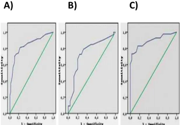
The ROC curves were carried out to investigate the cut-off NIHSS score that predicted artery occlusion in the patients with right- and left-hemisphere strokes. (Figure 2)
A) B) C)
Figure 2: The ROC curve of the cut-off NIHSS scores predicting artery occlusion in patients in the entire group (A), patients with right-hemisphere stroke (B) and patients with left-hemisphere stroke (C).
Table 2: The cut-off NIHSS score, the area under the curve (AUC), sensitivity, specificity and 95% confidence interval of the entire group, and patients with right- and left-hemisphere stroke.
The AUC (area under the curve) in the ROC curve was detected as >0.50 for both hemispheres. The cut-off NIHSS score predicting artery occlusion (patients with total occlusion in ICA distal, ICA supraclinoid, MCA M1 proximal and distal, MCA M2 superior and inferior division) was eight (sensitivity: 0,81 and specificity: 0,77 (At the same time the NIHSS score of the maximum specificity and sensitivity values was eight)). The cut-off NIHSS score of the left hemisphere was eight (sensitivity: 0,83 and specificity: 0,84). The cut-off NIHSS score of right hemisphere was detected as nine (sensitivity: 0,72 and specificity: 0,72). Using standard error and the AUC these two cut-off values were compared with a z-test (MedCalc® statistical software was used). The Z value was Z=1,805 (<1.96) and the P value was P=0.07 (>0.05), thus there was no statistically significant difference between the strength of the correlation of the cut-off NIHSS scores of the two hemispheres. Based on these findings it can be said that there was no statistically significant difference between the reliability of the cut-off NIHSS score for the two hemispheres in predicting an artery occlusion. (Table 2)
The Cut-off NIHSS Score Predicting Symptomatic Artery Stenosis
The patients were separated into two
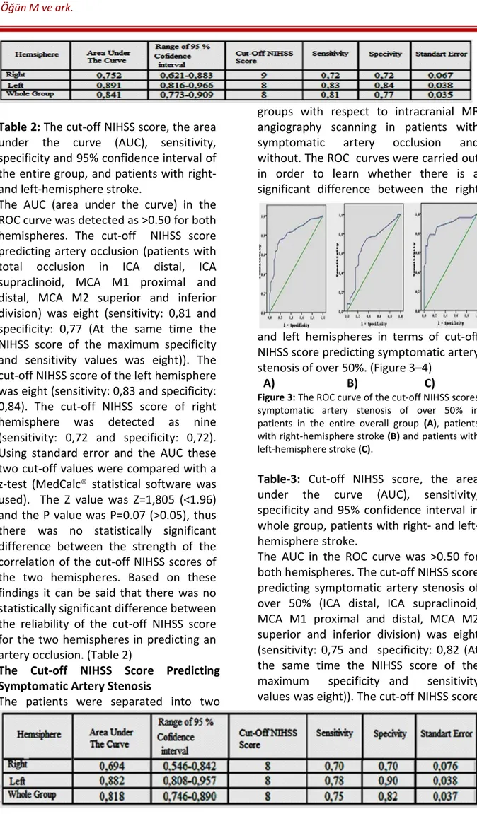
groups with respect to intracranial MR angiography scanning in patients with symptomatic artery occlusion and without. The ROC curves were carried out in order to learn whether there is a significant difference between the right
and left hemispheres in terms of cut-off NIHSS score predicting symptomatic artery stenosis of over 50%. (Figure 3–4)
A) B) C)
Figure 3: The ROC curve of the cut-off NIHSS scores symptomatic artery stenosis of over 50% in patients in the entire overall group (A), patients with right-hemisphere stroke (B) and patients with left-hemisphere stroke (C).
Table-3: Cut-off NIHSS score, the area under the curve (AUC), sensitivity, specificity and 95% confidence interval in whole group, patients with right- and left-hemisphere stroke.
The AUC in the ROC curve was >0.50 for both hemispheres. The cut-off NIHSS score predicting symptomatic artery stenosis of over 50% (ICA distal, ICA supraclinoid, MCA M1 proximal and distal, MCA M2 superior and inferior division) was eight (sensitivity: 0,75 and specificity: 0,82 (At the same time the NIHSS score of the maximum specificity and sensitivity values was eight)). The cut-off NIHSS score
0,78 and specificity: 0,90). Using standard error and the AUC of these two cut-off values were compared with a z-test (MedCalc® statistical software was used). The Z value was Z=2,24 (>1.96) and the P value was P=0.026 (<0.05), thus there was a statistically significant difference between the strength of the correlation of cut-off NIHSS scores between the two hemispheres. Based on these findings it can be assumed that the cut-off NIHSS score for the two hemispheres predicting symptomatic artery stenosis at over 50% is eight and it can be said that in the left hemisphere the reliability of this value is higher than in the right hemisphere.
DISCUSSION
The NIHSS is an important tool for clinical stroke studies and is a routine part of clinical practice for many neurologists who treat acute stroke patients. (5) It has been shown that the NIHSS score is a good predictor of a poor outcome following a stroke; (6) is superior to other simpler clinical stroke scales; (7) and is a powerful measure of the effectiveness of stroke treatment. Analysis of the essential structure of the NIHSS has shown that it closely represents the four primary clinical factors of right and left motor function plus right and left cortical function. (8) However, as mentioned in previous studies, the seven points of a possible maximum NIHSS score, 42 points, are directly related to language function (orientation questions = 2 points; commands = 2 points; aphasia = 3 points) and only two points related to neglect. (9) The lower the number of points for right cortical dysfunction is likely to relate to larger lesion volumes seen in patients with low NIHSS scores. Some studies have used an NIHSS score of <4 or <5 to exclude patients from treatment. (10) Based on these criteria, some patients with large right-hemisphere lesion volumes and with significant neurologic deficit could not be
excluded from the treatment resulting in poor functional outcomes.
This study indicated that:
1. There was no significant difference between the two hemispheres in terms of the cut-off NIHSS score predicting symptomatic artery occlusion.
2. The detected value of the cut-off NIHSS score is more reliable for the left hemisphere in terms of predicting symptomatic artery stenosis of over 50%. In our study, we detected the cut-off NIHSS score predicting artery stenosis as nine for the right hemisphere and as eight for the left hemisphere (also for the whole group). There was no statistically significant difference between the reliability of the cut-off NIHSS score of the two hemispheres predicting artery occlusion. The cut-off NIHSS predicting symptomatic artery stenosis of over 50% was eight for both hemispheres. It was determined that the value of the cut-off NIHSS score is more reliable for left hemisphere in terms of predicting symptomatic artery stenosis of over 50%. Urs Fischer et al. investigated in a study whether the NIHSS score is associated with the findings of arteriography performed within the first hours after ischemic stroke. The basilar artery, ICA and MCA were evaluated by DSA. They indicated that an NIHSS score ≥10 a vessel occlusion will likely be seen on the arteriography, and with a score ≥12 it will most probably be centrally located. (11) In a study carried out by Nakajima et al., 43 patients were evaluated by DSA within 12 hours of ischemic stroke onset and the cut-off NIHSS score predicting artery occlusion was 10. In this study, ICA and MCA were evaluated by DSA. A total of 96.9% of patients with NIHSS scores ≥10 displayed arterial occlusion and 63.6% of patients with NIHSS scores <10 displayed no arterial occlusion. (12)
In a study conducted by Maas et al., patients were evaluated by CT angiography within 12 hours of ischemic stroke onset. Patients with ACA and MCA were determined. The cut-off NIHSS score predicting artery occlusion was 10. However, in the case of an NIHSS score ≥10, the positive predictive value for artery occlusion was 81% but only 48% sensitivity for the majority of subjects with artery occlusion presenting with lower NIHSS scores. (13)
In our study, the patients with an isolated MCA stroke admitted within 12 hours after stroke onset were evaluated by MR angiography. The cut-off NIHSS score predicting artery occlusion for the entire study group (the patients with total occlusion in ICA distal, ICA supraclinoid, MCA M1 proximal and distal, MCA M2 superior and inferior division) was eight (sensitivity: 0,81 and specificity: 0,77). The cut-off NIHSS score in patients evaluated with left-sided stroke was an NIHSS of eight (83% sensitivity, specificity 84%), while in patients evaluated after with right-sided stroke cut-off NIHSS score was nine (sensitivity 72%, specificity 72%). Based on these findings, it can be said that there was no statistically significant difference between the reliability of the cut-off NIHSS score of the two hemispheres predicting artery occlusion. DSA is considered as the gold standard for evaluating cerebral artery occlusions. (20) Therefore, the reliability of determining artery occlusion and cut-off NIHSS score with studies performed using DSA should be higher than those our study that used MR angiography.
In addition to the abovementioned studies, in our study, the patients with symptomatic artery stenosis of over 50% were determined and the cut-off NIHSS score predicting this stenosis percentage was investigated. The cut-off NIHSS score of the two hemispheres predicting symptomatic artery stenosis of over 50%
was eight. It was confirmed that the value of the cut-off NIHSS score is more reliable for left hemisphere in terms of predicting symptomatic artery stenosis of over 50%. It was not investigated in previous studies that included researching the cut-off NIHSS score whether there was a difference between the right and left hemisphere in terms of these cut-off scores. Our study was the first to investigate if there was a difference between the cut-off NIHSS of the two hemispheres.
CONCLUSION
In this study; DWI MRI and intracranial MR angiography were performed on the 139 patients diagnosed with an ischemic stroke with isolated MCA infarction who were admitted to our hospital within 12 hours after stroke onset and the NIHSS scores were recorded during the acute period.
The patients were separated into two groups with respect to intracranial MR angiography scanning for patients with symptomatic artery stenosis and without. It was studied if there is a significant difference between the right and left hemispheres in terms of cut-off NIHSS score predicting symptomatic artery stenosis of over 50% and the following results were obtained:
The cut-off NIHSS score predicting artery stenosis was nine for the right hemisphere and eight for the left hemisphere. The cut-off NIHSS predicting symptomatic artery stenosis of over 50% was eight for both hemispheres. It was determined that the value of the cut-off NIHSS score is more reliable for the left hemisphere in terms of predicting symptomatic artery stenosis of over 50%.
These detected cut-off NIHSS scores can be used as a supportive finding, owing to predicting artery occlusion and artery stenosis of over 50% and to allow the patients to receive thrombolytic
imaging cannot be performed.
REFERENCES
1. Luker JA, Bernhardt J, Grimmer-Somers KA. Demographic and stroke-related factors as predictors of quality of acute stroke care provided by allied health professionals. J Multidiscip Healthc. 2011; 4: 247–259.
2. Boone M, Chillon JM, Garcia PY, Canaple S, Lamy C, Godefroy O, Bugnicourt JM. NIHSS and acute complications after anterior and posterior circulation strokes. Therapeutics and Clinal Risk Management 2012; 8: 87–93.
3. Dara C, Bang J, Gottesman RF, Hillis AE. Right Hemisphere Dysfunction is Better Predicted by Emotional Prosody Impairments as Compared to Neglect. J Neurol Transl Neurosci 2014: 2(1): 1037. 4. Kettunen J. Visual Neglect and Orienting Bias in Right Hemisphere Stroke Patients with and without Thrombolysis. 2013: 1455-1616
5. Kwah LK, Diong J. National Institutes of Health Stroke Scale (NIHSS) Journal of Physiotherapy. 2014: vol 60, issue 1, page 61
6- Queralt-Tomas L. Coordination strategies of care across stroke recovery: Proposals for nursing interventions in primary care. Clinical Nursing Studies 2015: Vol. 3. No. 2
7. Mansour OY, Megahed MM, Eman HS, Elganhy A. Acute ischemic stroke prognostication, comparison between Glasgow Coma Score, NIHSS scale and Full Outline of UnResponsiveness Score in intensive care unit. Alexandria Journal of Medicine,2014:10.002
8. Fischer U, Mono ML, Zwahlen M, Nedeltchev K, Arnold M, Galimanis A, Bucher S, Findling O, Meier N, Brekenfeld C, Gralla J, Heller R, Tschannen B, Schaad H, Waldegg G, Zehnder T, Ronsdorf A,
HP, the QABE investigators. Impact of thrombolysis on stroke outcome at 12 months in a population: The Bern Stroke Project. Stroke 2012; 43: 1039–1045 9- Uemura J, Kimura K, Inoue T, Shibazaki K, Sakamoto Y, Aoki J. The role of small vessel disease in re-exacerbation of stroke symptoms within 24 hours after tissue plasminogen activator infusion. Journal of Stroke and Cerebrovascular Diseases 2014; Volume 23, Issue 1, Pages 75–79
10. Golsari A, Cheng B, Sobesky J, Schellinger PD, Fiehler J, Gerloff C, Thomalla G. Stroke lesion volumes and outcome are not different in hemispheric stroke side treated with intravenous thrombolysis based on magnetic resonance imaging criteria. Stroke 2015; 46: 1004–1008
11. Heldner MR, Zubler C, Mattle HP, Schroth G, Weck A, Mono ML, Gralla J, Jung S, El-Koussy M, Lüdi R, Yan X, Arnold M, Ozdoba C, Mordasini P, Fischer U. National Institutes of Health Stroke Scale Score and vessel occlusion in 2.152 Patients with acute ischemic stroke. Stroke 2013; 44: 1153–1157
12. Cooray C, Fekete K, Mikulik R, Lees KR, Wahlgren N, Ahmed N. Threshold for NIH stroke scale in predicting vessel occlusion and functional outcome after stroke thrombolysis. International Journal of Stroke 2015: Volume 10, Issue 5, Pages 645–788,
13. Hansen CK, Christensen A, Ovesen C, Havsteen I, Christensen H. Stroke severity and incidence of acute large vessel occlusions in patients with hyper-acute cerebral ischemia: results from a prospective cohort study based on CT-angiography (CTA). International Journal of Stroke 2015: Volume 10, Issue 3, pages 336–342