Iranian Journal of Basic Medical Sciences
ijbms.mums.ac.irEvaluation of Δ
9
-tetrahydrocannabinol metabolites and oxidative
stress in type 2 diabetic rats
Zeynep Mine Coskun
1, 2*, Sema Bolkent
21 Department of Molecular Biology and Genetics, Faculty of Arts and Sciences, Istanbul Bilim University, Istanbul, Turkey 2 Department of Medical Biology, Faculty of Cerrahpasa Medicine, Istanbul University, Istanbul, Turkey
A R T I C L E I N F O A B S T R A C T Article type:
Original article Objective(s): The object of the study is to examine the effects of Δ
9-tetrahydrocannabinol (THC) against
oxidative stress in the blood and excretion of THC metabolites in urine of type 2 diabetic rats.
Materials and Methods: The control (n=8), THC control (n=6), diabetes (n=8) and diabetes + THC (n=7)
groups were created. Type 2 diabetes was induced by nicotinamide (NA, 85 mg/kg) + streptozotocin (STZ, 65 mg/kg). THC was administered intraperitoneally for seven days. The glutathione (GSH) level in erythrocytes and malondialdehyde (MDA) level, superoxide dismutase (SOD) and catalase (CAT) enzyme activities in plasma were measured. THC metabolites were analyzed in urine.
Results: The results showed that the erythrocyte GSH levels were significantly increased (P<0.05), but
plasma MDA levels were non-significantly decreased in diabetes group treated with THC when compared with the diabetes group. The CAT activity was non-significantly reduced and SOD was significantly increased (P<0.01) in the plasma of diabetes induced by THC in comparison with the diabetic group. The excretion of THC metabolites was higher in the urine of diabetes + THC rats as compared to the THC control rats.
Conclusion: These findings highlight that THC treatment may attenuate slightly the oxidative stress in
diabetic rats. The excretion rate of THC may vary in the type 2 diabetes mellitusstatus. Article history: Received: Jan 14, 2015 Accepted: Apr 16, 2015 Keywords: Diabetes mellitus Metabolite Oxidative stress Type 2 Urine Δ9-tetrahydrocannabinol
►
Please cite this article as:Coskun ZM, Bolkent S. Evaluation of Δ9-tetrahydrocannabinol metabolites and oxidative stress in type 2 diabetic rats. Iran J Basic Med Sci 2016; 19:154-158.
Introduction
Cannabis has been known to be the oldest
psychoactive plant for years. It is classified in the Cannabis genus, which is part of the Cannabacea
family. Cannabis sativa L. is the most common species (1). Δ9-tetrahydrocannabinol (THC) is the main psychoactive constituent identified in Cannabis sativa L. THC is the most notable cannabinoid among all phytocannabinoids (2).
THC is exposed to degradation and converted into its active and inactive metabolites that are conjugated with glucuronic acid, and excreted in urine. THC is converted to active metabolite, 11-hydroxy-Δ9-THC (11-OH-THC), and then converted to an inactive metabolite, 11-nor-9-carboxy- Δ9-THC (THC - COOH) (2, 3).
ElSohly and Slade (4) mention that C. sativa and its products have been used as medicinal agents. THC is a phytocannabinoid that is known to be the most widely-used illegal drug. Cannabinoids show a variety of therapeutic effects against chronic pain and muscle spasms, nausea and anorexia caused by HIV treatment, vomiting and nausea caused by
cancer chemotherapy as well as anorexia associated with weight loss caused by immune deficiency syndrome (5-7). Many studies report that THC provides protection against neuronal injury in a cell culture model of Parkinson disease and experimental models of Huntington disease, exhibits anti-oxidative action and mitigates the severity of the autoimmune response in an experimental model of diabetes (8-12).
The development and progression of diabetes mellitus and its complications arise out of increased oxidative damage (13). Kassab and Piwowar (14) report that the best-known pathways of diabetic complications include oxidative stress. It is well-known that type 2 diabetes (T2DM) is the most common form of diabetes mellitus (DM) and it is characterized by hyperglycemia and insulin resistance. The symptoms of T2DM are exhaustion, thirst, weight loss and frequent urination, blurred vision, frequent infections, etc. (15, 16).
The aims of the study presented in this paper were: (a) to explain the effects of THC on oxidative stress in T2DM treated with THC and (b) to determine the level of THC metabolites in the urine of diabetic and control rats induced by THC injection.
*Corresponding author: Zeynep Mine Coskun. Department of Molecular Biology and Genetics, Faculty of Arts and Sciences, Istanbul Bilim University,
Istanbul, Turkey, Department of Medical Biology, Faculty of Cerrahpasa Medicine, Istanbul University, Istanbul, Turkey. Tel: +90-2122136483; Fax: +90-2122723461; email: [email protected]
Materials and Methods
Experimental study
The study was carried out in accordance with the guidelines of the Local Ethic Committee of Animal Research (Istanbul University, HADYEK/103). For this study, 8–10 weeks-old male Sprague-Dawley rats were conducted by the Istanbul University, Experimental Medical Research Institute (DETAE). All the animals were housed in a temperature-controlled clean room with a 12 hr light/dark cycle and fed with tap water and standard chow ad libitum. The animals were selected randomly and arranged into four groups.
Group A: Control (n=8); the healthy animals received isotonic saline intraperitoneally (IP).
Group B: THC (n=6); 3 mg/kg/day THC (Lipomed THC-135-100LE, Switzerland) was administered to the animals for seven days.
Group C: Diabetes (n=8); the animals were injected with a single dose of streptozotocin (STZ) (65 mg/kg, Sigma-Aldrich, USA) dissolved in isotonic saline 15 min after the injection of nicotinamide (NA) (85 mg/kg, Sigma-Aldrich, USA) in isotonic saline (IP) (17). Blood glucose levels of the rats were measured 72 hr after the STZ + NA injection. The animals with blood glucose concentrations above 200 mg/dl were considered to be diabetic (18).
Group D: Diabetes + THC (n=7); the diabetic rats were treated with THC (3 mg/kg/day) for seven days.
The starting day for urine collection was considered to be day-0 following treatment with THC. The urine samples were collected on days-0, 7 and 14 after THC injection. The rats were anesthetized IP with ketamine-HCl (50 mg/kg, Pfizer, USA) + xylazine-HCl (10 mg/kg, Bayer, Canada). After the rats were sacrificed, their blood samples were collected from the right ventricle. The urine and plasma samples of animals were stored at -85 °C. Biochemical analysis
The reduced glutathione (GSH) level of erythrocytes was determined according to the method developed by Beutler et al (19) using metaphosphoric acid for protein precipitation and 5-5’-dithiobis (2-nitro benzoic acid (Sigma-Aldrich, USA) for color development at 412 nm. The GSH concentration in erythrocytes was expressed in nmol/mg hemoglobin (Hb) at 540 nm. The hemoglobin levels were determined via the cyanomethemoglobin method (20).
The lipid peroxidation was determined on the basis of the formation of malondialdehyde (MDA), which was estimated by using the thiobarbituric acid (TBA) method (21). The plasma samples were mixed thoroughly with trichloroacetic acid (TCA) (30 %) (Sigma-Aldrich, USA), TBA (0.75 %) (Merck, Germany) and 5 M HCl. The plasma samples in tubes
were placed in boiling water for 15 min and, centrifuged at 5000 rpm for 10 min. The supernatants were measured at 535 nm by the spectrophotometer. The plasma MDA levels were expressed as µmol/ml.
The superoxide dismutase (SOD) enzyme activity was ascertained by the method developed by Sun et al (22), which involved the inhibition of nitroblue tetrazolium (NBT) (Sigma-Aldrich, USA) reduction with xanthine/xanthine oxidase (Sigma-Aldrich, USA). The SOD activity was measured at 560 nm. The calculated SOD enzyme activity was expressed as SOD (U/ml) in plasma.
The catalase (CAT) enzyme activity in the plasma was determined via the Aebi’s method (23). The reaction mixture was made up of the sample, 50 mM phosphate buffered saline (PBS, pH 7.0) and 10 mM H2O2. The reduction rate of H2O2 was observed to be 240 nm at room temperature for 60 seconds. The CAT activity was expressed in U/ml.
THC metabolite analysis of rat urine
The metabolites of THC were identified in the urine samples of THC control rats and diabetic rats treated with THC. The analysis of urine samples was performed using a DRI® Cannabinoid Assay by
Hitachi 912 Chemistry Analyzer (Roche, USA). This assay is intended for the qualitative and
semiquantitative determination of cannabinoids in urine. The antibody/substrate reagent and enzyme conjugate reagent were added onto the urine sample of rats. The samples were measured at 340 nm. Statistical analysis
Statistical analysis was performed with non-parametric Kruskal - Wallis test. Then, comparisons between two groups were analysed using the Mann – Whitney U by means of the statistical package (SPSS version 21). All of the results were expressed as means ± standard error of the mean (SEM). P < 0.05 was considered to be statistically significant.
Results
Biochemical analysis
The levels of blood glucose indicated significant differences among the four groups on days-7, 14 and 21 (P<0.001). THC in diabetes group non-significantly reduced the blood glucose levels on days-14 and 21 (24).
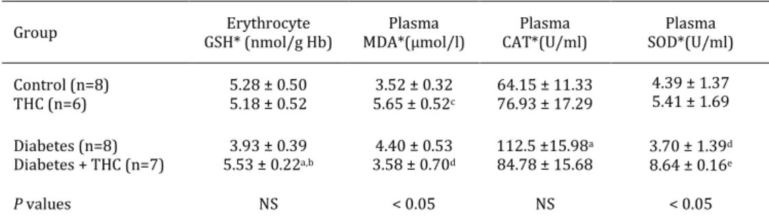
The erythrocyte GSH levels were almost similar in both control and THC control groups. The GSH levels in erythrocytes were non-significantly decreased in the diabetic rats as compared to the control rats. The GSH level was seen to have increased significantly in the diabetic rats treated with THC as compared to the diabetic rats (P< 0.05) (Table 1).
Table 1. Erythrocyte GSH, plasma MDA, CAT and SOD values in all groups of experimental rats
Group GSH* (nmol/g Hb) Erythrocyte MDA*(µmol/l) Plasma CAT*(U/ml) Plasma SOD*(U/ml) Plasma Control (n=8) THC (n=6) 5.28 ± 0.50 5.18 ± 0.52 5.65 ± 0.523.52 ± 0.32 c 64.15 ± 11.33 76.93 ± 17.29 4.39 ± 1.37 5.41 ± 1.69 Diabetes (n=8) Diabetes + THC (n=7) 5.53 ± 0.223.93 ± 0.39a,b 4.40 ± 0.53 3.58 ± 0.70d 112.5 ±15.98a 84.78 ± 15.68 3.70 ± 1.39 d 8.64 ± 0.16e P values NS < 0.05 NS < 0.05
*Mean±standard error of the mean (SEM); NS: Non-significant, THC: Δ9-tetrahydrocannabinol, GSH: reduced glutathione,
Hb: hemoglobin; MDA: malondialdehyde, CAT: catalase, SOD: superoxide dismutase
aP<0.05 versus control group; bP<0.05 versus diabetic group; cP<0.01 versus control group; dP<0.05 versus THC group; eP<0.01 versus
diabetic group
Table 2. The THC metabolites in urine of THC control and diabetes + THC groups of experimental rats
Group Day-0* (ng/ml) Day-7* (ng/ml) Day-14 *(ng//ml)
THC (n=6)
Diabetes + THC (n=7) 13.93 ± 6.183.80 ± 1.28 0.000.00 0.000.00
P values NS NS NS
*Mean ± standard error of the mean (SEM); NS: Non-significant, THC: Δ9-tetrahydrocannabinol
The plasma MDA level showed a significant difference among four groups (P<0.05). The MDA level in plasma was non-significantly increased in the diabetic group when compared with the control group. The MDA level in the THC control group was elevated when treated with THC for seven days. The MDA level was non-significantly lower in the diabetic rats treated with THC when compared with the diabetic animals (Table 1).
The plasma CAT enzyme activity in the THC control group showed an increase as compared to the control group. The CAT activity of plasma was significantly increased in the diabetic groups when compared with the control groups. However, a decrease was observed in these levels due to the supplementation of THC (Table 1).
The SOD enzyme activity of plasma showed a significant difference among four groups (P<0.05). In contrast with the CAT activity, the SOD activity showed a non-significant decrease in the diabetic group compared with the control group. In the THC control rats, the plasma SOD activity was non-significantly elevated as compared to the control animals. The plasma SOD enzyme activity was significantly increased in diabetes + THC group as compared to the diabetes group (P<0.01) (Table 1).
The analysis of Δ9-THC metabolites in the urine of
experimental rats
The day following the THC injection was accepted as day-0. The results of the analysis of THC metabolites are summarized in Table 2. It was observed that the THC metabolite level of urine
showed a non-significant increase in diabetic rats treated with THC as compared with the THC control group on day-0 (Figure 1). On day-7, no positive results were received in the rat urine THC control and diabetes + THC. Although, the level of THC metabolites was negative in THC control rats, only one rat showed a positive result (9.5 ng / ml) in the diabetes + THC group on day-14.
Discussion
In the present study, we examined the effects of THC on oxidative stress in plasma and the excretion of THC metabolites in the urine samples of type 2 diabetic rats.
Diabetes mellitus (DM) is characterized by insulinresistanceandhyperglycemia. Recent studies
Figure 1. The metabolite levels of tetrahydrocannabinol (THC) in urine on day-0 of experimental rats
indicate that oxidative stress may have a key role in the pathogenesis of T2DM (25, 26). Oxidative stress is an imbalance between the production of reactive oxygen species (ROS) and antioxidant levels of cells (27). ROS and lipid peroxide are known to be markers of oxidative stress. These markers are overproduced in T2DM, which is associated with oxidative stress.
THC has a wide area of use that includes appetite stimulants, analgesic, anti-pyretic, anti-emetic agents, etc. (28, 29). THC protects against oxidative stress by blocking the production of ROS [9]. In the study by Hampson et al (30), cannabidiol and THC were identified as antioxidants. They suggested that the anti-oxidative features of cannabidiol and THC may be preferential factors for their therapeutic use as neuroprotective agents.
GSH is known to provide defense against oxidative stress (31). The administration of THC did not change the erythrocyte GSH levels in normal rats. On the other hand, the decreased GSH levels in erythrocytes of diabetic animals showed an increase when treated with THC. The increased levels of lipid peroxidation during the progression of diabetes may have a role in tissue damage associated with diabetes (32). Malondialdehyde (MDA) is one of the most frequently used indicators of lipid peroxidation. In our previous study, it was reported that there was an increased lipid peroxidation in the stomach of diabetic rats (33). In our study, the plasma level of MDA increased with diabetes, non-statistically. THC slightly reduced the MDA level in plasma. Abdel-Salam et al (34) investigated the effects of Cannabis sativa extract on oxidative stress in mice. They reported that C. sativa extract (20 mg/kg) decreased the MDA levels and increased the GSH levels in brain tissue. These results show a concordance with our results of the erythrocyte GSH and plasma MDA levels. Both SOD and CAT enzymes are anti-oxidative defense system enzymes. The activity of CAT enzyme was increased while the SOD activity was decreased in the plasma of diabetic rats. Similar results were reported in the study by Shinde et al (13). They showed that the MDA level in serum increased while both SOD enzyme activity and GSH level were reduced in T2DM. We observed that the CAT activity decreased and the SOD activity was increased in the plasma of diabetes treated with THC as compared to diabetic rats. Finally, the oxidative damage in plasma was restored in diabetic rats through the administration of THC.
In the study on chronic cannabis users, the excretion of THC and 11-OH-THC were measured in urine for -3, -4, -7, -12 and -24 days after the cannabis cessation. It was seen that the metabolites were detectable in urine for at least -24 days (35).
It is known that THC accumulates in adipose and brain tissues (36). During the sacrification, we
observed that the adipose tissue was reduced with diabetes. In our previous study, we observed that the body weight was reduced significantly in diabetic rats as compared to control rats (24). For this reason, the metabolite level of the THC group was lower than that of the diabetes + THC group on day-0. In the THC group, there might be an accumulation of THC in the adipose tissue, so it may be inhibited from being excreted in urine.
THC positive result was indicated in the urine of only one rat among seven rats in the diabetes + THC group on day-14. In addition, we detected that the rat with THC positive urine was observed to have a higher rate of weight loss than the other rats in the diabetes + THC group. This may indicate that the accumulation of THC in adipose tissue decreased in parallel with the adipose tissue decrease in the diabetic rats treated with THC. Thus, the THC that was not accumulated may be excreted in the urine of diabetic rats. We did not find any positive metabolites of THC in the urine samples of rats on day-7. This may account for the increased loss of weight after day-7. The other probability, i.e. the metabolization of THC, may be higher with diabetic rats treated with THC when compared to THC control rats. It is suggested that higher levels of THC metabolite may be detected in the urine of diabetic individuals who take THC in comparison to healthy THC users.
Conclusion
The present study shows that the short-term administration of THC attenuated the oxidative stress in the rat model of T2DM. Thus, it may be suggested that THC could be used as a therapeutic agent against oxidative stress. In addition, the level of THC metabolites was increased in the urine of diabetic rats treated with THC. The level of THC metabolites in the urine may be associated with the weight loss caused by diabetes. Therefore, the level of THC metabolites may be lower in the urine of individuals with more adipose tissue among THC users.
Aknowledegment
The study was supported by the Scientific Research Projects Coordination Unit of Istanbul University, Project No. 7823. The results described in this paper were part of student thesis.
References
1. Hillig KW, Mahlberg PG. A chemotaxonomic analysis of cannabinoid variation in Cannabis (Cannabaceae). Am J Bot 2004; 91:961-975.
2. Kochanowski M, Kała M. Tetrahydrocannabinols in clinical and forensic toxicology. Przegl Lek 2005; 62:576-580.
3. Gieringer D, Rosenthal E, Carter GT. Marijuana medical handbook: Practical guide to the therapeutic uses of marijuana. Berkley, CA, USA: 2008.
4. ElSohly MA, Slade D. Chemical constituents of marijuana: The complex mixture of natural cannabinoids. Life Sci 2005; 78:539-548.
5. Borgelt LM, Franson KL, Nussbaum AM, Wang GS. The pharmacologic and clinical effects of medical cannabis. Pharmacotherapy 2013; 33:195-209.
6. Lutge EE, Gray A, Siegfried N. The medical use of cannabis for reducing morbidity and mortality in patients with HIV/AIDS. Cochrane Database Syst Rev 2013; 4:CD005175.
7. Reynolds TD, Osborn HL. The use of cannabinoids in chronic pain. BMJ Case Rep 2013; bcr2013010417. 8. Carroll CB, Zeissler ML, Hanemann CO, Zajicek JP. Δ⁹-tetrahydrocannabinol (Δ⁹-THC) exerts a direct neuroprotective effect in a human cell culture model of Parkinson's disease. Neuropathol Appl Neurobiol 2012; 38:535-547.
9. Chen J, Errico SL, Freed WJ. Reactive oxygen species and p38 phosphorylation regulate the protective effect of delta9-tetrahydrocannabinol in the apoptotic response to NMDA. Neurosci Lett 2005; 389:99-103. 10. Li X, Kaminski NE, Fischer LJ. Examination of the immunosuppressive effect of delta9-tetrahydro-cannabinol in streptozotocin-induced autoimmune diabetes. Int Immunopharmacol 2001; 1:699-712. 11. Moldzio R, Pacher T, Krewenka C, Kranner B, Novak J. Effects of cannabinoids Δ(9)-tetrahydrocannabinol, Δ(9)-tetrahydrocannabinolic acid and cannabidiol in MPP+ affected murine mesencephalic cultures. Phytomedicine 2012; 19:819-824.
12. Sagredo O, Pazos MR, Satta V, Ramos JA, Pertwee RG, Fernández-Ruiz J. Neuroprotective effects of phytocannabinoid-based medicines in experimental models of Huntington's disease. J Neurosci Res 2011; 89:1509-1518.
13. Shinde SN, Dhadke VN, Suryakar AN. Evaluation of oxidative stress in type 2 diabetes mellitus and follow-up along with vitamin E sfollow-upplementation. Indian J Clin Biochem 2011; 26:74-77.
14. Kassab A, Piwowar A. Cell oxidant stress delivery and cell dysfunction onset in type 2 diabetes. Biochimie 2012; 94:1837-1848.
15. Chen L, Magliano DJ, Zimmet PZ. The worldwide epidemiology of type 2 diabetes mellitus-present and future perspectives. Nat Rev Endocrinol 2011; 8:228-236.
16. Wolfsdorf J, Glaser N, Sperling MA. Diabetic ketoacidosis in infants, children, and adolescents. Diabetes Care 2006; 29:1150-1159.
17. Masiello P, Broca C, Gross R, Roye M, Manteghetti M, Hillaire-Buys D, et al. Experimental NIDDM: development of a new model inadult rats administered streptozotocin and nicotinamide. Diabetes 1998; 47:224–229.
18. Murugan P, Pari L. Influence of tetrahydrocurcumin on hepatic and renal functional markers and protein levels in experimental type 2 diabetic rats. Basic Clin Pharmacol Toxicol 2007; 101:241-245.
19. Beutler E, Duron O, Kelly BM. Improved method for the determination of blood glutathione. J Lab Clin Med 1963; 51:882-888.
20. Crosby WN, Munn JI, Furth FW. Standardizing a method for clinical hemoglobinometry. U S Armed Forces Med J 1954; 5:693- 703.
21. Ledwozyw A, Michalak J, Stepień A, Kadziolka A. The relationship between plasma triglycerides, cholesterol, total lipids and lipid peroxidation products during human atherosclerosis. Clin Chim Acta 1986; 155:275-283.
22. Sun Y, Oberley LW, Li Y. A simple method for clinical assay of superoxide dismutase. Clin Chem 1988; 34:497-500.
23. Aebi H. Catalase in vitro. Methods Enzymol 1984; 105:121–126.
24. Coskun ZM, Bolkent S. Oxidative stress and cannabinoid receptor expression in type-2 diabetic rat pancreas following treatment with Δ(9) -THC. Cell Biochem Funct 2014; 32:612-619.
25. Chang YC, Chuang LM. The role of oxidative stress in the pathogenesis of type 2 diabetes: from molecular mechanism to clinical implication. Am J Transl Res 2010; 2:316-331.
26. Niedowicz DM, Daleke DL. The role of oxidative stress in diabetic complications. Cell Biochem Biophys 2005; 43:289-330.
27. Kopáni M, Celec P, Danisovic L, Michalka P, Biró C. Oxidative stress and electron spin resonance. Clin Chim Acta 2006; 364:61-66.
28. Koch JE. Delta (9)-THC stimulates food intake in Lewis rats: effects on chow, high-fat and sweet high-fat diets. Pharmacol Biochem Behav 2001; 68:539-543. 29. Robson P. Therapeutic aspects of cannabis and cannabinoids. Br J Psychiatry 2001; 178:107-115. 30. Hampson AJ, Grilmaldi M, Axelrod J, Wink D. Cannabidiol and (-)Δ9-tetrahydrocannabinol are neuroprotective antioxidants. Proc Natl Acad Sci 1998; 95:8268-8273.
31. Nicotera P, Orrenius S. Role of thiols in protection against biological reactive intermediates. Adv Exp Med Biol 1986; 197:41–49.
32. Feillet-Coudray C, Rock E, Coudray C, Grzelkowska K, Azais-Braesco V, Dardavet D, et al. Lipid peroxidation and antioxidant status in experimental diabetes. Clin Chim Acta 1999; 284:31-43.
33. Coskun ZM, Sacan O, Karatug A, Turk N, Yanardag R, Bolkent S, et al. Regulation of oxidative stress and somatostatin, cholecystokinin, apelin gene expressions by ghrelin in stomach of newborn diabetic rats. Acta Histochem 2013; 115:740-747.
34. Abdel-Salam OME, Nada SA, Salem NA, Sayed El-Shamarka M, Omara E. Effect of Cannabis sativa on oxidative stress and organ damage after systemic endotoxin administration in mice. Comp Clin Pathol 2013; 23:1069-1085.
35. Lowe RH, Abraham TT, Darwin WD, Herning R, Cadet JL, Huestis MA. Extended urinary Delta9-tetrahydrocannabinol excretion in chronic cannabis users precludes use as a biomarker of new drug exposure. Drug Alcohol Depend 2009; 105:24-32. 36. Kreuz DS, Axelrod J. Delta-9-tetrahydrocannabinol: localization in body fat. Science 1973; 179:391-393.