ORIGINAL RESEARCH
Medicine Science 2018;7(3):604-9
The effect of caffeic acid phenethyl ester (CAPE) on hepatic histopathology and oxidative
stress in rats treated with malathion
Gokhan Nur1, Mehmet Tahir Husunet2, Izzettin Guler1, Ayla Deveci1, Evren Koc3, Ozlem Nur1, Pinar Aksu Kilicle4
1Gaziantep University, Islahiye Vocational School, Department of Veterinary, Gaziantep, Turkey 2Çukurova University, Faculty of Science and Literature, Department of Biology, Adana, Turkey 3Kafkas University, Faculty of Architecture and Engineering, Department of Bioengineering, Kars, Turkey
4Kafkas University, Faculty of Science and Literature, Department of Biology, Kars, Turkey
Received 09 March 2018; Accepted 22 March 2018
Available online 11.08.2018 with doi: 10.5455/medscience.2018.07.8833 Copyright © 2018 by authors and Medicine Science Publishing Inc. Abstract
Malathion is a broad-spectrum organophosphate insecticide used in the agricultural industry. Our study aimed to investigate the protective effects of caffeic acid phenethyl ester (CAPE) against malathion by examining histopathological and biochemical parameters. Forty Sprague-Dawley male rats were used in this study. Groups formed; group I (control), only 5 ml kg-1 oral corn oil; group II (malathion), 40 mg kg-1 malathion by gavage; group III (malathion+CAPE), intraperitoneal (i.p.) CAPE (10 μmol kg-1) followed by intragastric malathion (40 mg kg-1) after 1 hour; group IV (CAPE), intraperitoneal CAPE (10 μmol kg-1). At the end of the administrations, rats were anesthetised with ketamine/xylazine intraperitoneally and euthanasia was performed with cervical dislocation. Their intracardiac blood was drawn under anaesthesia, and after the euthanasia, their liver tissues were removed for histopathological analysis. In group II that was treated with malathion, cell infiltrations around the central and portal vein, degeneration and focal necrosis areas in the liver were detected. Although the histopathological findings observed in the malathion group were also encountered in group III (malathion+CAPE), lesions were less severe. In the malathion group II, total oxidant status (TOS) and total sialic acid (TSA) levels in plasma increased compared with the control group, while total antioxidant status (TAS) decreased compared with the control group. In the Malathion+CAPE group III, it was observed that biochemical parameters were closer to the control group. In malathion-treated rats, it was determined that CAPE reduced the degeneration in the liver and it could have a protective effect by partially correcting oxidative stress parameters. It was concluded based on our findings that malathion, which is an organophosphate compound, makes toxic effects in the liver, but that CAPE has a protective potential because it partially reduces the severity and frequency of histopathological lesions and it brings biochemical values closer to normal.
Keywords: CAPE, malathion, histopathology, total sialic acid, total oxidant/total antioxidant level
Medicine Science International Medical Journal
Introduction
The rapid increase of the world population and the fact that agricultural production fails to reach the sufficient production capacity have significantly increased the use of biological and chemical substances against the pests which cause a loss of production in this industry [1]. Those most commonly used pesticides include organophosphates, carbamates and chlorinated hydrocarbons. Organochlorines were used around the world very extensively. However, they have been prohibited because their biodegradation time in nature is longer than that of organophosphates [2]. The easy accessibility of pesticides and their low controllability in terms of use is indicated as the primary cause of poisonings. Chronic toxicity is caused by the effect of pesticide residues that lead to environmental pollution [3].
Coresponding Author: Gokhan Nur, Gaziantep University, Islahiye Vocational
School, Department of Veterinary, Gaziantep, Turkey
E-mail: [email protected]
Malathion was first commercialised in the 1950s, and it has been among the world’s best-selling broad spectrum organophosphate (OP) pesticides since the 1980s. Like other OPs, the main mode of actions of this insecticide is the inhibition of the acetylcholinesterase (AChE) activity in target tissues [4-7]. Misuse of organophosphate pesticides that affect the nervous system causes hundreds of thousands of poisonings and even deaths every year. The main treatments for such poisonings are based on immediate administration of atropine and acetylcholinesterase activators that are currently represented by mono- or bis-pyridinium aldoximes [8].
In a study with rats treated with acute malathion, this substance was found to have adverse effects on the biochemical parameters in the liver and other tissues of rats and cause oxidative stress, whereas CAPE was found to significantly reduce the harmful effects of malathion [9]. Measurement of the substances in the urine and amniotic fluids of Wistar rats orally treated with binary mixtures of 5 different insecticides including malathion showed a significant exposure of foetus not only to the insecticides but also to their
metabolites [10]. In a study investigating the effects of malathion and green tea on liver tissue and biochemical parameters, it was observed that the antioxidant content of the green tea reduced the malathion’s damage to liver tissue. It was also found that green tea significantly lower aspartate transaminase, alanine transaminase, alkaline phosphatase, total oxidant capacity and malondialdehyde concentrations and significantly increase the total antioxidant capacity [11].
In mice treated with subchronic malathion, acetylcholinesterase activity and testosterone levels decreased, apoptosis and necrosis were induced in spermatozoa, the reproductive performance of male mice diminished and their semen parameters changed [12]. An in vivo study investigating the effects of malathion on glucose metabolism regulation showed that malathion increased the biomarkers of insulin resistance and reduced insulin sensitivity indices [13]. In a study investigating the protective effect of N-acetyl-L-cysteine (NAC) against the toxic effects of malathion exposure in Wistar rats, leucocytosis and reduced hemoglobin (Hgb) content were determined in malathion treated rats when compared with the control group. In addition, with malathion administration, there was a significant increase in liver enzymes such as alanine aminotransferase, aspartate aminotransferase, alkaline phosphatase, lactate dehydrogenase and creatinine kinase. A decrease in acid phosphatase activity, protein and globulin levels was also observed. In the same study, it was reported that there was a defect in calcium, magnesium, phosphorus and iron contents due to malathion administration. NAC showed therapeutic effects against malathion toxicity, allowing HGB content and all liver enzymes to return to normal values [14].
In this study, CAPE was used as a protective agent against malathion. In studies conducted, CAPE was shown to exhibit antioxidant, antimicrobial, antiinflammatory, antiproliferative, cytostatic, antiviral, antibacterial, antifungal, antineoplastic, anticancerogenic, apoptosis inducing and immunomodulatory properties [7,15-24]. It was reported that CAPE is an active polyphenol in versatile therapeutic terms and an effective tumour suppressor agent in chemotherapy to reduce toxicities caused by chemotherapy [18,19]. In another study with rats, CAPE was shown to improve memory by influencing cholinergic signals. CAPE was therefore recommended as a potential therapeutic agent in disorders involving the cholinergic system [25].
The aim of this study is to investigate with histopathological and biochemical methods the protective effects of CAPE in rats against the harmful effects of malathion, which is widely used around the world and cause high toxicity in living organisms.
Material and Methods
Chemicals
All chemical substances obtained were trademarked. Malathion was purchased from Sigma-Aldrich (CAS No. 121-75-5). CAPE with 97% purity was purchased from Sigma-Aldrich (CAS No: 104594-70-9). Ketamine 10% (Inj. Deutch Farm Int.) and Xylazina 10% (Pro-Ser S.A.) were purchased commercially. Ethanol (CAS No. 64-17-5) used as solvent for CAPE was purchased from Merck. Animals
In the s,tudy, 40 adult male Sprague Dawley rats weighing
200-240 gr on average obtained from the Experimental Animals Implementation and Research Centre, Kafkas University, were used. Rats were selected at random and 4 groups were formed with 10 rats in each group (n=10). Throughout the experiment, the animals were kept at 25±2°C, a relative humidity of 55±10% and a 12-hour dark cycle. They were given food and drinking water ad libitum.
Experimental design
This study was carried out using laboratory facilities after obtaining ethics committee certificate numbered KAÜ-HADYEK/2017-093. For the groups formed with rats, administration of substances was carried out for 15 days according to the protocols. At the end of the study, rats were subjected to general anaesthesia by giving intraperitoneal ketamine 90 mg kg-1 i.p. xylazine 10 mg kg-1 and euthanasia was performed with cervical dislocation.
Group I (Control): Only 5 ml kg-1 of oral corn oil (malathion’s solver) was administered.
Group II (Malathion): 40 mg kg-1 malathion was administered via gavage.
Group III (Malathion+CAPE): Intraperitoneal administration of CAPE (10 μmol kg-1), followed by intragastric malathion (40 mg kg-1) 1 hour later.
Group IV (CAPE): Intraperitoneal administration of CAPE (10 μmol kg-1).
Intracardiac blood of the rats, which were sacrificed by euthanasia at the end of the treatment, was drawn. Liver tissues were taken in fixation solution for histological analysis.
Histological Analysis
At the end of the experiment period, liver tissues of rats sacrificed by cervical dislocation under general anaesthesia were fixed in 10% buffered formalin solution. After the fixation and routine tissue processing (graded alcohols, methyl benzoate and benzol processing), the tissues taken were embedded in paraffin and 5 μm serial sections were taken with microtome from the blocks to slides pre-coated with chrome alum gelatine (CAG). Histopathological changes were examined at light microscopic level by treating the sections with haematoxylin-eosin as a histological staining method [26].
Biochemical analysis
The total antioxidant level (TAS) measurement was determined by an automated measurement method based on the decolourization of the characteristic colour formed by the 2,2’azino-bis (3-ethylbenzothiazoline-6-sulfonic acid) (ABTS) radical with antioxidants in the sample added to the medium [27]. The results were calculated as mmol Trolox equivalent/L. The total oxidant status (TOS) measurement was made by the automated measurement method [28]. The oxidants in the sample have the task of converting the ferrous ion complex to ferric ion. Ferric ion (Fe3+) formed by oxidation of iron (Fe2+) to more stabilised form (Fe2O3) forms colour with xylenol orange in acidic environment. The intensity of the spectrophotometrically measured colour is related to the total amount of oxidant molecules present in the sample. The measurement was calibrated with hydrogen peroxide (H2O2) and the results were calculated as micromolar H2O2 equivalent (μmol H2O2 equiv./L) per litre. Total sialic acid (TSA) analysis was carried out colorimetrically using a spectrophotometer
(PowerWave XS, BioTek, USA) according to the method of Sydow [29] and given as mg dl-1.
Statistical analysis
Statistical analysis of the data obtained from the study was carried out in the SPSS package program (IBM SPSS Statistic 22). One-way analysis of variance (ANOVA) was used to determine whether there was a difference between the means of the experimental group and, if there was a difference between the means of the experimental group, the “Anova-Duncan” test was performed on the group means in order to determine the group or groups from which this difference was derived, and the value p<0.05 was considered statistically significant.
Results
Histological findings
After the fixation and tissue processing stages in the end of the study, the hepatic tissues taken were embedded in paraffin, and
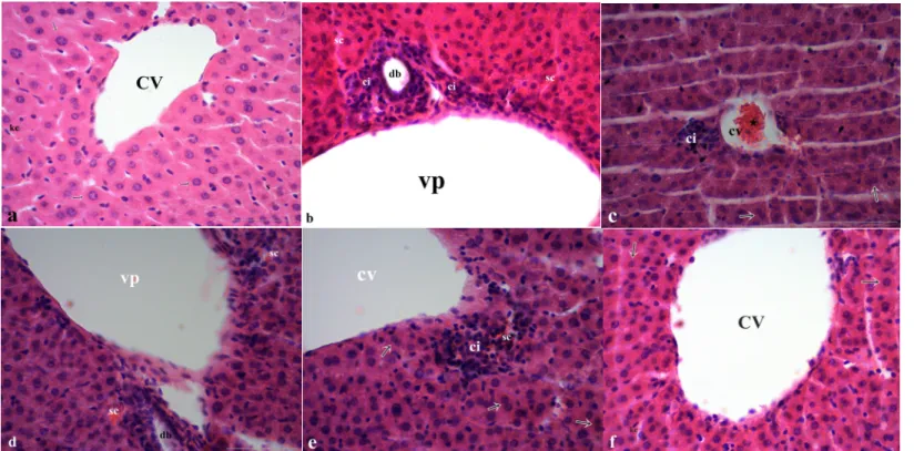
the 5 μm serial sections taken with a microtome from these blocks were examined at light microscopic level after staining with haematoxylin-eosin. Histopathological examinations of the sections obtained from the groups showed that the vena centralis and portal area appeared normal and that the hepatocyte sequence was regular in the control and CAPE groups. In the sections obtained from the malathion-treated group, hyperaemia was identified around the vein (central and portal), and cell infiltrations, necrotic and degenerative regions were determined around the central and portal vein.
In the group in which CAPE was administered as a protection against malathion, the same lesions as in the malathion group (central and portal vein congestion and surrounding infiltrations) were detected although the severity and frequency of lesions were diminished, (Table 1, Figure 1a, b, c, d, e, f).
Figure 1. The effect of CAPE (10 µmol/kg), corn oil (5 ml/kg) and malathion (40 mg/kg) either separately or 1 h ago malathion histology of liver section by using
hematoxylin and staining (H&E): (a) control group: hepatocytes (arrows), s: sinusoid, kc: kupffer cells, cv: central veins, (b, c) Malathion group: regenerative binuclear hepatocytes (arrows), ci: cell infiltrations, sc: sinusoidal congestion, vp: portal veins, db: ductus biliferi, (d, e) Malathion+CAPE group: sc: sinusoidal congestion, regenerative binuclear hepatocytes (arrows), ci: cell infiltrations, cv: central veins, vp: portal veins, db: ductus biliferi, (f) CAPE group: hepatocytes (arrows), kc: kupffer cells, cv: central veins, (bar: 50 µm).
Biochemical Findings
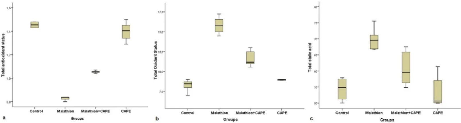
Total antioxidant, total oxidant and total sialic acid levels were measured in the obtained plasma samples. As a result of statistical comparison of biochemical data of the groups, there was no statistically significant difference between the control and CAPE groups in terms of total antioxidant capacity (p>0,05). But the statistical difference between the malathion group and all other groups was significant (p<0.05). Again, the difference between the group in which malathion and CAPE were administered together and all other groups was significant (p<0.05). In the evaluation of total oxidant capacity, the difference between the control and CAPE groups was insignificant (p>0.05) whereas the difference between the malathion group and all other groups was significant (p<0.05). Similarly, the difference between the malathion+CAPE group and all other groups was significant (p<0.05). Finally, when the groups were compared in terms of total sialic acid levels, no statistically significant difference was observed between malathion
and CAPE groups (p>0,05), while the malathion group and the malathion+CAPE group were statistically different both from each other and also from all other groups (p<0,05) (Table 2, Figure 2a, b, and c).
Table 2. TAS, TOS and TSA activities as a result of biochemical data obtained
from the groups
Figure 2. Box plot display of CAPE on TAS, TOS and TSA values in rats treated with subacute malathion
a. TAS, b. TOS, c. TSA levels
Discussion
The widespread use of organophosphate insecticides can lead to poisoning and even death in humans and many other living beings. Among pesticides, organophosphates are the most commonly used group around the world and cause the greatest number of poisoning cases globally.
The reason organophosphate pesticides are preferred over organochlorine compounds is because they are degraded faster [30]. However, it is known that these chemicals used are dangerous for the environment and public health [31]. Organophosphate compounds constitute 50% of the total poisoning rate among the pesticides [32]. Many of those used as insecticides are dimethoxy and diethoxy compounds. This group of pesticides contains the best known insecticides of malathion and diazinon. Because of this prevalence of use, malathion was chosen as the pesticide material of our study. Malathion toxicity is associated with acetylcholinesterase inhibition, oxidative stress induction, liver damage, and impaired renal function. In particular, detoxification of toxic substances or drugs entering the body via the oral route is performed in the liver [33]. Based on the histopathological examinations in our study, it was observed that the vena centralis and portal area appeared normal and that the hepatocyte sequence was regular in the control and CAPE groups. In the sections obtained from the malathion-treated group, hyperaemia was identified around the vein (central and portal), and cell infiltrations, necrotic
and degenerative regions were determined around the central and portal vein. In a study investigating the effects of malathion in rats, it was reported that a high dose of malathion caused damage to the liver tissues of rats [34]. Another study reported that malathion given for one month resulted in deterioration and damage of liver structure, and displayed histology of necrosis in many cells, and these findings are similar to our results. It was reported that malathion-exposed rats have increased vacuole formation in the liver, enlarged sinusoids, leukocytic infiltrates, and vaso-dilatation due to occlusion [35]. In another study, the livers of malathion-treated animals were reported to have mononuclear cell infiltration, bleeding, calcification, vacuolar degeneration, enlargement of sinusoids, vascular occlusion and necrosis [36]. All of the findings based on the malathion used in these studies are in parallel with the results of our study.
Reactive oxygen species (ROS) are produced in the cell in normal metabolic processes and transduction conditions, but also play an important role in pathophysiological processes. Biomolecules such as lipids, proteins and nucleic acids undergo degeneration by reactive oxygen species [37,38]. Total antioxidant, total oxidant and total sialic acid levels were measured in the plasma samples obtained from our study. In terms of total antioxidant capacity, there was no statistical difference between control and CAPE groups (p>0.05). The statistical difference between the malathion group and all other groups is significant (p<0.05). Again, the difference between the group in which malathion and CAPE were administered
together and all other groups was significant (p<0.05). Similarly, total oxidant capacity and total sialic profiles were parallel to total antioxidant capacity. Ekremoğlu reported that the total oxidant level increased due to increasing doses when malathion was administered acutely (24 hours) at different doses (100 mg kg-1, 200 mg kg-kg-1, 400 mg kg-1) [39]. In a study investigating the in vivo effects of malathion, Glutation-S-transferase (GST) and acetylcholinesterase (AChE) activities were evaluated in different organs of newborn rats. Accordingly, in adult female rats fed malathion, liver GST activity was increased about 2-fold, while AChE activity was reduced by more than 20% compared with the control. Similarly, liver and heart GST activities (0.14 and 0.035 U mg-1 protein, respectively) increased approximately 2-fold in neonatal rats, but a decrease was observed in brain GST activity (0.074 U mg-1 protein). Significant decreases were also observed in liver, brain, kidney and lung AChE activities (0.002, 0.035, 0.046, and 0.018 U/mg protein, respectively) compared with the control [40]. In an in vivo study with dichlorvos, the total antioxidant levels in the dichlorvos group were significantly lower than the other groups (p<0.05), whereas the total antioxidant values in the CAPE groups were significantly higher than the dichlorvos+CAPE group (p<0.05). In the same study, total oxidant values were significantly lower in the control and CAPE groups than in the dichlorvos group. No significant difference was observed between the other groups [9]. In a study investigating the protective effect of CAPE in malathion-treated rats, it was reported that malathion disrupts the enzyme activity of AChE, GP (glycogen phosphorylase) and HK (hexokinase) but improves AChE, GP and HK activities of the group in which malathion was given with CAPE [41]. In another study investigating the acute protective effect of CAPE against poisoning by chlorpyrifos-ethyl (CPF), an organophosphate compound, paraoxonase (PON1) activity and TAS levels were reduced while oxidative stress index and TOS levels were found to increase in the context of a CPF-induced intoxication. CAPE was reported to have a protective effect by reducing the oxidative stress caused by CPF [16]. In one study, malathion was reported to reduce the expression of catalase and superoxide dismutase-2 from antioxidant enzymes, thus suppressing the antioxidant system [42]. One study reported that the amount of malondialdehyde (MDA) increased and glutathione (GSH) levels decreased with increasing reactive oxygen species after tetramethrin administration. In the same study, reduction of MDA and increase in GSH levels were observed due to the free radical scavenging properties of CAPE by administration of CAPE with tetramethrin [15].
Conclusion
In conclusion the current study determined that malathion caused cell infiltrations, degeneration and focal necrosis in liver tissue, while CAPE reduced the severity of histopathological lesions caused by malathion. At the same time, it was observed that malathion increased total oxidant and sialic acid level and decreased total antioxidant compared with the control group. On the other hand, CAPE was observed to reduce the harmful effects of malathion and bring biochemical parameters closer to the control group. It was found that in rats administered CAPE together with malathion, CAPE reduced the degeneration in the liver and could have a protective effect by partially correcting the oxidative stress parameters. These data show that malathion, an organophosphate compound, has a toxic effect on the liver and
CAPE, an important antioxidant molecule, may have a protective effect on the antioxidant system of the organism against the oxidative stress caused by organophosphate pesticides, and that intake of CAPE and other antioxidant molecules in advance has a protective potential against cases of poisoning among those people who are engaged in agriculture.
Competing interests
The authors declare that they have no competing interest Financial Disclosure
The financial support for this study was provided by the investigators themselves. Ethical approval
Before the study, permissions were obtained from local ethical committee.
References
1. Nur G, Deveci MA, Kırpık MA, ark. Sürdürülebilir üretim yaklaşımı: Ekolojik tarım. Kafkas Üniv Fen Bil Enst Derg. 2016;9:3-8.
2. Moses V, Peter JV. Acute intentional toxicity: endosulfan and other organochlorines. Clin Toxicol (Phila). 2010;48:539-44.
3. Kesavachandran CN, Fareed M, Pathak MK, et al. Adverse health effects of pesticides in agrarian populations of developing countries. Rev Environ Contam Toxicol. 2009;200:33-52.
4. Nili-Ahmadabadi A, Pourkhalili N, Fouladdel S, et al. On the biochemical and molecular mechanisms by which malathion induces dysfunction in pancreatic islets in vivo and in vitro. Pestic Biochem Physiol. 2013;106:51-60. 5. Bogen KT, Singhal A. Malathion dermal permeability in relation to dermal
load: Assessment by physiologically based pharmacokinetic modeling of in vivo human data. J Environ Sci Health B. 2017;52:138-46.
6. Deveci HA, Ünal S, Karapehlivan M, et al. Effects of glyphosate (herbicide) on serum paraoxonase activity, high density lipoprotein, total antioxidant and oxidant levels in Kars creek transcaucasıan barbs (Capoeta capoeta [Guldenstaedt, 1773]). Fresen Environ Bull. 2017;26:3514-8.
7. Deveci HA, Karapehlivan M. Chlorpyrifos-induced parkinsonian model in mice: Behavior, histopathology and biochemistry. Pestic Biochem Physiol. 2018;144:36-41.
8. Gorecki L, Korabecny J, Musilek K, et al. SAR study to find optimal cholinesterase reactivator against organophosphorous nerve agents and pesticides. Arch Toxicol. 2016;90:2831-59.
9. Alp H, Aytekin İ, Atakişi O, et al. The effects of caffeic acid phenethyl ester and ellagic acid on oxidative stress created by acute malathion toxicity in rat. Atatürk Üniversitesi Vet Bil Derg. 2011;6:117-24.
10. Bossi R, Vinggaard AM, Taxvig C, et al. Levels of pesticides and their metabolites in wistar rat amniotic fluids and maternal urine upon gestational exposure. Int J Environ Res Public Health. 2013;10:2271-81.
11. Raoofi R, Jahromi HK, Jahromi ZK, et al. Antioxidant effects of green-tea on biochemical and histopathological changes of liver in male rats poisoned by malathion insecticide. Int J Med Res Health Sci. 2016;5:361-70.
12. Selmi S, Tounsi H, Safra I, et al. Histopathological, biochemical and molecular changes of reproductive function after malathion exposure of prepubertal male mice. RSC Adv. 2015;5:13743-75.
13. Lasram MM, Bouzid K, Douib IB, et al. Lipid metabolism disturbances contribute to insulin resistance and decrease insulin sensitivity by malathion exposure in wistar rat. Drug Chem Toxicol. 2015;38:227-34.
14. Lasram MM, Douib IB, Bouzid K, et al. Effects of N-acetyl-L-cysteine, in vivo, against pathological changes induced by malathion. Toxicol Mech Methods. 2014;24:294-306.
15. Nur G, Deveci HA, Ersan Y, et al. Protective role of caffeic acid phenethyl ester against tetramethrine-induced toxicity in mice. Med-Science. 2016;5:972-8.
16. Deveci HA, Karapehlivan M, Kaya İ, et al. Akut klorprifos-etil zehirlenmesine karşı kafeik asit fenetil ester’in koruyucu etkisi. Ankara Üniv Vet Fak Derg. 2015;62:255-60.
17. Murtaza G, Karim S, Akram MR, et al. Caffeic acid phenethyl ester and therapeutic potentials. BioMed Res Int. 2014;2014:145342.
18. 1Ozturk G, Ginis Z, Akyol S, et al. The anticancer mechanism of caffeic acid phenethyl ester (CAPE): review of melanomas, lung and prostate cancers. Eur Rev Med Pharmacol Sci. 2012;16:2064-8.
19. Akyol S, Ozturk G, Ginis Z, et al. In vivo and in vitro antineoplastic actions of caffeic acid phenethyl ester (CAPE): Therapeutic perspectives. Nutr Cancer. 2013;65515-26.
20. Hepsen IF, Bayramlar H, Gultek A, et al. Caffeic acid phenethyl ester to inhibit posterior capsule opacification in rabbits. J Cataract Refract Surg. 1997;23:1572-6.
21. Natarajan K, Singh S, Burke TR Jr, Grunberger D, Aggarval BB. Caffeic acid phenethyl ester is a potent and specific inhibitor of activation of nuclear transcription factor NFkappaB. Proc Natl Acad Sci. 1996;93(17):9090-9095. 22. Mirzeova OK, Calder PC. The effects of propolis and its components on
eicosanoid production during the inflammatory response. Prostaglandins Leukot Essent Fatty Acids. 1996;55:441-9.
23. Hepşen İF, Tilgen F, Er H. Propolis: Tıbbi özellikleri ve oftalmolojik kullanımı. Turgut Özal Tıp Merk Derg. 1996;3:386-91.
24. Sud’ina GF, Mirzoeva OK, Pushkareva MA, et al. Caffeic acid phenethyl ester as a lipoxygenase inhibitor with antioxidant properties. FEBS Lett. 1993;329:21-4.
25. Anwar J, Spanevello RM, Thomé G, et al.. Effects of caffeic acid on behavioral parameters and on the activity of acetylcholinesterase in different tissues from adult rats. Pharmacol Biochem Behav. 2012;103:386-94. 26. Luna LG. Manual of histologic staining methods of armed forces institute of
pathology. 3rd Edition. MC. Graw. Hill Book Comp. London, 1968. 27. Erel O. A novel automated direct measurement method for total antioxidant
capacity using a new generation, more stable abts radical cation. Clin Biochem. 2004;37:277-85.
28. Erel O. A new automated colorimetric method for measuring total oxidant status. Clin Biochem. 2005;38:1103-11.
29. Sydow G. A simplified quick method for determination of sialic acid in serum. Biomed Biochim Acta. 1985;44:1721-3.
30. John S, Kale M, Rathore N, et al. Protective effect of vitamin E in dimethoate
and malathion induced oxidative stress in rat erythrocytes. J Nutr Biochem. 2001;12:500-4.
31. Ojha A, Reuben AC, Sharma D. Solid Waste Management in Developing Countries through Plasma Arc Gasification- An Alternative Approach. APCBEE Proc. 2012;1:193-18.
32. Karami-Mohajeri S, Abdollahi M. Toxic influence of organophosphate, carbamate, and organochlorine pesticides on cellular metabolism of lipids, proteins, and carbohydrates: A systematic review. Hum Exp Toxicol. 2011;30(9):1119-40.
33. Lasram MM, Lamine AJ, Dhouib IB, et al. Antioxidant and antiinflamatory effects of N-acetylcystein against malathion-induced liver damages and immuntoxicity in rats. Life Sci. 2014;107:50-8.
34. Tos-Luty S, Obuchowska-Przebirowska D, Latuszynska J, et al. Dermal and oral toxicity of malathion in rats. Ann Agric Environ Med. 2003;10:101-6. 35. Al-Attar AM. Physiological and histopathological investigations on the
effects of α-lipoic acid in rats exposed to malathion. J Biomed Biotechnol. 2010;2010:203503.
36. Kalender S, Uzun FG, Durak D, et al. Malathion-induced hepatotoxicity in rats: The effects of vitamins C and E. Food Chem Toxicol. 2010;48:633-8. 37. Deveci HA, Nur G, Kukurt A. Biochemical and histopathological changes of
babesiosis in naturally infected sheep in Gaziantep region. Fresen Environ Bull. 2017;26:4883-9.
38. Deveci HA, Kükürt A, Nur G, et al. Serum paraoxonase activity and total sialic acid in sheep with foot and mouth disease. Med Weter. 2018;74:199-202.
39. Ekremoğlu M. Malathionun, rat serumunda oksidan ve antioksidan sistem, inflamatuar belirteçler ve bazı metabolik biyomoleküller üzerine etkilerinin incelenmesi. Doktora Tezi, Gazi Üniversitesi, Sağlık Bilimleri Enstitüsü, Ankara, 2016.
40. Tümur S, Önal S, Karabay NÜ, et al. In vivo effects of malathion on Glutathione-S-Transferase and acetylcholinesterase activities in various tissues of neonatal rats. Turk J Zool. 2003;27:247-52.
41. Rezg R, Mornagui B, El-Fazaa S, et al. Caffeic acid attenuates malathion induced metabolic disruption in rat liver, involvement of acetylcholinesterase activity. Toxicol. 2008;250:27-31.
42. Bakır B, Erdağı D, Yıldız SE et al. Immunohistochemical examination on the effects of malathion and Onosma nigricaule (Boraginaceae) on the catalase (CAT) and superoxide dismutase-2 (Mn-SOD) in renal tissues of mice. Ankara Üniv Vet Fak Derg. 2017;64:125-30.