lithium disilicate glass ceramic after various
surface treatments
H. B. Kara*
1, O. Kara
2, G. Sayin
3, U. Cakan
1and A. N. Ozturk
3The aim of this study is to evaluate the influence of various surface treatments on the surface
roughness of a lithium disilicate glass ceramic. A total of 25 discs of lithium disilicate glass ceramic
were prepared. Specimens were divided into five groups: (1) no treatment, (2) air abrasion, (3) acid
etching, (4) Nd:YAG laser irradiation and (5) Er:YAG laser irradiation. Digital images from the
surfaces were obtained by means of an atomic force microscope in tapping mode. Surface
roughness was measured in 10610 mm boxes. Surface roughness data were analysed by one way
analysis of variance. The air abrasion group (550¡212 nm) had a significantly higher mean surface
roughness value than the other groups. The acid etching group (392¡157 nm) had a significantly
higher mean roughness value than the control group (228¡105 nm). Surface roughness of lithium
disilicate glass ceramic was not increased by any of the laser irradiation treatments.
Keywords: Laser, Surface roughness, Ceramic, Dental materials
Introduction
All ceramic restorations have gained popularity among dentists and patients because of their favourable aes-thetics and durability in conservative tooth preparations.1 The increasing demand for metal free fixed partial den-tures has fuelled the development of all ceramic materials with optimised mechanical properties.2 In order to im-prove strength and durability over glass ceramics, the addition of reinforcement crystals such as lithium disili-cate (LiSi2O5) in the glass matrix of the ceramics is
considered. A lithium disilicate glass ceramic core, ve-neered with a sintered glass ceramic, provides higher strength and allows fabrication of short span fixed restorations.3 The adhesive technology used for fixing current all ceramic restorations involves the use of resin based luting materials.4,5Some authors postulate that the advantages of adhesion of all ceramic crowns and bridges are due to improved retention, better marginal adapta-tion and fracture resistance.6
New self-adhesive cements that rely on single step application have been proposed for luting ceramic based restorations.7–9 These are characterised by a simplified application procedure and avoidance of the time consum-ing rinsconsum-ing step.10 When using these cements, microme-chanical retention seems to become a crucial factor in their bonding performance.9The internal surface of the ceramic
restoration must be prepared to optimise micromechanical retention of the cement into the ceramic microroughness. Surface treatment of porcelain increases the surface area and creates microporosities on the surface while enhancing the potential for mechanical retention of the cement.11
Different conditioning systems have been proposed to increase surface roughness and promote micromechanical retention.12,13 Air abrasion is abrasion with airborne particles of aluminium oxide using a chair side device. This procedure increases the surface area and surface energy for adhesion of resin cements and promotes micro-mechanical retention. It also decreases surface tension, thereby enabling optimal wetting of silanes or adhesive media.14Furthermore, the silane coupling agents present bifunctional characteristics, promoting a chemical interac-tion between the silica in the glass phase of ceramics and the methacrylate groups of the resin through siloxane bonds.15 Hydrofluoric (HF) acid selectively attacks the glass matrix, producing a retentive surface for micro-mechanical bonding. However, decreasing glass content, thus increasing the mechanical strength by exposing crystalline content, results in an acid resistant ceramic, whereby any type of acid treatment produces insufficient surface changes for adequate bonding to resin.16Various applications of dental lasers on dental materials have also been proposed for surface modification, such as forming a glazed surface layer on ceramics, the removal of resin composite filling materials, laser welding of ceramics and metal alloys, including titanium, and increasing the corrosion resistance of metal alloys.17
Atomic force microscopy (AFM) characterises the surface topography of different materials with near atomic resolution, requires minimal preparation of specimens and is capable of evaluating the surface three-dimensionally
1Department of Prosthodontics, Faculty of Dentistry, Istanbul Medipol
University, Istanbul, Turkey
2Department of Prosthodontics, Faculty of Dentistry, Bezm-i Alem
University, Istanbul, Turkey
3Department of Prosthodontics, Faculty of Dentistry, Selcuk University,
Konya, Turkey
Experimental
Twenty-five disc shaped wax (IQ Sculpturing Wax; YETI Dentalprodukte, Engen, Germany) patterns in 10 mm diameter and 2 mm thickness were prepared using a custom made metal mould for the fabrication of lithium disilicate based all ceramic discs. The wax patterns were invested in IPS Empress e.max investment and eliminated in a burnout furnace (Dentsply Vulcan A-550 Burnout Furnace). By heating the refractory die at the same time, the IPS Empress e.max ingots (lot no. R85375 IPS e.max Press, Ivoclar Vivadent, Schaan, Liechtenstein) and the alumina plunger were heated at 3uC min21to 850uC and
held for 90 min. After the procedure described, the investment, plunger and ingot were transferred to a furnace (EP 500; Ivoclar-Vivadent, Schaan, Liechtenstein) that increased the temperature to 1180uC and automati-cally pressed the melted ingot to the mould. After pressing and cooling to room temperature, the specimens were divested with 50 mm glass beads at 2 bar pressure, ultra-sonically cleaned in a special liquid (Invex liquid; Ivoclar-Vivadent) for 10 min, washed in running water and dried. No glaze was applied to the ceramic surface of the discs. The bonding surface of each disc was polished using silicon carbide paper (grit 300 and 400) on a rotating metallographic polishing device (Isomet 1000, Buehler, Lake Bluff, IL, USA) under water cooling. The surfaces were cleaned with ethanol and dried carefully in air before surface treatment. After the finishing procedures, the discs were subjected to ultrasonic treatment (Biosonic UC 50, Coltene Whaledent, OH, USA) in distilled water to re-move any surface residues and dried. Thereafter, the ceramic discs were randomly divided into five groups, according to the surface treatments applied:
(i) group I (control): no surface treatment was applied to this group
(ii) group II (air abrasion): air abrasion was carried out with 50 mm aluminium oxide particles (Korox; Bego, Bremen, Germany) at a pressure of 2?8 bar, from a distance of 10 mm, perpen-dicular to the treated surface for 20 s; specimens were rinsed after air abrasion and then dried with oil free compressed air
(iii) group III (HF acid etching): treatment surfaces of ceramic discs were etched with 9% HF acid (lot no. B74V5 Ultradent Porcelain Etch, Ultradent Dental Products, UT, USA) for 60 s; the etching gel was neutralised using a neutralising powder (calcium carbonate, CaCO3and sodium
carbo-nate, Na2CO3) for 5 min; the neutralising powder
was used for irradiation of the ceramic surfaces; a contact hand piece (R14) (1?3 mm in dia-meter) with an integrated spray nozzle was placed perpendicular to the ceramic surface at a 1 mm distance, and the entire ceramic area was manually scanned with water cooling; the laser parameters were as follows: 500 mJ (pulse energy), 10 W (power), MSP mode (100 ms pulse length), 20 Hz (pulses per second) and 37?68 J cm22(energy density).
Atomic force microscopy examination
Five ceramic surfaces per group were evaluated under AFM (NTEGRA Solaris, NTMDT, Russia). Digital images were obtained in air. A 0?01–0?025 V cm gold doped (Au doped) silicon tip (40 nm) was used in semicontact mode. Changes in vertical position provided the height of the images and were registered as bright and dark regions. A constant tip sample ‘tap’ was maintained through a constant oscillation amplitude (set point amplitude). Five 25625 mm digital images were acquired for each surface and recorded with a slow scan rate (1 Hz). In each image, four randomised boxes 10610 mm were created. Nanoroughness (Ra) was automatically assessed as a numeric value (in nano-metres) with specific software (Nova plus 1?1?0?1773), and mean image roughness was calculated. Each image was considered as a statistical unit; sample size per group was n520.
Statistical analysis
Surface roughness Radata were analysed with one way
analysis of variance. Roughness was the dependent variable, while surface treatment was the influencing factor. The level of significance was set at p,0?05.
Results
Mean roughness Ra(nm) and standard deviations are
presented in Table 1. One way analysis of variance indicated that surface roughness was significantly affected by the surface treatment methods (p,0?05). The results of the Tukey’s honestly significant difference test comparing the roughness values between the groups are presented in Table 1. The air abrasion group had a significantly higher mean roughness value (p,0?05) than the other groups. The acid etching group had a significantly higher mean roughness value than the control group (p,0?05). No significant difference was found between the control and laser irradiation (Er:YAG and Nd:YAG) groups (p.0?05)
(Table 1). The difference between acid etching and laser irradiation groups was not statistically significant (p.0?05) (Table 1).
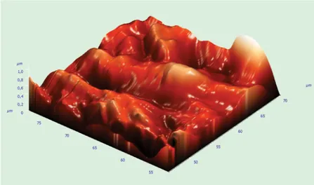
Atomic force microscopy images of the ceramic surfaces treated with the different techniques are presented in Figs. 1–5. Laser irradiated (Er:YAG, Nd:YAG) surfaces showed moderate irregularity with peaks and valleys, and showed less roughness than air abraded surfaces. Acid etched surfaces were slightly rougher than laser irradiated surfaces, and the peaks were higher and wider than those of the laser irradiated surfaces. The air abrasion group had the most distinct sharp peaks among the groups.
Discussion
Self-adhesive cements have been proposed for luting ceramic based restorations.7,9The adhesive properties of these cements are claimed to be based upon acidic monomers that demineralise and infiltrate the tooth substrate, resulting in micromechanical retention. Secon-dary reactions have been suggested to provide chemical adhesion to hydroxyapatite, a feature currently only proven for glass ionomers.21Intaglio surfaces of ceramic based restorations require pretreatment, such as air abra-sion, acid etching or tribochemical silica coating, to pro-duce a micromechanical retentive ceramic surface.9,22,23 Several pretreatment procedures, such as acid etching and laser irradiation, have been reported and used clinically.24–26 Atomic force microscopy analysis is a commonly used technique to observe surface details. It has many advantages, such as providing detailed surface information with high resolution and providing quanti-tative information (dimensions, profile, roughness, peri-odicity) related to the surface.27 In the present study, AFM analysis was used quantitatively to demonstrate the morphologic changes of lithium disilicate ceramic after different surface treatments and to interpret these with the roughness analysis.
Although many studies evaluated the effect of lasers on hard tissues,28–35their effect on ceramic surfaces has
1 Image (AFM) of untreated IPS Empress e.max surface
Table 1 Surface roughness Ra values of each treatment
group (means¡SD)/nm
Treatment group
Roughness value
Ra(mean¡SD) Post hoc
Air abrasion (a) 550 (¡212)
HF acid etching (b) 392 (¡157) a.b,c,d,e*
Nd:YAG (c) 303 (¡103) b.d*
Er:YAG (d) 300 (¡91)
Control (e) 228 (¡105)
*p,0?01.
not been thoroughly investigated. The null hypothesis of the present study was accepted, since air abrasion and HF acid etching significantly increased the surface roughness of lithium disilicate based core ceramic compared to untreated surfaces, while Er:YAG and Nd:YAG laser irradiation did not.
Micromechanical retention was achieved by rough-ening the ceramic surface with 50 mm grain sized pure aluminium oxide particles. Surface roughening increases the total bonding area as well as the wettability of the ceramic with the resin substrate.17 Soderholm and Shang15stated that air abrasion was the most effective surface treatment for In-Ceram Alumina ceramic. Dilber et al.19showed that air abrasion provides sufficient bond strength. However, excessive air abrasion induces chip-ping or a significant loss of ceramic material and is not recommended for cementation of silica based and feldspathic ceramic restorations.36,37In the present study, air abraded specimens showed the highest surface rough-ness, and AFM images of air abraded specimens showed a non-uniform surface with projections dotted with pores. In addition, Ersu et al.22 demonstrated that the air abrasion provides rougher surfaces than laser irradiation and acid etching, which is in agreement with our study.
HF acid dissolves glassy components of ceramics and creates a microporous irregular surface, thereby rough-ening the surface area and facilitating penetration of the
resin into the etched ceramic surface.19,23 Previous studies38,40 demonstrated that HF acidzsilane was consistently the most effective ceramic surface treat-ment, although it was not always significantly better than the other treatments that involved roughening and silane application.41 In the present study, there were significant differences between the HF acid etching and control groups. HF acid is well recognised to have hazardous effects in vivo; it is harmful and irritating to soft tissues.42Szep et al.43reported that HF acid leaves an amorphous precipitate of fluoride on tooth struc-tures, which may negatively influence caries resistance and the bonding interaction that acid etching creates. Most adhesively bonded restorations are etched using HF acid in the lab before being cemented in the mouth. They are then thoroughly rinsed in water, and a neutralising gel is used to reduce any harmful risk. HF acid etching is therefore in practical use throughout dentistry.
Laser treatments may form conchoidal tears on the ceramic surface, which are assumed to be a typical effect of surface warming, and these tears may contribute to mechanical retention between the resin composite and ceramics. On the other hand, the harmful effect of internal tensions due to steep local temperature changes in heating and cooling phases of laser irradiation has been reported.22 Nd:YAG laser irradiation has been
3 Image (AFM) of IPS Empress e.max surface treated with HF acid etching
proposed for surface modification of ceramics.16Osorio et al.9evaluated the effect of different surface treatments (sandblasting, Nd:YAG laser irradiation, Rocatec sys-tem) on In-Ceram Alumina roughness. In their AFM study, 2 W (100 mJ5pulse at 15 Hz) output power was selected for laser irradiation, and similar topographic AFM images were found for all surface treatments. Akyıl et al.44 reported that HF acid etching provides higher shear bond strength than Nd:YAG laser (1 W, 100 mJ per pulse at 10 Hz) etching. Kara et al.45 reported that there were no significant differences in the shear bond strength of a composite resin to ceramic after Nd:YAG laser irradiation (2 W, 100 mJ5pulse at 20 Hz) and 5%HF acid etching. In the present study, 2 W output power (100 mJ5pulse at 20 Hz) was used for laser irradiation, and the difference between HF acid etching and Nd:YAG laser irradiation groups was not statistically different.
The extent of the superficial changes on the ceramic surface depends on the energy density of the laser radiation as well as on the type of irradiated ceramic.41 Go¨kc¸e et al.25 reported that the shear bond strength after laser treatment at 300 mJ was higher than after treatment at 600–900 mJ, and the reason for the low bond strengths observed after laser treatment at high power settings could have been related to the observed heat damaged layer. This layer might be poorly attached to the infralayers of the substrate, while the outermost layer of the substrate remained strongly bonded to the silane and luting agents. Sorensen et al.12 found that laser treated surfaces (10 W output power, 500 mJ per pulse at 20 Hz) were not significantly different from HF acid treated IPS Empress 2 ceramic surfaces. In the present study, Er:YAG laser irradiation of a IPS Empress e.max ceramic surface at 10 W output power, 500 mJ per pulse at 20 Hz, did not lead to adequate roughening of the surface because of the composition of the ceramic and its reflectance. Shallow irregularities due to erosion and melting were observed, but no fissures or cracks were observed on the surface after Er:YAG laser irradiation using AFM. Even though the topological features of the acid etched surface (Fig. 3) were more morphologically developed than the laser irradiated samples (Figs. 4 and 5), the difference between surface roughness values of HF acid etching and Nd:YAG laser irradiation groups was not statistically different.
The differences of surface treatment modalities hereby evaluated for their potential in providing surface roughness on lithium disilicate glass ceramic might be important in obtaining proper bond strength between ceramic and teeth. There were some limitations in this study. Only one type of ceramic material (IPS Empress e.max) was evaluated. Differences in the composition and microstructure of different ceramic materials may affect the surface texture and bond strength between the ceramics and resin cement. In addition, the effects of different HF acid concentrations and application periods have not been evaluated. Further studies are required to evaluate the effects of different power settings and different laser applications on the surface roughness of different ceramic surfaces.
Conclusions
Based on the results of the statistical analysis and the AFM evaluations, the following conclusions could be drawn.
1. Air abrasion created rougher surfaces than the other surface treatment methods (acid etching, Er:YAG and Nd:YAG laser irradiation; p,0?05).
2. There were no significant differences between surface roughness values of Nd:YAG laser irradiation, Er:YAG laser irradiation and the non-treated group (p,0?05).
3. The acid etching method created rougher surfaces than the non-treated control group (p,0?05).
Acknowledgement
The authors would like to thank Dr Seden Akan for generous contribution to the statistical analysis of the study.
References
1. M. O¨ zcan, H. Nijhuis and L. Valandro: ‘Effect of various surface conditioning methods on the adhesion of dual-cure resin cement with MDP functional monomer to zirconia after thermal aging’, Dent. Mater., 2008, 27, 99–104.
2. S. Rosenstiel, M. Land and J. Fujimoto: (2006) ‘Contemporary fixed prosthodontics’, 4th edn, 909–925; 2006, St Louis, Elsevier Health Sciences.
3. W. Ho¨land. M. Schweiger, V. M. Rheinberger and H. Kappert: ‘Bioceramics and their application for dental restoration’, Appl. Ceram., 2009, 108, (6), 373–380.
10. C. Besnault, J. P. Attal, D. Ruse and M. Degrange: ‘Self etching adhesives improve the shear bond strength of a resin-modified glass-ionomer cement to dentin’, J. Adhes. Dent., 2004, 6, (1), 55– 59.
11. G. A. Borges, A. M. Sophr, M. F. de Goes, L. Correr and D. C. N. Chan: ‘Effect of etching and air borne particle abrasion on the microstructure of different dental ceramics’, J. Prosthet. Dent., 2003, 89, 479–488.
12. J. A. Sorensen, M. J. Engelman, T. J. Torres and S. P. Avera: ‘Shear bond strength of composite resin to porcelain’, Int. J. Prosthodont., 1991, 4, 17–23.
13. M. Aida, T. Hayakawa and K. Mizukawa: ‘Adhesion of composite to porcelain with various surfaces conditions’, J. Prosthet. Dent., 1995, 73, 464–470.
14. E. P. Plueddemann: ‘Adhesion through silane coupling agents’, J. Adhes., 1970, 2, 184–201.
15. K. J. Soderholm and S. W. Shang: ‘Molecular orientation of silane at the surface of colloidal silica’, J. Dent. Res, 1993, 72, 1050–1054. 16. D. J. Wood, N. L. Bubb, B. J. Millar and S. M. Dunne: ‘Preliminary investigation of a novel retentive system for hydro-fluoric acid etching resistant dental ceramics’, J. Prosthet. Dent., 1997, 78, 275–280.
17. H. B. Kara, E. Dilber, O. Koc, A. N. Ozturk and M. Bulbul: ‘Effect of different surface treatments on roughness of IPS Empress 2 ceramic’, Lasers Med. Sci., 2012, 27, 267–272.
18. A. C. P. de Freitas, L. C. Espejo, S. B. Botta, F. de Sa Teixeira, M. A. A. Cerqueira Luz, N. Garone-Netto, A. B. Matos and M. C. B. da Silveira Salvadori: ‘AFM analysis of bleaching effects on dental enamel microtopography’, Appl. Surf. Sci., 2010, 256, 2915– 2919.
19. E. Dilber, T. Yavuz, H. B. Kara and A. N. Ozturk: ‘Comparison of the effects of surface treatments on roughness of two ceramic systems’, Photomed. Laser Surg., 2012, 30, 308–314.
20. R. P. Sanches, C. Otani, A. J. Damiao and W. Miyakawa: ‘AFM characterization of bovine enamel and dentine after acid-etching’, Micron, 2009, 40, 502–506.
21. J. De Munck, M. Vargas, K. Van Landuyt, K. Hikitaa, P. Lambrechts and B. Van Meerbeek: ‘Bonding of an autoadhesive luting materials to enamel and dentin’, Dent. Mater., 2004, 20, 963–971.
22. B. Ersu, B. Yuzugullu, A. R. Yazici and S. Canay: ‘Surface roughness and bond strengths of glass-infiltrated alumina-ceramics prepared using various surface treatments’, J. Dent., 2009, 37, 848– 856.
23. M. Ozcan and P. K. Vallittu: ‘Effect of surface conditioning methods on the bond strength of luting cement to ceramics’, Dent. Mater., 2003, 19, 725–731.
24. M. Aida, T. Hayakawa and K. Mizukawa: ‘Adhesion of composite to porcelain with various surface conditions’, J. Prosthet. Dent., 1995, 73, 464–470.
25. B. Go¨kc¸e, B. O¨ zpinar, M. Du¨ndar, E. C¸o¨mlekoglu, B. H. Sen and M. A. Gu¨ngo¨r: ‘Bond strengths of all-ceramics: acid vs laser etching’, Oper. Dent., 2007, 32, 173–178.
32. V. Armengol, O. Laboux, P. Weiss, A. Jean and H. Hamel: ‘Effects of Er:YAG and Nd:YAP laser irradiation on the surface roughness and free surface energy of enamel and dentin: an in vitro study’, Oper. Dent., 2003, 28, 67–74.
33. K. I. Delme and R. J. De Moor: ‘Scanning electron microscopic evaluation of enamel and dentin surfaces after Er:YAG laser preparation and laser conditioning’, Photomed. Laser Surg., 2007, 25, 393–401.
34. S. Usumez, M. Orhan and A. Usumez: ‘Laser etching of enamel for direct bonding with an Er,Cr:YSGG hydrokinetic laser system’, Am. J. Orthod. Dentofacial Orthop., 2002, 122, 649–656. 35. L. E. Rodriguez Vilchis, R. Contreras Bulnes, O. F. Olea Mejia,
I. Sanchez Flores and C. Centeno Pedraza: ‘Morphological and structural changes on human dental enamel after Er:YAG laser irradiation: AFM, SEM, and EDS evaluation’, Photomed. Laser Surg., 2011, 29, 493–500.
36. M. B. Blatz, A. Sadan and M. Kern: ‘Resin–ceramic bonding: a review of the literature’, J. Prosthet. Dent., 2003, 89, 268–274. 37. M. Kern and V. P. Thompson: ‘Sandblasting and silica coating of a
glass-infiltrated alumina ceramic: volume loss, morphology, and changes in the surface composition’, J. Prosthet. Dent., 1994, 71, 453–461.
38. A. al Edris, A. al Jabr, R. L. Cooley and N. Barghi: ‘SEM evaluation of etch patterns by three etchants on three porcelains’, J. Prosthet. Dent., 1990, 64, 734–739.
39. A. H. Suliman, E. J. Swift Jr and J. Perdigao: ‘Effects of surface treatment and bonding agents on bond strength of composite resin to porcelain’, J. Prosthet. Dent., 1993, 70, 118–120.
40. J. W. Thurmond, W. W. Barkmeier and T. M. Wilwerding: ‘Effect of porcelain surface treatments on bond strengths of composite resin bonded to porcelain’, J. Prosthet. Dent., 1994, 72, 355–359. 41. P. Shiu, W. C. De Souza-Zaroni, P. Eduardo Cde and M. N.
Youssef: ‘Effect of feldspathic ceramic surface treatments on bond strength to resin cement’, Photomed. Laser Surg., 2007, 25, 291– 296.
42. K. Kamada, K. Yoshida and M. Atsuta: ‘Effect of ceramic surface treatments on the bond of four resin luting agents to a ceramic material’, J. Prosthet. Dent., 1998, 79, 508–513.
43. S. Szep, T. Gerhardt, H. W. Gockel, M. Ruppel, D. Metzeltin and D. Heidemann: ‘In vitro dentinal surface reaction of 9.5% buffered hydro fluoric acid in repair of ceramic restorations: a scanning electron microscopic investigation’, J. Prosthet. Dent.., 2000, 83, 668–674.
44. M. S. Akyıl, A. Yılmaz, O. F. Karaaliog˘lu and Z. Y. Duymus¸: ‘Shear bond strength of repair composite resin to an acid-etched and a laser-irradiated feldspathic ceramic surface’, Photomed. Laser Surg., 2010, 28, 539–545.
45. H. B. Kara, A. N. Ozturk, F. Aykent, O. Koc and B. Ozturk: ‘The effect of different surface treatments on roughness and bond strength in low-fusing ceramics’, Lasers Med. Sci., 2011, 26, 599– 604.