Research Article
Neuroendocrinology 2020;110:258–270
MCH Neuron Activity Is Sufficient for
Reward and Reinforces Feeding
Pelin Dilsiz
aIltan Aklan
a, cNilufer Sayar Atasoy
a, cYavuz Yavuz
bGizem Filiz
aFulya Koksalar
aTayfun Ates
aMerve Oncul
aIlknur Coban
aEdanur Ates Oz
aUtku Cebecioglu
aMuhammed Ikbal Alp
aBayram Yilmaz
bDeniz Atasoy
a, caDepartment of Physiology, School of Medicine, Regenerative and Restorative Medical Research Center (REMER), Istanbul Medipol University, Istanbul, Turkey; bDepartment of Physiology, School of Medicine, Yeditepe University, Istanbul, Turkey; cDepartment of Pharmacology, Iowa Neuroscience Institute, Roy J. and Lucille A. Carver College of Medicine, University of Iowa, Iowa City, IA, USA
Received: December 12, 2018 Accepted after revision: May 31, 2019 Published online: June 3, 2019
DOI: 10.1159/000501234
Keywords
Melanin-concentrating hormone · Reward · Optogenetics · Chemogenetics · Glucose homeostasis
Abstract
Background: Melanin-concentrating hormone
(MCH)-ex-pressing neurons have been implicated in regulation of en-ergy homeostasis and reward, yet the role of their electrical activity in short-term appetite and reward modulation has not been fully understood. Objectives: We investigated short-term behavioral and physiological effects of MCH neu-ron activity manipulations. Methods: We used optogenetic and chemogenetic approaches in Pmch-cre transgenic mice to acutely stimulate/inhibit MCH neuronal activity while probing feeding, locomotor activity, anxiety-like behaviors, glucose homeostasis, and reward. Results: MCH neuron ac-tivity is neither required nor sufficient for short-term appe-tite unless stimulation is temporally paired with consump-tion. MCH neuronal activation does not affect short-term lo-comotor activity, but inhibition improves glucose tolerance and is mildly anxiolytic. Finally, using two different operant
tasks, we showed that activation of MCH neurons alone is sufficient to induce reward. Conclusions: Our results confirm diverse behavioral/physiological functions of MCH neurons and suggest a direct role in reward function.
© 2019 S. Karger AG, Basel
Introduction
Lateral hypothalamic (LH) melanin-concentrating
hormone (MCH)-expressing neurons have long been
im-plicated in energy homeostasis, reward, and sleep-wake
regulation. Intracerebroventricular (i.c.v.)
administra-tion of MCH causes an acute increase in food intake and
its long-term infusion promotes weight gain, while MCH
receptor antagonists have an anti-obesity effect [1–3].
Overexpression of the Pmch gene causes mild weight
gain, whereas ablation of the MCH peptide or its receptor
Pelin Dilsiz, Iltan Aklan, and Nilufer Sayar Atasoy contributed equally to this work.
causes leanness and resistance to high-fat-diet-induced
obesity [4–7]. Similarly, mice with ablation of MCH
neu-rons display an improved glucose tolerance and late onset
leanness [8, 9].
Although the mechanism through which MCH
neu-rons may influence appetite is not clear, a role in
connect-ing homeostatic needs with reward function has been
suggested [10–14]. Consistently, ex vivo MCH neuronal
activity is increased by glucose [15, 16], and this
activa-tion is suggested to be involved in reward funcactiva-tion as well
as peripheral glucose homeostasis [10, 16, 17]. Despite
this, it is not clear whether MCH neuronal activity can
acutely change short-term appetite since much of the
ma-nipulations involve congenital ablations of MCH
neu-rons, their expressed genes, or pharmacological
adminis-tration of MCH receptor agonists and antagonists. Few
studies have addressed the role of electrical activity
changes of MCH neurons, yielding variable results
de-pending on the organism and time of stimulation [10, 17,
18], and no acute effect of inhibition has been reported.
Using both optogenetics and chemogenetics, we
investi-gated behavioral and physiological effects of acute MCH
neuronal activity manipulations. We showed that
chang-es of MCH neuronal activity are neither required nor
suf-ficient to alter short-term appetite (measured here by
food intake) unless the stimulation is time locked with
consumption. Furthermore, we discovered that MCH
neuronal activation alone is rewarding independently of
any oral or post-ingestive cues.
Materials and Methods
Mice
Mice were housed at 22–24 ° C on a 12-h light (06:00) and dark
(18:00) cycle with ad libitum access to water and standard mouse chow unless otherwise noted. Cre recombinase-expressing line
Tg(Pmch-cre)1Lowl/J (Jackson Labs Stock 014099) [16] were
back-crossed with C57BL/6 (Jackson Labs Stock 000664) for mainte-nance. Behavioral experiments were conducted with 8- to 10-week-old male and female mice.
Recombinant Adeno-Associated Viral Vectors and Virus Production
Recombinant adeno-associated virus production was performed as previously described [19]. The Cre-dependent recombinant ade-no-associated viral vectors (rAAV) plasmids used in this study were purchased from Addgene (http://www.addgene.org/) and are as fol-lows: rAAV2/1-CAG-FLEX-tdTomato (1013 genomic copies/mL),
rAAV2/1-EF1a-DIO-hM3D(Gq)-mCherry (6 × 10¹² genomic
cop-ies/mL), rAAV2/8-EF1a-DIO-hM4D(Gi)-mCherry (1011 genomic
copies/mL), and
rAAV2/1-EF1a-FLEX-hChR2(H134R)-eYFP-WPRE-HGHpA (1.87 × 10¹⁴ genomic copies/mL).
Stereotaxic Viral Injections and Optical Fiber Implantation
Intracranial injections were performed as described previously [20]. Briefly, mice (P30 to P40) were anaesthetized with isoflurane in the stereotaxic instrument (David Kopf Instruments, Tujunga, CA, USA). Before drilling the skull for injection, the scalp was in-cised carefully and a total of 600 nL of intracranial injection per side was performed with a pulled glass pipette with a tip diameter of 20–40 μm (Wiretrol; Drummond Scientific, Broomall, PA, USA). rAAV was injected at coordinates around the LH (bregma: –1.30 mm, midline: ±1.00 mm, and dorsal surface [relative to the brain surface]: –4.90 and 4.70 mm, 300 nL at each Z-position/side, total 1.2 µL/mouse). For chemogenetic stimulation assays, the scalp was stitched together and the mice were allowed at least 2 weeks for recovery and transgene expression.
For in vivo photostimulation assays, ferrule capped optical fi-ber (200-µm core diameter, NA = 0.50, ThorLabs) was implanted over the LH and fixed with dental cement over the skull. The fiber tip coordinates are: x: ±1.00, y: –1.300, and z: –4.500 (online suppl. Fig. 1E; for all online suppl. material, see www.karger.com/ doi/10.1159/000501234). The mice were allowed 3 weeks for post-operative recovery. For post hoc histological evaluation of optoge-netic activation, the animals were photostimulated for 30 min im-mediately after which they were sacrificed and transcardially per-fused with 4% PFA.
Electrophysiology
P50-P60 mice were deeply anaesthetized with isoflurane and decapitated. The brains were obtained and placed in an ice-cold cutting solution including: 234 mM sucrose, 28 mM NaHCO3, 7
mM dextrose, 2.5 mM KCl, 7 mM MgCl2, 0.5 mM CaCl2, 1 mM
so-dium ascorbate, 3 mM sodium pyruvate, and 1.25 mM NaH2PO4,
aerated with 95% O2/5% CO2. Coronal slices (300 μm thick)
con-taining the LH area were sectioned using a vibratome (Leica VT1000S) and placed in artificial cerebrospinal fluid containing: 119 mM NaCl, 25 mM NaHCO3, 11 mMD-glucose, 2.5 mM KCl,
1.25 mM MgCl2, 2 mM CaCl2, and 1.25 mM NaH2PO4, aerated with
95% O2/5% CO2 for 30 min at room temperature. MCH neurons
were identified with an mCherry fluorescent tag and loose seal patched using electrodes with 4- to 5-MΩ tip resistances using ar-tificial cerebrospinal fluid as the intracellular solution and 5 μM
clozapine added during recording.
Food Intake Studies
The animals were single housed following postoperative recov-ery and handled for 3 days. The mice were fed ad libitum. By the beginning of the light cycle, food consumption was monitored for 2 h for baseline. For DREADD activation experiments 0.01 mg/kg clozapine or saline was administered i.p., and the food intake was measured by weighing for 2 more hours. For a subset of experi-ments (Fig. 2; online suppl. Fig. 1) 3 mg/kg CNO was used. For light cycle manipulations, the premanipulation 2-h food con-sumption was taken as baseline. For dark onset experiments, acute chemogenetic activation and inhibition were performed at the be-ginning of the dark cycle with ad libitum-fed Pmch-cre mice. Con-sumption during the same circadian period of the previous day was used for baseline comparison, also with saline injections. Similar timelines were used for optogenetic experiments except that the CLZ/CNO injection was replaced by photostimulation at 20 Hz applied over the LH through a 473-nm diode laser (Doric Lenses Inc., QC, Canada). The pulse protocol used throughout this study
was 20-Hz, 10-ms pulses for 1 s, repeated every 4 s; photostimula-tion took 2 h. For all studies, the food intake was measured manu-ally and the spilled amount, which accumulated below the mesh-wired floor of the cage floor, was subtracted.
Glucose and Insulin Tolerance Tests
The blood glucose concentrations of 16-h food-deprived MCHhM3Dq and MCHhM4Di mice were recorded by an automatic
glucose monitor (t = 0 min; Accu-Check Performa Nano; Roche Diagnostics, Germany). The blood was obtained from the tail vein. Thirty minutes after the first measurement, CNO (3 mg/kg) or sa-line was administered i.p.. At the 60-min time point, 20% glucose (10 mL/kg) [21] was injected i.p. and blood glucose concentrations were measured at 90, 120, 150 min.
For the insulin tolerance test, blood glucose levels of ad libitum-fed MCHhM3Dq and MCHhM4Di mice were measured and, 30 min
later, i.p. injection of CNO (3 mg/kg) or saline was performed. In-sulin (0.75 IU/kg; Humulin R; Eli Lilly, Indianapolis, IN, USA) was administrated i.p. at the 60-min time point. Blood glucose concen-trations were measured at 90, 120, and 150 min. The first blood glucose recordings were obtained at 09:00 for both ITT and GTT.
Self-Stimulation, Open Field, and Elevated Plus Maze
Nose Poke Self-Stimulation Assay
Two nose poke ports were located on opposite sides of Coul-bourn Habitest test cages (CoulCoul-bourn Instruments, Allentown, PA, USA). By the beginning of the dark cycle, MCHChR2 and
MCHtdTom mice had been acclimatized in test cages for 3
consecu-tive days, 1 h each. The number of times the mice poked each port was recorded. The less preferred operandum was paired with a 20-Hz photostimulation of 10 s per entrance (10-ms pulses of 15 mW, 473 nm) on the test day. During the test period, the animals were allowed 1 h for 2 days with active and inactive nose poke ports. Nose poke counts were monitored by Graphic State software and recorded by a CCD camera.
Lever Press Self-Stimulation Assay
Habitest test cages were supplied with 2 levers placed on opposite sides. MCHChR2 and MCHtdTom mice were acclimatized in test cages
for 1 h for 3 consecutive days. During the acclimatization period, the mice were monitored to count the number of times they pressed each lever. The less preferred operandum was paired with a 20-Hz photostimulation of 10 s per entrance (10-ms pulses of 15 mW, 473 nm) on the test day. The mice were allowed 1 h on test days for 2 days with active and inactive levers. Lever press counts were dis-played by Graphic State software and recorded by a CCD camera.
Food Consumption-Paired MCH Stimulation Assay
MCHChR2 andMCHtdTom mice were placed in Coulbourn
Habitest cages (Coulbourn Instruments) with one automated feeder installed. The mice were acclimatized for automated pellet delivery for 3 days and consumption of 20-mg pellets (Bio-Serv, Flemington, NJ, USA) was recorded. The testing session took 1 day (24 h), in which 20-Hz photostimulation was applied for 15 s each time the mouse removed a pellet from the feeder for consumption.
Open Field Analysis
For open field (OF) analysis, MCHChR2 and MCHtdTom mice
were acclimatized to the testing room before testing. For acclima-tization, following the postoperative recovery period, the mice
were transferred to the testing room; each mouse had an individ-ual cage. For 3 days, the mice were acclimatized to the testing room and to the cages without handling. The operator handled the mice for another 3 days in order to reduce any stress. The animals were placed in an OF test chamber (40 × 40 × 40 cm) and stimulated with the pulse protocol or CLZ/CNO during the test, which lasted 30 min [22, 23]. The assay was traced with a CCD camera and ANY-maze software.
Elevated Plus Maze
An elevated plus maze (EPM) with 2 open arms (5 × 35 cm) and 2 closed arms (5 × 35 × 20 cm) was elevated for 35 cm. MCHChR2
and MCHtdTom mice were habituated in the test room for an hour.
The mice were placed in the center of the maze, facing forward to-ward the open arm, and were photostimulated (or chemogeneti-cally activated by CLZ/CNO just prior to the test) and monitored for 5 min [24–26]. The tests were recorded with a CCD camera and analyzed with ANY-maze software.
Immunohistochemistry and Imaging
The animals were photostimulated for 30 min and transcardi-ally perfused with 4% paraformaldehyde in 0.1 M phosphate buffer fixative (pH 7.4). The brains were removed and post-fixed with the same solution for 4 h. Brain sections (75 μm) were sliced with a vibratome and washed in 0.1 M phosphate-buffered saline with Triton X-100 (0.1% PBST). Brain sections were blocked in 5% nor-mal goat serum/PBST for 1 h at room temperature and incubated overnight at +4 ° C in blocking solution containing the primary
antibody (anti-cFos, 1:5,000; Cell Signaling). The slices were then rinsed with PBST 3 times, incubated with the secondary antibody (Alexa Flour 488, 1:500; Invitrogen) for 1 h in room temperature, and washed with PBST 3 more times. The brain sections were transferred to microscope slides and mounted with Fluoromount (Sigma F4680) for imaging. Brain images were collected by confo-cal microscopy (Carl Zeiss, Thornwood, NY, USA).
Results
MCH Neurons Are neither Sufficient nor Required for
Acute Appetite for Chow Food
Given the extensive literature on MCH neurons and
long-term body weight regulation, we evaluated whether
acute manipulation of MCH neuronal activity has any
in-fluence on short-term appetite regulation. To achieve this,
we used DREADDs chemogenetic activity manipulation
tools. We transduced MCH neurons in pmch-cre reporter
mice with bilateral LH injections of cre-dependent
che-mogenetic activator rAAV2-DIO-hM3Dq-mCherry or
inhibitor rAAV2-DIO-hM4Di-mCherry viruses. We then
i.p. delivered the clozapine ligand to acutely activate or
inhibit MCH neurons (Fig. 1a–f). Neither activation nor
inhibition of MCH neurons had any significant impact on
the short-term food intake in the day time and at dark
on-set (Fig. 1g, h; online suppl. Fig. 1a–d).
2.0 1.5 1.0 0.5 0 Food intake, g tdTom hM3Dq hM4Di g Saline CLZ 2.0 1.5 1.0 0.5 0 Food intake, g tdTom hM3Dq hM4Di h Saline CLZ 8 6 4 2 0 Frequency, Hz Base CLZ f e b 1 min CLZ c 1 min CLZ
*
3 2 1 0 Frequency, Hz Base CLZ*
dEF1a hM4Di-mCherry WPRE
a
EF1a hM4Di-mCherry WPRE
Fig. 1. MCH neuron activity is neither necessary nor sufficient for chow food appetite. a Schematic diagram of hM3Dq- mCherry expression in MCH neurons in Pmch-cre mice. b Rep-resentative image of transduced MCH neurons in the LH. Scale bar, 500 µm. c Left: representative loose seal recordings from an MCHhM3Dq-mCherry neuron showing increased spontaneous action
currents upon bath perfusion of clozapine (CLZ, 5 µM). Right:
Quantification of change in the action current firing rate before and after clozapine perfusion (n = 4 neurons, mean ± SEM: base = 1.05 ± 0.35 Hz, CLZ = 1.97 ± 0.40 Hz; p = 0.0462, one-tailed t test).
d Schematic diagram of hM4Di-mCherry expression in MCH neurons in Pmch-cre mice. e Representative image of transduced MCH neurons in the LH. Scale bar, 500 µm. f Left: representative loose seal recordings from an MCHhM4Di-mCherry neuron showing
suppression of spontaneous action currents upon bath perfusion of CLZ (5 µM). Right: quantification of change in the action current firing rate before and after CLZ perfusion (n = 6 neurons, mean ± SEM: base = 2.45 ± 0.97 Hz, CLZ = 0.03 ± 0.03 Hz; p = 0.024, one-tailed t test; GABAA blocker picrotoxin, 50 µM, was added to the
bath to increase the baseline spontaneous firing rate). Summary graph of daytime (g, n = 11 tdTom mice, n = 11 hM3Dq-mCherry
mice, n = 8 hM4Di-mCherry mice; mean ± SEM for each bar in g: tdTom saline = 0.28 ± 0.04, tdTom CLZ = 0.29 ± 0.06, hM3Dq sa-line = 0.11 ± 0.02, hM3Dq CLZ = 0.16 ± 0.05, hM4Di sasa-line = 0.20 ± 0.07, and hM4Di CLZ = 0.26 ± 0.06; two-way ANOVA, in-teraction F[2, 27] = 0.2597, p = 0.7731; effect of treatment F[1, 27] = 1.051, p = 0.3143; effect of virus injection F[2, 27] = 4.496, p = 0.0206; saline vs. CLZ Sidak’s adjusted p values: p > 0.9999 [tdTom], p = 0.7883 [hM3Dq], and p = 0.7785 [hM4Di]) and dark onset (h, n = 11 tdTom mice, n = 9 hM3Dq-mCherry mice, n = 5 hM4Di-mCherry mice; mean ± SEM for each bar in g: tdTom saline = 1.28 ± 0.09, tdTom CLZ = 1.10 ± 0.09, hM3Dq sa-line = 0.84 ± 0.18, hM3Dq CLZ = 1.04 ± 0.12, hM4Di sasa-line = 1.17 ± 0.12, hM4Di CLZ = 0.98 ± 0.23; two-way ANOVA, interaction F[2, 22] = 1.877, p = 0.1768; effect of treatment F[1, 22] = 0.3212,
p = 0.5767; effect of virus injection F[2, 22] = 1.809, p = 0.1874;
saline versus CLZ Sidak’s adjusted p values: p = 0.5124 [tdTom],
p = 0.5291 [hM3Dq], and p = 0.7739 [hM4D]) food intake in
re-sponse to saline or CLZ (0.01 mg/kg) administration in ad libitum-fed Pmch-cre mice expressing tdTomato, hM3Dq-mCherry, or hM4Di-mCherry in MCH neurons. Each bar represents 2 h of food intake. * p < 0.05.
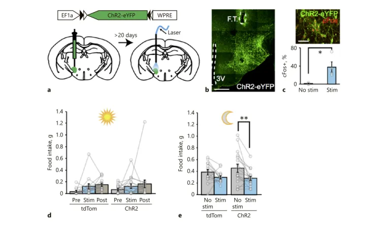
It is estimated that activated MCH neurons may fire
at rates of up to 10–40 Hz in vivo [27, 28]. To ensure that
chemogenetic activation did not underestimate a
pos-sible role of MCH neuron activity on feeding, we also
used optogenetic stimulation as an alternative approach.
For this, we transduced MCH neurons with
rAAV2-DIO-ChR2-eYFP virus for cre-dependent Channelrho
-dopsin-2 expression and placed an optical fiber over the
LH (Fig. 2a–c; online suppl. Fig. 1e). Similar to the
che-mogenetic activation, optogenetic stimulation of MCH
neurons at 20 Hz did not cause any significant increase
in food intake (Fig. 2d, e; online suppl. Fig. 1f). On the
contrary, the dark onset food consumption was slightly
decreased in MCH neuron-stimulated mice. These
re-sults suggest that, under these conditions, MCH
neuro-nal activity is not sufficient to drive the consumption of
chow food and is dispensable for acute regulation of
ap-petite.
1.4 1.2 1.0 0.8 0.6 0.4 0.2 0 1.4 1.2 1.0 0.8 0.6 0.4 0.2 0 Food intake, g Pre tdTom ChR2 ChR2 Stim Post Pre Stim Postd
Food intake, g
tdTom No
stimStim NostimStim
e 80 40 0 cFos+, % No stim Stim c b
*
**
aEF1a ChR2-eYFP WPRE
>20 days Laser
Fig. 2. Optogenetic stimulation of MCH neurons does not increase acute food intake. a Schematic representation of ChR2-eYFP ex-pression in MCH neurons in Pmch-cre mice and their photostim-ulation. b Representative photomicrographs showing ChR2-eY-FP-expressing MCH neurons in the LH. Scale bar, 250 µm. FT, fiber track. c Top: representative image of ChR2-eYFP-transduced MCH neurons with cFos expression (red) in response to 20 Hz photostimulation. Scale bar, 100 µm. Bottom: quantification of the percentage of cFos-expressing MCHChR2-eYFP neurons in
stimu-lated (Stim) vs. unstimulatied (No stim) mice. p = 0.0222 (one-tailed t test), mean ± SEM in %: No stim = 1.03 ± 1.03, Stim = 37.5 ± 11,53. Summary graph of daytime (d, n = 14 tdTom mice,
n = 17 ChR2-eYFP mice, mean ± SEM in g: tdTom pre-Stim =
0.034 ± 0.01, Stim = 0.12 ± 0.05, post-Stim = 0.15 ± 0.03; ChR2 pre-Stim = 0.07 ± 0.02, Stim = 0.12 ± 0.04, post-Stim = 0.17 ± 0.07;
Sidak’s adjusted p values for pre-Stim vs. Stim: p = 0.5390 [tdTom],
p = 0,8408 [ChR2], two-way ANOVA: interaction, F[2, 58] =
0.08368, p = 0.9198; effect of stimulation, F[2, 58] = 4.204, p = 0.0197; and effect of viral injections F[1, 29] = 0.1525, p = 0.6990) food intake before, during, and after photostimulation, and dark cycle (e, n = 14 tdTomato mice, n = 17 ChR2-eYFP mice, mean ± SEM in g: tdTom No stim = 0.39 ± 0.05, Stim = 0.30 ± 0.03; ChR2 No stim = 0.46 ± 0.07, Stim = 0.28 ± 0.03; Sidak’s adjusted p values for No stim vs. Stim: p = 0.2485 [tdTom], p = 0,0040 [ChR2]; 2-way ANOVA: interaction, F[1, 29] = 1.299, p = 0.2637; effect of stimu-lation, F[1, 29] = 11.76, p = 0.0018; and effect of viral injections F[1, 29] = 0.1864, p = 0.6692) food intake with or without photo-stimulation of MCH neurons in ad libitum-fed Pmch-cre mice. Each bar represents 2 h of food intake. * p < 0.05, ** p < 0.01.
Effect of Acute MCH Neuronal Activity
Manipulations on Glucose Homeostasis, Short-Term
Locomotor Activity, and Anxiety
Ex vivo recordings suggest that the activity of MCH
neurons is sensitive to the extracellular glucose
concen-tration, and genetic manipulations of MCH neurons can
profoundly influence glucose homeostasis [15–17]. To
investigate whether acute changes in MCH neuron
activ-ity have a role in glucose regulation, we stimulated and
inhibited MCH neurons chemogenetically as described
above while monitoring glucose tolerance and insulin
sensitivity. In agreement with a recent report, acute
che-250 200 150 100 50 0 Blood glucose, mg/dL 0 60 120 Time, min S/C I hM4Di c 300 200 100 0 Blood glucose, mg/dL 0 60 120 Time, min S/C G hM4Di d**
180 120 60 0 Blood glucose, mg/dL 0 60 120 Time, min S/C IhM3Dq a 300 200 100 0 Blood glucose, mg/dL 0 60 120 Time, min S/C G hM3Dq b Saline CNOFig. 3. Effect of MCH neuron activity manipulations on glucose homeostasis. a Insulin tolerance plot for MCH-specific hM3Dq-mCherry-expressing mice (n = 9 hM3Dq mice, two-way ANOVA, interaction F[5, 80] = 0.2218, p = 0.9522; effect of time, F[5, 80] = 61.89, p < 0.0001; effect of CNO treatment F[1, 16] = 0.04625, p = 0.8324). b glucose tolerance plot for hM3Dq-mCherry mice (n = 6 hM3Dq mice, two-way ANOVA, interaction F[5, 50] = 0.3099,
p = 0.9047; effect of time, F[5, 50] = 53.34, p < 0.0001; effect of CNO
treatment F[1, 10] = 0.217, p = 0.6513). c insulin tolerance plot for MCH-specific hM4Di-mCherry-expressing mice (n = 6 hM4Di
mice, two-way ANOVA, interaction F[5, 50] = 2.113, p = 0.0792; effect of time, F[5, 50] = 65.98, p < 0.0001; effect of CNO treatment F[1, 10] = 0.5847, p = 0.4621). d Glucose tolerance test in MCH-specific hM4Di-mCherry-expressing mice (n = 6 hM4Di mice, two-way ANOVA, interaction F[5, 50] = 4.052, p = 0.0036; effect of time, F[5, 50] = 40.75, p < 0.0001; effect of CNO treatment F[1, 10] = 0.5603, p = 0.4714; Sidak’s adjusted p = 0.0014 at 90 min, sa-line vs. CNO) in response to i.p. sasa-line or CNO administration. S, saline; C, CNO; I, insulin; G, glucose.** p < 0.01.
mogenetic stimulation of MCH neurons did not cause
significant changes in glucose tolerance or insulin
sensi-tivity (Fig. 3; online suppl. Fig. 2a–d) [17]. Conversely,
acute chemogenetic inhibition of MCH neurons
im-proved both glucose tolerance and insulin sensitivity
(Fig. 3; online suppl. Fig. 2e–h). Consistent with previous
reports that used genetic manipulations [16, 17], these
results suggest that inhibition of MCH neuronal activity
improves whole-body glucose metabolism.
Alterations in energy expenditure, in the form of
loco-motor activity, have been suggested to contribute to body
weight changes seen in genetic MCH pathway
manipula-tions. While MCH receptor-1 knockout mice or MCH
neuron-ablated mice are hyperactive [8, 29], infusion of
MCH or MCHR1 agonists had no effect on locomotion [2,
30], suggesting a discrepancy between genetic and
phar-macologic approaches. Therefore, we evaluated the effect
of short-term manipulations of MCH neuronal activity on
locomotor activity. Neither chemogenetic silencing nor
optogenetic activation had any significant effect on
loco-motion (Fig. 4c–e, h–j), which is different from the results
of a recent study examining acute chemogenetic
activa-tion of MCH neurons [17]. However, it must be noted that
we focused on the short-term effects of unilateral
activa-tion, whereas the former study analyzed a period of 8 h in
which the entire MCH neuron population was activated.
Finally, given the contradictory reports about the effects
of pharmacologic and genetic MCH pathway
manipula-tions on anxiety-like behavior [31–34], we probed the
influ-ence of acute MCH neuron activity changes on this
behav-ioral domain. We evaluated the effect of bilateral
chemo-genetic activation/inhibition and unilateral optochemo-genetic
activation of MCH neurons in OF and EPM tasks. We did
not observe any significant difference in the OF test (Fig. 4a–
j; online suppl. Fig. 3a, b) for either MCH inhibition or
ac-tivation. On the other hand, optogenetic activation reduced
the average speed in the open arm of the EPM (Fig. 4k–n;
online suppl. Fig. 3c, d), whereas chemogenetic inhibition
significantly increased the number of entries into the open
arm, suggesting that suppression of the MCH neuron
out-put may have an anxiolytic effect (Fig. 4o–r). Collectively,
these experiments reveal that MCH neuron electrical
activ-ity manipulations can rapidly modulate glucose
homeosta-sis and anxiety-like behavior without a significant effect on
the short-term locomotor activity.
MCH Neuron Activation during Feeding Increases
Chow Food Consumption
Our results suggest that acute stimulation of MCH
neurons is not sufficient to drive food intake. This is in
striking contrast to the behavior observed upon
activa-tion of other orexigenic populaactiva-tions such as arcuate
nu-(For figure see next page.)
Fig. 4. Effect of MCH neuron activity manipulations on anxiety-like behaviors and short-term locomotor activity. a–d OF test for mice with ChR2-eYFP-expressing MCH neurons. a Schematic di-agram describing the OF assay during 20 Hz photostimulation of LH. The time spent (b, mean ± SEM in s: tdTom = 78.29 ± 24.8, ChR2 = 75.2 ± 20.6, tdTom vs. ChR2 unpaired two-tailed t test,
p = 0.9265), the total distance (c, mean ± SEM in m: tdTom =
58.92 ± 6.89, ChR2 = 55.89 ± 7.45, tdTom vs. ChR2 unpaired two-tailed t test, p = 0.7698), and the average speed (d, mean ± SEM in m/s: tdTom = 0.033 ± 0.004, ChR2 = 0.031 ± 0.004, tdTom vs. ChR2 unpaired two-tailed t test, p = 0.7771) were similar between tdTomato and ChR2 mice (n = 8 tdTomato, n = 7 ChR2-eYFP mice). e Mean heat maps and representative traces for each group are shown. Blue dots mark the beginning and red dots mark the end points of the mice’s positions. f–i OF test for mice with hM-4Di-mCherry-expressing MCH neurons. f Schematic diagram de-scribing the OF assay followed by i.p. CNO administration. The time spent (g, mean ± SEM in s: tdTom = 75.05 ± 16.80, hM4Di = 122.86 ± 22.42, tdTom vs. hM4Di unpaired two-tailed t test, p = 0.1385), the total distance (h, mean ± SEM in m: tdTom = 56.03 ± 4.83, hM4Di = 57.50 ± 4.06, tdTom vs. hM4Di unpaired two-tailed
t test, p = 0.8193), and the average speed (i, mean ± SEM in m/s:
tdTom = 0.031 ± 0.0027, hM4Di = 0.032 ± 0.0023, tdTom vs. hM-4Di unpaired two-tailed t test, p = 0.7829) were similar between tdTomato and hM4Di-mCherry mice (n = 8 tdTomato, n = hM4Di
12 mice). j Mean heat maps and representative traces for each group are shown. Blue dots mark the beginning and red dots mark the end points of the mice’s positions. k–m EPM test during MCH neuron stimulation. The number of entries to the open arms (k, mean ± SEM: tdTom = 9.29 ± 2.40, ChR2 = 7.00 ± 1.67, tdTom vs. ChR2 unpaired two-tailed t test, p = 0.4388) and the time spent in the open arms (l, mean ± SEM in s: tdTom = 29.00 ± 8.22, ChR2 = 66.86 ± 19.06, tdTom vs. ChR2 unpaired two-tailed t test, p = 0.1068) were comparable between the ChR2 and tdTomato groups, whereas the average speed in the open arms (m, mean ± SEM in m/s: tdTom = 0.020 ± 0.0035, ChR2 = 0.011 ± 0.0026, tdTom vs. ChR2 unpaired two-tailed t test, p = 0.04557) was significantly de-creased in the ChR2 group (n = 7 tdTomato, n = 8 ChR2-eYFP mice). n Mean heat maps for each group. o–q EPM test in MCH-specific hM4Di- or tdTomato-expressing mice. The number of en-tries to the open arms was significantly increased in the hM4Di group (o, mean ± SEM: tdTom = 5.75 ± 1.06, hM4Di = 10.00 ± 1.41, tdTom vs. hM4Di unpaired two-tailed t test, p = 0.04252), while the time spent in the open arms (p, mean ± SEM in s: tdTom = 65.00 ± 32.93, hM4Di = 79.54 ± 21.15, tdTom vs. hM4Di unpaired two tailed t test, p = 0.7007) and the average speed in the open arms (q, mean ± SEM in m/s: tdTom = 0.017 ± 0.0030, hM-4Di = 0.019 ± 0.0032, tdTom vs. hMhM-4Di unpaired two-tailed t test,
p = 0.6246) were similar in the 2 groups (n = 8 tdTomato, n = 12
cleus AgRP neurons and LH
vGATneurons [20, 35].
How-ever, if MCH neurons have a reinforcing role during
feeding rather than simply increasing the level of hunger,
they would be expected to become active during or
short-ly after consumption. Given their implication in food
re-ward, we reasoned that pairing the activation of MCH
neurons with chow food consumption might increase
the reward value of the consumed pellet, leading to
over-eating. To test this possibility, we paired the optogenetic
stimulation of MCH neurons with chow pellet removal
200 150 100 50 0 Time in center, s tdTom ChR2 b a 100 75 50 25 0 Total distan ce, m tdTom ChR2 c 0.05 0.04 0.03 0.02 0.01 0 Average speed, m/s tdTom ChR2 tdTom ChR2 d e 0 s 5 min 32 s 300 200 100 0 Time in center, s tdTom hM4Di g f 80 60 40 20 0 Total distan ce, m tdTom hM4Di h 0.05 0.04 0.03 0.02 0.01 0 Average speed, m/s tdTom hM4Di tdTom hM4Di i j 0 s 5 min 32 s 25 20 15 10 5 0 Entries, n tdTom ChR2 k 150 100 50 0 Time spent, s tdTom ChR2 l 0.04 0.03 0.02 0.01 0 Average speed, m/s tdTom ChR2 tdTom ChR2 m n 0 s 2 min24 s 25 20 15 10 5 0 Entries, n tdTom hM4Di o 300 200 100 0 Time spent, s tdTom hM4Di p 0.04 0.03 0.02 0.01 0 Average speed, m/s tdTom hM4Di tdTom hM4Di q r 0 s 2 min32s 4
from the feeder to ensure that MCH activation occurs
during consumption. It takes approximately 15 s to
con-sume a 20-mg chow pellet; therefore, we adjusted the
MCH stimulation to the 15-s time period following the
pellet removal (Fig. 5a). Unlike continuous stimulation,
pellet consumption-paired stimulation of MCH neurons
significantly increased the total number of chow pellets
consumed over a 24-h period (Fig. 5b; online suppl.
Fig. 4). Taken together, these results suggest that MCH
neuron activation alone may not be involved in food
seeking or elevation of the hunger state but, when paired
with consummatory activity, they may have role in
rein-forcing feeding.
MCH Neuronal Activation Is Sufficient for Reward
without Food
Our results showed that, when simultaneously paired
with a consummatory event, MCH neuron activation
could increase chow consumption. It has been suggested
that MCH neuronal activation can add reward value only
when oral sweet cues are present [10]. However, our
re-sults suggest that, even with nonsweet chow,
consump-tion can also be increased by MCH activaconsump-tion. To test
whether oral/postoral cues are required at all, we further
investigated the reward value of direct MCH stimulation
using non-food-related tasks of nose poke and lever press
self-stimulation. For this, we coupled brief optogenetic
activation of ChR2-expressing MCH neurons (10 s) with
nose poke or lever press operant tasks. We also placed an
inactive nose poke port/lever to test the specificity of
ac-tivation of the laser-coupled port/lever, respectively
(Fig. 6a, d). For both nose poke and lever press operant
tasks, MCH activation significantly increased port entries
and lever press events over the inactive instrument,
sug-gesting that MCH activation alone is sufficient to
gener-ate a reward effect even in the absence of oral or
posting-estive cues (Fig. 6).
Discussion
Here we evaluated the short-term behavioral and
physiological effects of MCH neuronal electrical activity
manipulations. We report that acute changes in MCH
neuronal activity are not sufficient to alter feeding
behav-ior, unless the stimulation is delivered during the feeding
event. In addition, inhibition of MCH neurons improves
glucose tolerance and reduces anxiety-like behavior, but
neither activation nor inhibition affects the short-term
locomotor activity. Furthermore, using 2 different
instru-mental tasks, we showed that activation of MCH neurons
alone is rewarding.
Diverse Physiological and Behavioral Functions of
MCH Neurons
Our results on MCH neuronal activity-dependent
changes in glucose tolerance and anxiety are consistent
with earlier genetic and cell type-specific ablation studies
[8, 16]. Rapid improvement of glucose tolerance upon
MCH inhibition suggests that developmental
compensa-tion or other circuit reorganizacompensa-tions upon genetic
manip-ulation do not account for the observed effect. Similarly,
a weak anxiolytic effect of MCH neuronal inhibition,
which was predicted from genetic ablation studies but not
observed with pharmacologic administration of MCHR1
antagonists, supports a role of MCH in the stress axis.
MCH Neurons and Reward
The mechanisms that underlie MCH neuronal
regula-tion of metabolism and reward are not completely
under-stood. For short-term appetite regulation, our results
in-dicate that MCH neuronal activity is neither sufficient to
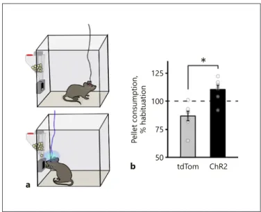
a
125 100 75 50 Pellet consumption, % habituation
tdTom ChR2
b
*
Fig. 5. MCH neuron photoactivation during feeding increases chow food consumption. a Schematic representation of the food-coupled MCH-photostimulation assay performed with Pmch-cre mice expressing tdTomato or ChR2-eYFP in MCH neurons. b
Summary graph of the change in pellet consumption relative to a nonstimulated habituation day upon 20 Hz self-stimulation (15 s/ pellet) of LH (n = 7 tdTomato mice, n = 7 ChR2-eYFP mice, mean ± SEM: % of habituation: tdTom = 86.87 ± 4.30, ChR2 + 110.67 ± 3.72, tdTomato vs. ChR2-eYFP: unpaired two-tailed t test, * p = 0.0013).
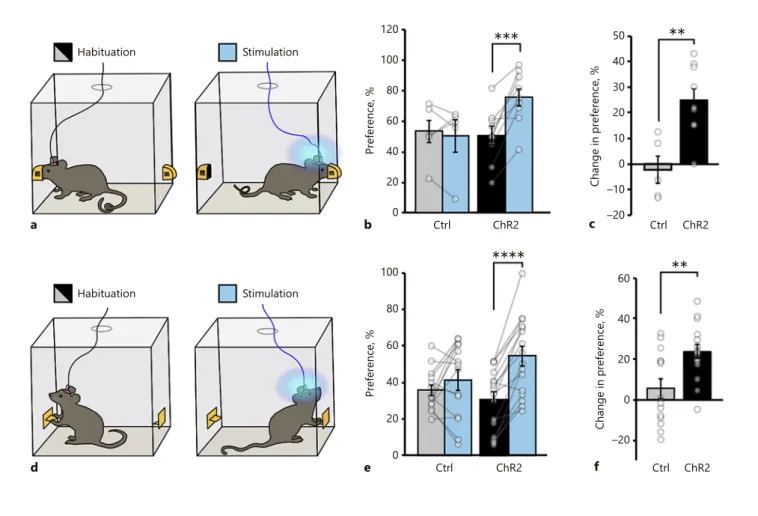
****
***
60 40 20 0 –20 Change in pre fere nce, % Ctrl ChR2 f**
60 40 100 80 20 0 Preference, % Ctrl ChR2 e d Habituation Stimulation 50 40 30 20 10 0 –10 –20 Change in pre fere nce, % Ctrl ChR2 c**
60 40 120 100 80 20 0 Preference, % Ctrl ChR2 b a Habituation StimulationFig. 6. Activation of MCH neurons is sufficient for a reward even in the absence of food consumption. a–c Schematic diagram de-scribing the nose poke assay. a Mice were initially observed for nose-poke port preference. Photostimulation (20 Hz; 10 s photo-stimulation/port entry) was paired with the less preferred one. The preference for the laser-coupled operandum was significantly in-creased on stimulation days compared both to the tdTomato group and the habituation days (b), and the change in preference was significantly higher in the ChR2 group compared to the tdTo-mato animals (mean ± SEM: control habituation = 52.66 ± 8.70, control stimulation = 50.34 ± 10.44, ChR2 habituation = 50.68 ± 6.02, ChR2 stimulation = 75.56 ± 5.74, n = 5 tdTomato mice, n = 9 ChR2-eYFP mice; two-way ANOVA, interaction F[1,12] = 13.46,
p = 0.0032; effect of trial F[1,12] = 9.258, p = 0.0102; effect of viral
injections F[1,12] = 1.366, p = 0.2652; Sidak’s adjusted p values:
p = 0.9784 [tdTomato vs. ChR2-eYFP, habituation], p = 0.0508
[tdTomato vs. ChR2-eYFP, stimulation], p = 0.0002 [habituation vs. stimulation, ChR2-eYFP], and p = 0.9118 [habituation vs. stim-ulation, tdTomato, ** p < 0.01 *** p < 0.001]). c The change in preference was significantly greater in the ChR2-eYFP group com-pared to the tdTomato group (mean ± SEM: control = –2.32 ± 5.34,
ChR2 = 24.88 ± 4.64, n = 5 tdTomato mice, n = 9 ChR2-eYFP mice; tdTomato vs. ChR2-YFP p = 0.003213, unpaired two-tailed t test, ** p < 0.01). d–f Lever press assay. Mice were initially observed for normal lever press frequencies, and then the less preferred lever was coupled with a laser for the stimulation day (d). Summary graph showing the increase in preference for the laser coupled le-ver on the stimulation day in ChR2-eYFP-transduced Pmch-cre mice only (e); mean ± SEM: control habituation = 35.81 ± 2.98, control stimulation = 41.41 ± 5.52, ChR2 habituation = 30.68 ± 4.19, ChR2 stimulation = 54.37 ± 5.36, n = 14 tdTomato mice, n = 16 ChR2-eYFP mice, 2-way ANOVA, interaction F(1,28) = 9.864,
p = 0.0040; effect of trial F(1,28) = 25.89, p < 0.00001; effect of viral
injections F(1,28) = 0.4332, p = 0.5158; Sidak’s adjusted p values:
p = 0.6878 (tdTomato vs. ChR2-eYFP, habituation), p = 0.1068
(tdTomato vs. ChR2-eYFP, stimulation), p = 0.3490 (habituation vs. stimulation, tdTomato, p < 0.00001; habituation vs. stimula-tion, ChR2-eYFP, **** p < 0.0001). The change in preference was significantly greater compared to that in the tdTomato group. f Mean ± SEM: control = –5.60 ± 4.80, ChR2 = 23.69 ± 3.38, (n = 14 tdTomato mice, n = 16 ChR2-eYFP mice, tdTomato vs. ChR2-YFP
initiate nor necessary to sustain food intake, suggested by
the inability to effect overall consumption by
chemoge-netic activation or inhibition during both day time and
dark onset. Indeed, contrary to our expectations, the dark
cycle food consumption was slightly reduced in mice
which were subjected to continuous MCH neuron
pho-tostimulation (20 Hz). However, if activation is time
locked during consumption, MCH neurons appear to
in-crease the food intake, suggesting a reinforcing role for
ongoing consumption (Fig. 5). It is unlikely that this
in-crease is driven by a delay in satiety signals since
contin-ued stimulation does not result in an overall increase in
feeding (Fig. 2; online suppl. Fig. 1f).
Earlier results using 2 bottle preference assays in which
MCH activation was paired with noncaloric sugar
drink-ing resulted in an inability to display a natural preference
toward caloric sugar [10]. Use of a 2-bottle assay with
wa-ter, however, did not result in a preference for the
MCH-activated one, leading to the suggestion that, when paired
with sweet oral cues, MCH neuron activation can ensue
a reward function that is normally derived from calories.
Our results from operant task self-stimulation, however,
suggest that in the absence of a sweet taste or even in the
absence of any oral/post oral cues MCH activation can
still ensue reward. It is unlikely that the discrepancy
be-tween our results and the previous report could stem
from the use of distinct transgenic Pmch-cre drivers, since
both lines appear to label >
80% of all MCH neurons.
However, the longer stimulation durations used in this
study (1 s per 5 licks vs. 15 s per 20-mg pellet or 10 s per
operant activation) likely uncovered the sufficiency of
MCH neuron activity for reward even in the absence of
any other food- or taste-related stimuli.
The reward effects of LH stimulations have been
previ-ously noted in the GABAergic subpopulation, whereas
glutamate-expressing neurons have been shown to be
aversive [35, 36]. Our results extend these findings and
show that, despite being mostly glutamatergic [11], MCH
neurons can also induce self-stimulation. In agreement
with the reward function, MCH axons have been
local-ized to the VTA and the striatum, where MCHR1 is
high-ly expressed [10, 37]. Rather than directhigh-ly stimulating the
motivation to eat, as seen with ARC
AgRPand LH
vGATrons, our results support a reinforcing role for MCH
neu-rons. This is consistent with earlier reports showing that
rats given an MCHR1 antagonist displayed reduced lever
press for sucrose solution, MCH knockout rats displayed
a reduced operant response to fat, and MCH
neuron-ab-lated mice were unable distinguish caloric versus
nonca-loric sweets [10, 38, 39].
Surprisingly, it was the feeding-paired stimulation, but
not continuous activation, that increased pellet
consump-tion. Continuous activation even in dark onset, during
which mice consume abundantly, did not yield an
addi-tional increase in the feeding response but indeed
de-creased it. One interpretation could be that under the
continuous stimulation regime animals might be unable
to discern which of their actions, i.e., feeding, nesting, or
grooming, is causing the reward effect and therefore
can-not reinforce that specific action. On the other hand, if
MCH neuronal stimulation is time restricted to a specific
action, e.g., pellet consumption, nose poke, or lever press,
only then does a significant increase in preference for that
action occur. This is consistent with a reinforcing role of
MCH neurons rather than simply an increase in appetite.
Limitations of This Study
Our approach was based on addressing the short-term
effects of acute MCH neuron activity manipulations,
spe-cifically focusing on energy homeostasis, stress, and
re-ward. Overall, our results support a role for MCH neurons
in amplification of consumption by reinforcing feeding
only when activated in a specific temporal pattern.
How-ever, our study does not address whether such activation
occurs during feeding, and it remains to be seen how in
vivo MCH neuron activity dynamics respond to feeding
of different types of diets. The reduced response to
he-donically active food in MCH-compromised animal
mod-els suggests that MCH activation might take place during
palatable food consumption [10, 38, 39]. However,
endog-enous activity dynamics of MCH neurons have only been
studied during sleep-wake regulation and novelty
expo-sure but not in the context of feeding [27, 28].
Our negative results on locomotor activity changes
should be interpreted cautiously since we only evaluated
the effect of daytime manipulations and we only focused
on the first 30 min. It is likely that prolonged observation
upon bilateral activation, as reported recently [17], may
unveil a likely contribution. In addition, MCH
neuron-ab-lated mice showed locomotor activity differences only in
the dark period [8], consistent with a time-sensitive effect.
Similarly, although we only used bilaterally infected
MCH
hM4Dimice, our loss-of-function approach is limited
by the penetrance of viral transduction, and therefore
pos-sible effects of inhibition on behaviors for which a small
fraction of uninfected MCH neurons is sufficient, might
have been underestimated. Nevertheless, our transduction
rate was sufficient to observe significant impact of
inhibi-tion on glucose regulainhibi-tion and anxiety-like behaviors
(Fig. 2, 3).
Finally, our study does not address which
MCH-neu-ron derived signaling molecule governs reward function.
Recent studies using MCH neuron-selective ablation of
MCH peptide or vGlut2 expression (2 major signaling
molecules secreted by MCH neurons) have arrived at
dif-ferent conclusions with respect to sugar reward function,
likely reflecting partial overlap between these signaling
pathways [11, 12].
Collectively, consistent with genetic approaches, our
results support diverse behavioral and physiological
func-tions of MCH neurons. We revealed that involvement in
short-term appetite regulation might have strict temporal
requirements and, while their acute inhibition promotes
glucose tolerance and reduces anxiety-like behaviors,
ac-tivation is sufficient to induce reward.
Acknowledgement
We would like to thank to Gurkan Ozturk and Ekrem Musa Ozdemir for their help with imaging facilities and animal care, as well as Kamal Rahmouni and Huxing Cui for critical reading of this paper.
Statement of Ethics
All animal generation, care, maintenance, and experimental procedures conform to the internationally accepted standards and were approved by the Istanbul Medipol University (IMU) Animal Care and Use Committee.
Disclosure Statement
The authors declare no competing financial interests.
Funding Sources
This work was supported by EMBO IG grant No. 2539 and in part by Scientific and Technological Research Council of Turkey (TUBITAK) grant No. 214S085 to D.A.
Author Contributions
P.D.: animal care, intracranial virus injections, behavioral periments, imaging, and paper preparation. I.A.: behavioral ex-periments and paper preparation. N.S.A.: virus preparation, be-havioral experiments, data analysis, figure preparation, and paper preparation. Y.Y., T.A., and M.O.: electrophysiological recordings. G.F. and F.K.: confocal imaging. I.C.: intracranial virus injections and ferule placement. E.A.O., M.I.A., U.C., and B.Y.: technical help. D.A.: study conception and design, analysis and interpreta-tion of data, and drafting of this article.
References
1 Qu D, Ludwig DS, Gammeltoft S, Piper M, Pelleymounter MA, Cullen MJ, et al. A role for melanin-concentrating hormone in the central regulation of feeding behaviour. Na-ture. 1996 Mar;380(6571):243–7.
2 Gomori A, Ishihara A, Ito M, Mashiko S, Mat-sushita H, Yumoto M, et al. Chronic intra-cerebroventricular infusion of MCH causes obesity in mice. Melanin-concentrating hor-mone. Am J Physiol Endocrinol Metab. 2003 Mar;284(3):E583–8.
3 Gehlert DR, Rasmussen K, Shaw J, Li X, Ar-dayfio P, Craft L, et al. Preclinical evaluation of melanin-concentrating hormone receptor 1 antagonism for the treatment of obesity and depression. J Pharmacol Exp Ther. 2009 May; 329(2):429–38.
4 Ludwig DS, Tritos NA, Mastaitis JW, Kulkarni R, Kokkotou E, Elmquist J, et al. Melanin-con-centrating hormone overexpression in trans-genic mice leads to obesity and insulin resis-tance. J Clin Invest. 2001 Feb;107(3):379–86. 5 Shimada M, Tritos NA, Lowell BB, Flier JS,
Maratos-Flier E. Mice lacking melanin-con-centrating hormone are hypophagic and lean. Nature. 1998 Dec;396(6712):670–4.
6 Chen Y, Hu C, Hsu CK, Zhang Q, Bi C, Asni-car M, et al. Targeted disruption of the mela-nin-concentrating hormone receptor-1 re-sults in hyperphagia and resistance to diet-induced obesity. Endocrinology. 2002 Jul; 143(7):2469–77.
7 Marsh DJ, Weingarth DT, Novi DE, Chen HY, Trumbauer ME, Chen AS, et al. Melanin-concentrating hormone 1 receptor-deficient mice are lean, hyperactive, and hyperphagic and have altered metabolism. Proc Natl Acad Sci USA. 2002 Mar;99(5):3240–5.
8 Whiddon BB, Palmiter RD. Ablation of neu-rons expressing melanin-concentrating hor-mone (MCH) in adult mice improves glucose tolerance independent of MCH signaling. J Neurosci. 2013 Jan;33(5):2009–16.
9 Alon T, Friedman JM. Late-onset leanness in mice with targeted ablation of melanin con-centrating hormone neurons. J Neurosci. 2006 Jan;26(2):389–97.
10 Domingos AI, Sordillo A, Dietrich MO, Liu ZW, Tellez LA, Vaynshteyn J, et al. Hypotha-lamic melanin concentrating hormone neu-rons communicate the nutrient value of sug-ar. eLife. 2013 Dec;2:e01462.
11 Schneeberger M, Tan K, Nectow AR, Parolari L, Caglar C, Azevedo E, et al. Functional anal-ysis reveals differential effects of glutamate and MCH neuropeptide in MCH neurons. Mol Metab. 2018 Jul;13:83–9.
12 Sherwood A, Wosiski-Kuhn M, Nguyen T, Holland PC, Lakaye B, Adamantidis A, et al. The role of melanin-concentrating hormone in conditioned reward learning. Eur J Neuro-sci. 2012 Oct;36(8):3126–33.
13 Sherwood A, Holland PC, Adamantidis A, Johnson AW. Deletion of melanin concen-trating hormone receptor-1 disrupts overeat-ing in the presence of food cues. Physiol Be-hav. 2015 Dec;152:402–7.
14 Sclafani A, Adamantidis A, Ackroff K. MCH receptor deletion does not impair glucose-conditioned flavor preferences in mice. Physiol Behav. 2016 Sep;163:239–44. 15 Burdakov D, Gerasimenko O, Verkhratsky A.
Physiological changes in glucose differentially modulate the excitability of hypothalamic melanin-concentrating hormone and orexin neurons in situ. J Neurosci. 2005 Mar;25(9): 2429–33.
16 Kong D, Vong L, Parton LE, Ye C, Tong Q, Hu X, et al. Glucose stimulation of hypotha-lamic MCH neurons involves K(ATP) chan-nels, is modulated by UCP2, and regulates pe-ripheral glucose homeostasis. Cell Metab. 2010 Nov;12(5):545–52.
17 Hausen AC, Ruud J, Jiang H, Hess S, Varbanov H, Kloppenburg P, et al. Insulin-dependent activation of MCH neurons impairs locomo-tor activity and insulin sensitivity in obesity. Cell Rep. 2016 Dec;17(10):2512–21. 18 Noble EE, Hahn JD, Konanur VR, Hsu TM,
Page SJ, Cortella AM, Liu CM, Song MY, Su-arez AN, Szujewski CC, Rider D, Clarke JE, Darvas M, Appleyard SM, Kanoski SE: Con-trol of feeding behavior by cerebral ventricu-lar volume transmission of melanin-concen-trating hormone. Cell Metab 2018;28:55–68. 19 Mathews LC, Gray JT, Gallagher MR, Snyder
RO. Recombinant adeno-associated viral vec-tor production using stable packaging and producer cell lines. Methods Enzymol. 2002; 346:393–413.
20 Atasoy D, Betley JN, Su HH, Sternson SM. Deconstruction of a neural circuit for hunger. Nature. 2012 Aug;488(7410):172–7.
21 Steculorum SM, Ruud J, Karakasilioti I, Backes H, Engström Ruud L, Timper K, et al. AgRP neurons control systemic insulin sensi-tivity via myostatin expression in brown adi-pose tissue. Cell. 2016 Mar;165(1):125–38. 22 Seibenhener ML, Wooten MC. Use of the
open field maze to measure locomotor and anxiety-like behavior in mice. J Vis Exp. 2015 Feb;6(96):e52434.
23 Lopatina O, Yoshihara T, Nishimura T, Zhong J, Akther S, Fakhrul AA, et al. Anxiety- and depression-like behavior in mice lacking the CD157/BST1 gene, a risk factor for Par-kinson’s disease. Front Behav Neurosci. 2014 Apr;8:133.
24 Lister RG. The use of a plus-maze to measure anxiety in the mouse. Psychopharmacology (Berl). 1987;92(2):180–5.
25 Walf AA, Frye CA. The use of the elevated plus maze as an assay of anxiety-related be-havior in rodents. Nat Protoc. 2007;2(2):322– 8.
26 Carola V, D’Olimpio F, Brunamonti E, Man-gia F, Renzi P. Evaluation of the elevated plus-maze and open-field tests for the assessment of anxiety-related behaviour in inbred mice. Behav Brain Res. 2002 Aug;134(1-2):49–57. 27 González JA, Iordanidou P, Strom M,
Ada-mantidis A, Burdakov D. Awake dynamics and brain-wide direct inputs of hypothalamic MCH and orexin networks. Nat Commun. 2016 Apr;7(1):11395.
28 Hassani OK, Lee MG, Jones BE. Melanin-concentrating hormone neurons discharge in a reciprocal manner to orexin neurons across the sleep-wake cycle. Proc Natl Acad Sci USA. 2009 Feb;106(7):2418–22.
29 Smith DG, Tzavara ET, Shaw J, Luecke S, Wade M, Davis R, et al. Mesolimbic dopa-mine super-sensitivity in melanin-concen-trating hormone-1 receptor-deficient mice. J Neurosci. 2005 Jan;25(4):914–22.
30 Shearman LP, Camacho RE, Sloan Stribling D, Zhou D, Bednarek MA, Hreniuk DL, et al. Chronic MCH-1 receptor modulation alters appetite, body weight and adiposity in rats. Eur J Pharmacol. 2003 Aug;475(1-3):37–47. 31 Smith DG, Davis RJ, Rorick-Kehn L, Morin
M, Witkin JM, McKinzie DL, et al. Melanin-concentrating hormone-1 receptor modu-lates neuroendocrine, behavioral, and corti-colimbic neurochemical stress responses in mice. Neuropsychopharmacology. 2006 Jun; 31(6):1135–45.
32 Roy M, David NK, Danao JV, Baribault H, Tian H, Giorgetti M. Genetic inactivation of melanin-concentrating hormone receptor subtype 1 (MCHR1) in mice exerts anxiolytic-like behavioral effects. Neuropsychopharma-cology. 2006 Jan;31(1):112–20.
33 Adamantidis A, Thomas E, Foidart A, Tyhon A, Coumans B, Minet A, et al. Disrupting the melanin-concentrating hormone receptor 1 in mice leads to cognitive deficits and altera-tions of NMDA receptor function. Eur J Neu-rosci. 2005 May;21(10):2837–44.
34 Monzón ME, De Barioglio SR. Response to novelty after i.c.v. injection of melanin-con-centrating hormone (MCH) in rats. Physiol Behav. 1999 Nov;67(5):813–7.
35 Jennings JH, Ung RL, Resendez SL, Stamata-kis AM, Taylor JG, Huang J, et al. Visualizing hypothalamic network dynamics for appeti-tive and consummatory behaviors. Cell. 2015 Jan;160(3):516–27.
36 Stamatakis AM, Van Swieten M, Basiri ML, Blair GA, Kantak P, Stuber GD. Lateral Hypo-thalamic Area Glutamatergic Neurons and Their Projections to the Lateral Habenula Regulate Feeding and Reward. J Neurosci. 2016 Jan;36(2):302–11.
37 Chee MJ, Pissios P, Maratos-Flier E. Neuro-chemical characterization of neurons ex-pressing melanin-concentrating hormone re-ceptor 1 in the mouse hypothalamus. J Comp Neurol. 2013 Jul;521(10):2208–34.
38 Mul JD, la Fleur SE, Toonen PW, Afrasiab-Middelman A, Binnekade R, Schetters D, et al. Chronic loss of melanin-concentrating hor-mone affects motivational aspects of feeding in the rat. PLoS One. 2011 May;6(5):e19600. 39 Karlsson C, Zook M, Ciccocioppo R, Gehlert
DR, Thorsell A, Heilig M, et al. Melanin-con-centrating hormone receptor 1 (MCH1-R) an-tagonism: reduced appetite for calories and suppression of addictive-like behaviors. Phar-macol Biochem Behav. 2012 Sep;102(3):400–6.

![Fig. 3. Effect of MCH neuron activity manipulations on glucose homeostasis. a Insulin tolerance plot for MCH-specific hM3Dq-mCherry-expressing mice (n = 9 hM3Dq mice, two-way ANOVA, interaction F[5, 80] = 0.2218, p = 0.9522; effect of time, F[5, 80]](https://thumb-eu.123doks.com/thumbv2/9libnet/5426433.103436/6.892.65.829.97.719/activity-manipulations-homeostasis-insulin-tolerance-specific-expressing-interaction.webp)