ORIGINAL ARTICLE
Effect of triclosan-coated sutures on surgical site infections in pilonidal
disease: prospective randomized study
Naciye Cigdem Arslan1 &Gulsen Atasoy2&Tansu Altintas3&Cem Terzi4
Accepted: 27 July 2018 / Published online: 30 July 2018 # Springer-Verlag GmbH Germany, part of Springer Nature 2018
Abstract
Purpose The aim of this study is to investigate the effect of triclosan-coated sutures on surgical site infections after wide excision and primary closure for pilonidal disease.
Methods One hundred seventy-seven patients were randomized into two groups: 91 in control and 86 in triclosan groups. In the control group, 1/0 monofilament polypropylene retention sutures, 3/0 polyglactin subcutaneous sutures, and 3/0 polypropylene skin sutures were used. In the triclosan group, 1/0 triclosan-coated monofilament polydioxanone, 3/0 triclosan-coated polyglactin, and 3/0 triclosan-coated monofilament polydioxanone were used. Postoperative care and follow-up was made by a surgeon according to Centers for Disease Control guideline. Surgical site infection rates between groups were compared. Secondary outcomes were seroma and wound dehiscence.
Results Seroma was seen in 30 (16.9%) patients: 20 (23.3%) in the triclosan group and 10 (10.9%) in the control group (p = 0.030). Thirteen (7.3%) patients had superficial wound dehiscence: 5 (5.5%) patients in the control group and 10 (11.6%) patients in the triclosan group (p = 0.116). Overall surgical site infection (SSI) rate was 15.8% (n = 28): 19 (20.8%) patients in the control group and 9 (10.5%) patients in the triclosan group (p = 0.044). Healing was observed on mean 17.8 ± 6.7 days. Primary and secondary healing rates and time to healing were similar between groups.
Conclusion Triclosan-coated sutures decreased surgical site infection rate but had no effect on time to healing in pilonidal disease. Seroma and wound dehiscence were more common in triclosan groups. Randomized trials are needed to clear the effect of triclosan-coated sutures on postoperative wound complications.
Keywords Surgical site infection . Pilonidal disease . Triclosan . Wound infection . Antibacterial sutures
Introduction
Pilonidal disease (PD) is a common condition characterized with inflammation and epithelized sinuses caused by moving hair towards the gluteal sulcus. The presentation varies be-tween asymptomatic hair pits and chronic fistulas with severe infection. Conservative and surgical treatments both have high wound complication and recurrence rates which causes con-siderable work day losses and increased healthcare costs [1, 2]. Surgical site infection (SSI) after PD surgery has been reported up to 38% and related with increased recurrence rate [3,4].
During recent years, in addition to well-known precautions such as parenteral antimicrobial prophylaxis and preoperative bathing, new issues are focused to prevent SSI. These are glycemic control, maintaining normothermia, and oxygena-tion [5]. The effect of antimicrobial-coated biomaterials is still controversial. Many randomized trials on the effect of
* Naciye Cigdem Arslan [email protected] Gulsen Atasoy [email protected] Tansu Altintas [email protected] Cem Terzi [email protected] 1
Department of General Surgery, Medipol University, Birlik mah Bahceler cd 5, 34230 Istanbul, Turkey
2 Department of General Surgery, Gokceada State Hospital,
17760 Canakkale, Turkey
3
Department of General Surgery, Kanuni SS Research Hospital, 34303 Istanbul, Turkey
4
Department General Surgery, Bogazici Academy of Clinical Sciences, 34357 Istanbul, Turkey
triclosan-coated sutures in various surgical procedures have been published [6–8]; however, there are scarce data focusing on PD [9]. Recent meta-analyses showed moderate-quality evidence and robust effect that triclosan-coated sutures reduce SSI and costs [10–12]. The wound classifications and surgical procedures including abdominal, vascular, colorectal, head and neck, cardiac, and breast surgery are quite heterogeneous in these studies.
As PD is one of the most complicating procedures of sur-gical practice, we aimed to investigate the effect of triclosan-coated sutures on PD surgery.
Patients and methods
The study was approved by Dokuz Eylul University Ethics Committee (approval no. 20101193-17). The patients were detailly informed about the protocol and written consent was received. Patients older than 18 years old who underwent wide excision and primary closure for PD were included in the study. Exclusion criteria were immunosuppression, antibiotherapy and/or infection history within 1 week before surgery, acute abscess, recurrent PD, different procedures oth-er than wide excision and primary closure, use of drain, and postoperative administration of antibiotics (Fig.1).
The severity of PD was determined according to Chavoin classification [13]: grade I: asymptomatic hair pits at midline, grade II: drainage from midline sinuses, grade III: acute ab-scess, and grade IV: chronic drainage and painful swelling with midline and lateral sinuses. Chavoin grade III patients were initially treated with abscess drainage and oral antibi-otics; elective surgery was planned at least 1 week after antibiotherapy was stopped. When collecting data, Chavoin classifications were recorded based on the symptoms on pre-sentation; thus, those patients were recorded as Chavoin grade III, but they did not have acute abscess at the time of surgery.
Study groups and randomization
Between January 2011 and January 2013, 222 patients were assessed for PD. Patients were tiered into two groups using block randomization at 1:1 ratio. The surgeon (NCA, TA, CT) was not blinded to the randomization as he/she used and rec-ognized the sutures. Another surgeon (GA) performed the postoperative care and assessment of the surgical site. In the control group, conventional suture materials were used. In the study (triclosan) group, triclosan-coated sutures were used. The primary end-point of the study was to compare SSI rates between study and triclosan groups. Secondary end-points were comparing wound dehiscence without infection and seroma rates.
Surgery
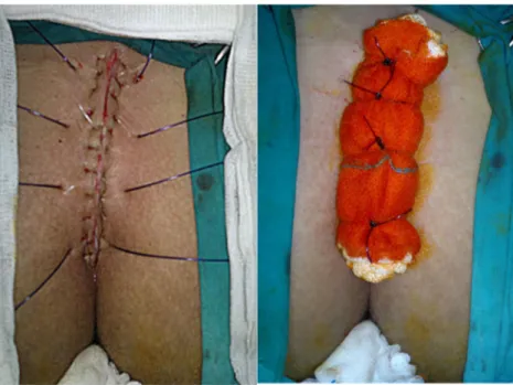
Rectal enema was applied to all the patients 2 h before oper-ation. Under general or locoregional anesthesia, patients were positioned in prone. Intravenous 1.5 g cefuroxime axetil and 500 mg metronidazole were administered 30 min before inci-sion. The buttocks were retracted with bandages. Hair was shaved with razor on the operation table (Fig.2a). An elliptical incision as small as possible but covering all the pits and sinuses was made (Fig.2b). Pilonidal cyst was excised with clean surgical borders (Fig.2c). The bandages were removed (Fig.2d). Retention sutures were prepared, and subcutaneous tissue was closed with interrupted sutures. Skin was closed, and retention sutures were tied over a compression gauze (Fig.3). Skin sutures were seated paramedian and retention sutures were median on the gauze.
Except suture materials, all the steps of surgery were sim-ilar in all patients. In the control group, the retention sutures were 1/0 monofilament polypropylene (Prolene®, Ethicon, USA), subcutaneous sutures were 3/0 polyglactin (Vicryl®, Ethicon, USA), and skin sutures were 3/0 polypropylene (Prolene®, Ethicon, USA). In the triclosan group, 1/0 triclosan-coated monofilament polydioxanone (PDS Plus®, Ethicon, USA) was used for retention, 3/0 triclosan-coated polyglactin (Vicryl Plus®, Ethicon, USA) was used for sub-cutaneous tissue, and 3/0 triclosan-coated monofilament polydioxanone (PDS Plus®, Ethicon, USA) was used for skin closure.
Follow-up and definition of complications
All the patients were discharged on the same day after surgery. Antibiotics were not continued. Outpatient visits were per-formed by a surgeon (GA) on postoperative days 1, 3, 7, 15, and 30. At the first visit, dressings were changed. On postop-erative day 3, dressings were removed, and the wound was left open. Hair removal during at least 2 years with razor or dep-ilation gel was recommended to all patients. The retention sutures were removed on postoperative day 7 and skin sutures were removed on day 14 in uneventful patients.
Surgical site infection
Centers for Disease Control guideline was used to define SSI [5].
Superficial SSI Infection is associated with skin and subcuta-neous tissue within 30 days after surgery and at least one of the following:
1. Purulent drainage with or without culture confirmation 2. Positive tissue or fluid culture
Fig. 1 Flow diagram of the study
Fig. 2 Preparation of the surgical site and excision of the sinus
3. Incision opened by the surgeon with at least one of pain, tenderness, swelling, redness and heat—unless culture is negative
4. Diagnosis of superficial incisional infection made by a surgeon or attending physician
Deep SSI Infection associated with deep tissues (e.g., fascia, muscle) within 30 days after surgery and at least one of the following:
1. Purulent drainage from the deep incision
2. A deep incision spontaneously dehisces or opened by the surgeon with at least one of fever, pain, tenderness—un-less culture is negative
3. Radiologically or histologically detected abscess or deep infection
4. Diagnosis of deep incisional infection made by a surgeon or attending physician
Superficial wound dehiscence
Superficial wound dehiscence includes superficial or partial separation of the wound edges without any signs of infection. Spontaneous separation of the entire incision is described as c o m p l e t e d e h i s c e n c e a n d i n c l u d e d i n d e e p S S I (abovementioned).
Seroma
Seroma includes fluid collection in subcutaneous tissue with-out any signs of infection. When SSI, wound dehiscence, or
seroma was observed, cultures were taken. If there were se-vere signs of infection (high fever, leukocytosis, wide celluli-tis around the incision), empirical antibiotics were adminis-tered. Otherwise, local wound care was performed and antibiotherapy was decided after culture results. Surgical site infections were treated with daily dressings or negative pres-sure wound therapy and debridement in operative room when needed. The wound was primarily closed or left for secondary healing. When complete wound dehiscence occurred, primary closure was performed. If the dehiscence was partial/small, the patient was followed up with daily changed dressings. Seroma was aspirated and followed up in outpatient visits. Complete healing was recorded as the postoperative day on which the sutures were removed for uneventful patients. For complicat-ed patients, removal of the sutures or healing without any necomplicat-ed of dressing/care was accepted as complete healing.
Statistical analysis
Continuous variables were expressed as means and standard deviation, and categorical variables as frequencies and per-centages. Differences between groups were tested by chi-square, Fisher exact, and independent sample t tests. p < 0.05 was accepted as significant.
Results
Among 222 patients, 177 met the inclusion criteria. Ten pa-tients were excluded after randomization due to protocol vio-lence. Finally, 91 patients in the control group and 86 patients in the study group were included in the analysis. Flow dia-gram of the study is given in Fig.1.
Fig. 3 Closure of the wound and compression dressing tied with retention sutures
The mean age was 25.6 ± 5.9 years, 155 (87.6%) patients were male, and 22 (12.4%) were female. The mean BMI was 25.9 ± 2.7 kg/m2. The mean duration of the symptoms was 17.1 ± 20.2 months. Twenty-eight (15.8%) patients had family history of PD and 6 (3.4%) had comorbidities. Smoking his-tory was positive in 79 (44.6%) patients. Ten (5.6%) of the procedures were under general anesthesia. The mean opera-tive time was 40.1 ± 12.4 min. The mean distance between the anus and distal edge of the incision was 3.7 ± 1.3 cm. The mean number of sinuses was 2.1 ± 0.9. Groups were similar in terms of demographic, clinical, and surgical characteristics (Tables1and2).
Seroma was seen in 30 (16.9%) patients: 20 (23.3%) in the triclosan group and 10 (10.9%) in the control group (p = 0.030). These patients were followed up with aspiration once in 3 days and primary healing was achieved at usual time in all. Thirteen (7.3%) patients had superficial wound dehis-cence. There were 5 (5.5%) patients in the control and 10 (11.6%) patients in the triclosan group with wound dehis-cence, but the difference was not statistically significant (p = 0.116). Complete spontaneous dehiscence of the entire inci-sion was seen in one patient each in both groups; these were accepted as deep SSI. There was no correlation between length of incision and wound dehiscence incidence; the mean length of wound was 48.5 ± 17.9 in patients without dehis-cence and 47.6 ± 13.9 in patients with dehisdehis-cence (p = 0.863). Patients with complete dehiscence underwent primary closure after negative culture was seen; others were followed up with dressings.
The overall SSI rate was 15.8% (n = 28). In the control group, 19 (20.8%) patients had SSI, and this was significantly higher when compared with 9 (10.5%) patients with SSI in the
triclosan group (p = 0.044). Healing was observed on mean 17.8 ± 6.7 days. Primary and secondary healing rates and time to healing were similar between groups (Table3). The most common microorganism isolated from the cultures was Escherichia coli. In contrast with the control group, there was no Staphylococcus aureus and Pseudomonas aeruginosa infection in the triclosan group (Table4). The patients with SSI were treated with debridement and daily dressings (n = 22) and negative pressure wound therapy (n = 6). Four pa-tients (2 in the control and 2 in the triclosan groups) with leukocytosis and high fever were administered oral antibiotherapy.
Table 1 Demographic and clinical characteristics of the patients Control group
(n = 91)
Triclosan group (n = 86)
p
Age (years, mean ± SD) 25.5 ± 5.5 25.8 ± 6.5 0.733
Sex 0.113
Male 76 79
Female 15 7
BMI (kg/m2, mean ± SD) 26.2 ± 2.8 25.6 ± 2.6 0.128
Duration of the symptoms (months, mean ± SD) 19.8 ± 21 14.2 ± 18.9 0.063 Family history (+) 14 14 1.000 Smoking (+) 38 41 0.453 Chavoin classification 0.198 I 10 7 II 65 56 III 11 10 IV 8 10
SD standard deviation, BMI body mass index
Table 2 Surgical characteristics of the patients Control group (n = 91) Triclosan group (n = 86) p
Length of the specimen (cm, mean ± SD)
5.1 ± 1.8 5.6 ± 2.1 0.324 Width of the specimen
(cm, mean ± SD)
3.2 ± 1 3.4 ± 0.8 0.512 Depth of the specimen
(cm, mean ± SD)
2.4 ± 0.6 2.6 ± 0.7 0.438 Distance between anus and
distal edge of the incision (cm, mean ± SD) 3.6 ± 1.2 3.7 ± 1.3 0.445 Number of sinuses (mean ± SD) 2.1 ± 1.1 2.02 ± 0.8 0.852 Anesthesia 0.527 General 5 5 Locoregional 86 81 Operative time (min, mean ± SD) 40.1 ± 12.5 40.8 ± 12.6 0.778 SD standard deviation
Table 3 Comparison of complications between control and study groups Control group (n = 91) Triclosan group (n = 86) p Seroma 10 (10.9%) 20 (23.2%) 0.030 Wound dehiscence 5 (5.5%) 10 (11.6%) 0.116 Superficial 4 9 Deep 1 1
Surgical site infection 19 (20.8%) 9 (10.4%) 0.044
Superficial 18 8 Deep 1 1 Primary healing 70 69 0.715 Secondary healing 21 17 0.469 Time to healing (days, mean ± SD) 17.9 ± 6.4 17.6 ± 7.1 0.689 SD standard deviation
Discussion
Pilonidal disease is a chronic inflammatory condition. Both the natural history of the disease and wound com-plications after treatment eventuate in a substantial mor-bidity [14]. Several conservative options including simple drainage of the sinus, crystallized phenol, laser ablation, and endoscopic video-assisted debridement have been suggested; however, surgery remains the main definitive treatment [1, 15–18]. Unfortunately, major complication rates and healthcare costs are still high after surgery [2]. Older age, male gender, obesity, presence of chronic
gastrointestinal conditions, midline closure techniques, smoking, and lack of antibiotic prophylaxis have been considered responsible for SSI and recurrence after PD surgery [4,19].
During recent years, efforts to prevent SSI have been fo-cused on antibacterial-coated biomaterials. Several random-ized trials and meta-analyses showed the favorable effect of triclosan-coated sutures on SSI [6, 7, 11, 12, 20] and healthcare costs [10]. A substantial part of these studies re-ported a decrease SSI rate, but the available evidence is moderate/low quality and many studies had conflict of inter-ests, thus further studies are needed. The most recent meta-analysis by Jonge et al. [12] analyzed 21 randomized trials including 6462 patients and concluded that triclosan-coated sutures are effective in reducing SSI and the effect is robust with moderate-quality evidence. This meta-analysis included a wide range of surgical procedures such as colorectal, head and neck, cardiac, vascular, and breast surgery. The only study on PD surgery included in this meta-analysis is the present study (it was cited from a poster presentation).
In 2016, Karip et al. [9] published the results of 106 Karydakis flap repair. Poliglecaprone and triclosan-coated poliglecaprone sutures were compared. They reported 10 (19.2%) cases with SSI in the conventional suture group and 12 (22.2%) cases in the triclosan group (p > 0.05). Wound dehiscence rates were 15.5% and 18.5% in the conventional and triclosan groups (p > 0.05). They observed two recur-rences both in the triclosan group. The overall SSI rate in this study is 20.7% and can be considered relatively high for Karydakis procedure [21,22]. Our overall SSI rate was com-patible with that of the literature and our historic series of wide excision and primary closure [23]. We observed a significant decrease—more than 2-fold—on SSI rate with triclosan-coated sutures. Seroma was significantly more common in the triclosan group. Moreover, higher wound dehiscence rate in the triclosan group was remarkable, and even this difference was not statistically significant. Time to healing was not dif-ferent between groups. The reason for not achieving the ex-pected decrease in time to healing in the triclosan group may be associated with high wound dehiscence rates. In the triclo-san group, the SSI rate was decreased but in other respects, wound complications without infection caused a delay in healing. These findings may depend on the use of non-spouse sutures for retention and skin. In the control group, interrupted polypropylene was used for retention and skin s u t u re s . U n f o r t u n a t e l y, we u s e d tr i c l o sa n - c o a t e d polydioxanone as coated polypropylene was not commercial-ly available. In a recent meta-anacommercial-lysis, subgroup anacommercial-lysis showed that conventional and coated polydioxanone sutures had no difference in reducing SSI in abdominal surgery; the favorable effect of coated sutures was observed in polyglactin sutures [24]. Secondary outcomes of the same meta-analysis reported no significant difference in length of hospital stay;
Table 4 Details of the patients with surgical site infection Patient SSI diagnose
(postoperative day)
Sex Age Wound culture
Control group
1 7 F 28 Escherichia Coli (E. coli) 2 7 M 19 Staphylococcus epidermidis
(S. epidermidis) 3 7 M 20 E. coli, Bacteroides fragilis
(B. fragilis) 4 7 M 32 E. coli 5 9 M 19 Staphylococcus aureus (S. aureus) 6 5 M 30 E. coli 7 7 M 29 Pseudomonas aeruginosa (P. aeruginosa) 8 7 M 26 E. coli 9 7 M 24 E. coli 10 5 M 25 S. epidermidis 11 7 M 42 E. coli, B. fragilis 12 5 M 22 S. epidermidis 13 4 M 24 E. coli, B. fragilis 14 6 M 26 E. coli 15 5 M 28 S. aureus 16 8 F 21 E. coli 17 7 M 19 P. aeruginosa 18 7 M 33 E. coli 19 15 F 20 E. coli Triclosan group 1 3 M 29 E. coli 2 7 F 27 E. coli 3 7 M 18 S. epidermidis 4 9 M 22 E. coli, B. fragilis 5 7 M 24 E. coli 6 7 M 32 S. epidermidis 7 6 M 21 S. epidermidis 8 7 M 29 E. coli, B. fragilis 9 6 M 26 E. coli F female, M male
however, healthcare costs were decreased with triclosan-coated sutures. We did not perform a cost analysis. Apart from this, limited sample size and lack of long-term follow-up for recurrence were the limitations of our study.
Triclosan has an effective antimicrobial activity which has been shown to be safe in several in vivo and in vitro studies, with no adverse effect wound healing [25,26]. Clinical studies with triclosan-coated sutures are very heterogeneous including various procedures. In a newly published review, the decrease in the SSI rate observed in abdominal surgery was not seen in cardiac and breast procedures [6]. In our series, we observed a reduction in SSI, but wound dehiscence rate was unsatisfactory. The good results for triclosan-coated sutures were mostly report-ed from abdominal surgery trials and valid for coatreport-ed polyglactin sutures. Most of the wounds tested were linear wounds which are expected to heal well. In our patients, the tension of the wound edges was relatively high which could explain high dehiscence rate conformably with a previous double-blind randomized study of breast augmentation [27].
Conclusion
Triclosan-coated sutures reduce SSI rate but has no effect on time to healing after wide excision and primary closure for PD. Increased seroma and wound dehiscence rates should be taken into consideration. Further randomized studies focused on specific procedures are needed to clear the effect of triclosan-coated sutures.
Compliance with ethical standards
The study was approved by Dokuz Eylul University Ethics Committee (approval no. 20101193-17). The patients were detailly informed about the protocol and written consent was received.
References
1. Stauffer VK, Luedi MM, Kauf P, Schmid M, Diekmann M, Wieferich K, Schnüriger B, Doll D (2018) Common surgical pro-cedures in pilonidal sinus disease: a meta-analysis, merged data analysis, and comprehensive study on recurrence. Sci Rep 8(1):1– 28
2. Lo JJ, Cooper JN, Halleran DR, Deans KJ, Minneci PC (2018) High rate of major morbidity after surgical excision for pilonidal disease. 19(X):1–5
3. Petersen S, Koch R, Stelzner S, Wendlandt T-P, Ludwig K (2002) Primary closure techniques in chronic pilonidal sinus: a survey of the results of different surgical approaches. Dis Colon Rectum 45(11):1458–1467
4. Fahrni GT, Vuille-Dit-Bille RN, Leu S, Meuli M, Staerkle RF, Fink L, Dinçler S, Muff BS (2016) Five-year follow-up and recurrence rates following surgery for acute and chronic pilonidal disease: a survey of 421 cases. Wounds 28(1):20–26
5. O’Hara LM, Thom KA, Preas MA (2018) Update to the Centers for Disease Control and Prevention and the Healthcare Infection
Control Practices Advisory Committee guideline for the prevention of surgical site infection (2017): a summary, review, and strategies for implementation. Am J Infect Control 46(6):602–609
6. Guo J, Pan LH, Li YX, Di Yang X, Li LQ, Zhang CY, Zhong JH (2016) Efficacy of triclosan-coated sutures for reducing risk of sur-gical site infection in adults: a meta-analysis of randomized clinical trials. J Surg Res 201(1):105–117
7. Sandini M, Mattavelli I, Nespoli L, Uggeri F, Gianotti L (2016) Systematic review and meta-analysis of sutures coated with triclo-san for the prevention of surgical site infection after elective colo-rectal surgery according to the PRISMA statement. Medicine 95:35 8. Sprowson AP, Jensen C, Parsons N, Partington P, Emmerson K, Carluke I, Asaad S, Pratt R, Muller S, Ahmed I, Reed MR (2018) The effect of triclosan-coated sutures on the rate of surgical site infection after hip and knee arthroplasty: a double-blind random-ized controlled trial of 2546 patients. Bone Jt J 100B(3):296–302 9. Karip AB, Çelik K, Aydın T, Yazıcılar H, İşcan Y, Ağalar C,
Memişoğlu K (2016) Effect of triclosan-coated suture and antibiot-ic prophylaxis on infection and recurrence after Karydakis flap repair for pilonidal disease: a randomized parallel-arm double-blinded clinical trial. Surg Infect 17(5):583–588
10. Leaper DJ, Edmiston CE, Holy CE (2017) Meta-analysis of the potential economic impact following introduction of absorbable antimicrobial sutures. Br J Surg 104(2):e134–e144
11. Daoud FC, Edmiston CE, Leaper D (2014) Meta-analysis of pre-vention of surgical site infections following incision closure with triclosan-coated sutures: robustness to new evidence. Surg Infect 15(3):165–181
12. de Jonge SW, Atema JJ, Solomkin JS, Boermeester MA (2017) Meta-analysis and trial sequential analysis of triclosan-coated su-tures for the prevention of surgical-site infection. Br J Surg 104(2): e118–e133
13. Quinodoz PD, Chilcott M, Grolleau JL, Chavoin JP, Costagliola M (1999) Surgical treatment of sacrococcygeal pilonidal sinus disease by excision and skin flaps: the Toulouse experience. Eur J Surg 165(11):1061–1065
14. Clothier PR, Haywood IR (1984) The natural history of the post anal (pilonidal) sinus. Ann R Coll Surg Engl 66(3):201–203 15. Emiroǧlu M, Karaali C, Esin H, Akpinar G, Aydin C (2017)
Treatment of pilonidal disease by phenol application. Turkish J Surg 33(1):5–9
16. Karakas E, Steinfeldt T, Gockel A, Mangalo A, Sesterhenn A, Bartsch DK (2014) Transoral parathyroid surgery—a new alterna-tive or nonsense? Langenbeck's Arch Surg 399(6):741–745 17. Mutus HM, Aksu B, Uzun E, Gulcin N, Gercel G, Ozatman E,
Durakbasa CU, Okur H (2018) Long-term analysis of surgical treat-ment outcomes in chronic pilonidal sinus disease. J Pediatr Surg 53(2):293–294
18. Kuckelman JP (2018) Pilonidal disease: management and definitive treatment. Dis Colon Rectum 61(7):775–777
19. Al-Khayat H, Al-Khayat H, Sadeq A, Groof A, Haider HH, Hayati H, Shamsah A, Zarka ZA, Al-Hajj H, Al-Momen A (2007) Risk factors for wound complication in pilonidal sinus procedures. J Am Coll Surg 205(3):439–444
20. Diener MK, Knebel P, Kieser M, Schüler P, Schiergens TS, Atanassov V, Neudecker J, Stein E, Thielemann H, Kunz R, Von Frankenberg M, Schernikau U, Bunse J, Jansen-Winkeln B, Partecke LI, Prechtl G, Pochhammer J, Bouchard R, Hodina R, Beckurts KTE, Leißner L, Lemmens HP, Kallinowski F, Thomusch O, Seehofer D, Simon T, Hyhlik-Dürr A, Seiler CM, Hackert T, Reissfelder C, Hennig R, Doerr-Harim C, Klose C, Ulrich A, Büchler MW (2014) Effectiveness of triclosan-coated PDS plus versus uncoated PDS II sutures for prevention of surgical site infection after abdominal wall closure: the randomised con-trolled PROUD trial. Lancet 384(9938):142–152
21. Sewefy AM, Hassanen A, Atyia AM, Saleh SK (2017) Karydakis flap with compressing tie-over interrupted sutures without drain versus standard karydakis for treatment of sacrococcygeal pilonidal sinus disease. Dis Colon Rectum 60(5):514–520
22. Karydakis GE (1992) Easy and successful treatment of pilonidal sinus after explanation of its causative process. Aust N Z J Surg 62(5):385–389
23. Terzi C, Canda AE, Unek T, Dalgic E, Fuzun M (2005) What is the role of mechanical bowel preparation in patients with pilonidal sinus undergoing surgery? Prospective, randomized, surgeon-blinded trial. World J Surg 29(11):1465–1471
24. Wu X, Kubilay NZ, Ren J, Allegranzi B, Bischoff P, Zayed B, Pittet D, Li J (2017) Antimicrobial-coated sutures to decrease surgical site infections: a systematic review and meta-analysis. Eur J Clin Microbiol Infect Dis 36(1):19–32
25. Rothenburger S, Spangler D, Bhende S, Burkley D (2002) In vitro antimicrobial evaluation of coated VICRYL* plus antibacterial su-ture (coated Polyglactin 910 with triclosan) using zone of inhibition assays. Surg Infect 3(Suppl 1):S79–S87
26. Ford HR, Jones P, Gaines B, Reblock K, Simpkins DL (2005) Intraoperative handling and wound healing: controlled clinical trial comparing coated VICRYL® plus antibacterial suture (coated Polyglactin 910 suture with triclosan) with coated VICRYL® su-ture (coated Polyglactin 910 susu-ture). Surg Infect.Fall 6(3):313–321 27. Deliaert AE, Van den Kerckhove E, Tuinder S, Fieuws S, Sawor JH, Meesters-Caberg MA, van der Hulst RR (2009) The effect of triclosan-coated sutures in wound healing. A double blind randomised prospective pilot study. J Plast Reconstr Aesthet Surg 62(6):771–773