INJECTABLE ALGINATE / DICALCIUM PHOSPHATE CEMENT COMPOSITES FOR BONE TISSUE ENGINEERING
Saliha BİLGİN
T.C
Karabuk University Institute of Graduate Programs Department of Biomedical Engineering
Prepared as Master Thesis
Assist.Prof.Dr. Ammar Zeidan Ghailan ALSHEMARY
KARABUK June 2020
ii
I certify that in my opinion the thesis submitted by Saliha BİLGİN titled
“INJECTABLE ALGINATE / DICALCIUM PHOSPHATE CEMENT
COMPOSITES FOR BONE TISSUE ENGINEERING” is fully adequate in scope and in quality as a thesis for the degree of Master of Science.
Assist. Prof. Dr. Ammar Zeidan Ghailan ALSHEMARY ... Thesis Advisor, Department of Biomedical Engineering
Prof. Dr. Zafer EVİS ...
Thesis Co-Advisor, Department of Engineering Sciences, METU
This thesis is accepted by the examining committee with a unanimous vote in the Department of Biomedical Engineering as a Master of Science thesis. June 25, 2020
Examining Committee Members (Institutions) Signature
Chairman : Prof. Dr. İdris KABALCI (KBU) ...
Member : Assist.Prof.Dr. Ammar Zeidan Ghailan ALSHEMARY (KBU)………...
Member : Prof. Dr. Zafer EVİS (METU) ………..
Member : Prof. Dr. Fatma KANDEMİRLİ (KASU) ...
Member : Assist. Prof. Dr. Hacı Mehmet KAYILI (KBU) ………
The degree of Master of Science by the thesis submitted is approved by the Administrative Board of the Institute of Graduate Programs, Karabuk University.
Prof. Dr. Hasan SOLMAZ ...
iii
I hereby declare that all information in this document has been obtained and presented in accordance with academic rules and ethical conduct. I also declare that, as required by these rules and conduct, I have fully cited and referenced all material and results that are not original to this document.
iv
ABSTRACT
M. Sc. Thesis
INJECTABLE ALGINATE / DICALCIUM PHOSPHATE CEMENT COMPOSITES FOR BONE TISSUE ENGINEERING
Saliha BİLGİN
Karabük University Institute of Graduate Programs The Department of Biomedical Engineering
Thesis Advisor:
Assist. Prof. Dr. Ammar Zeidan Ghailan ALSHEMARY
Co-Advisor: Prof. Dr. Zafer EVİS
June 2020, 60 pages
Biocompatible dicalcium phosphate (DCP) cement have been widely used for fixing orthopaedic implants. In this thesis, it was aimed to investigate the impact of sodium alginate (SA) addition of different amounts on microstructural, mechanical, and biological properties of DCP cement. β-Tricalcium phosphate (β-TCP) was prepared using a microwave-assisted wet precipitation method. The lattice parameters of the obtained particles determined from X-ray diffraction (XRD) patterns, were in good match with a standard phase of β-TCP. Field emission scanning electron microscopy (FESEM) examination revealed that the particles were in globular shape. Furthermore, all functional groups of β-TCP were also detected in Fourier-transform infrared spectroscopy (FTIR) spectra. A pure phase of DCP cement was prepared
v
using monocalcium phosphate monohydrate (MCPM) / β-TCP powder mixture mixed with a calculated amount of water. SA/DCP cement composites were prepared by dissolving different amounts of SA into the liquid phase (water) to obtain different final concentrations (0.5%, 1.0%, 2.0% and 3.0%). The prepared cements were characterized with XRD, SEM, FTIR and Thermogravimetric analysis (TGA) techniques. XRD results showed that pure DCP and SA/DCP cements were in good match with Monetite phase. SEM results confirmed that addition of SA inhibited growth of DCP particles. Setting time and injectability behaviour were increased significantly upon increasing SA amount into DCP cements. In vitro biodegradation analysis was evaluated using Simulated body fluid (SBF) over 21 days at 37 ºC. The highest cumulative weight loss (%) in SBF was observed for 2.0% SA/DCP (about 26.52%) after 21 days of incubation. Amount of Ca2+ ions released in SBF increased with the addition of SA. DCP and SA/DCP cements showed the highest mechanical strength after 3 days of incubation in SBF and declined with prolonged immersion periods. In vitro cell culture studies were conducted using Dental pulp stem cells (DPSCs). Viability and morphology of cells incubated in extracts media of DCP and SA/DCP discs after 24 h incubation was studied with MTT assay and fluorescence microscopy imaging, respectively. All prepared cements were cytocompatible and viability of cells incubated in extracts of cements was higher than observed in the control group. Based on the outcomes, SA/DCP bone cements have a promising future to be used as a bone filler.
Key Words: Biomedical biomaterials; Dicalcium phosphate cement; Sodium
alginate; Setting time; Mechanical properties; In vitro cell culture analysis.
vi
ÖZET
Yüksek Lisans Tezi
KEMİK DOKU MÜHENDİSLİĞİ İÇİN ENJEKTE EDİLEBİLİR ALJİNAT / DİKALSİYUM FOSFAT ÇİMENTO KOMPOZİTLERİ
Saliha BİLGİN
Karabük Üniversitesi Lisansüstü Eğitim Enstitüsü Biyomedikal Mühendisliği Anabilim Dalı
Tez Danışmanı:
Dr.Öğr.Üyesi Ammar Zeidan Ghailan ALSHEMARY
İkinci Danışmanı: Prof. Dr. Zafer EVİS Haziran 2020, 60 sayfa
Biyouyumlu dikalsiyum fosfat (DCP) çimentolar ortopedik implantları sabitlemek için yaygın olarak kullanılmaktadır. Bu tezde farklı miktarlarda sodyum aljinat (SA) ilavesinin DCP çimentolarının mikroyapısal, mekanik ve biyolojik özellikleri üzerindeki etkisini araştırma amaçlanmıştır. Beta-trikalsiyum fosfat (β-TCP) mikrodalga destekli ıslak çökeltme yöntemi kullanılarak hazırlanmıştır. Elde edilen parçacıkların X-ışını kırınımından (XRD) belirlenen latis parametreleri, standart bir β-TCP fazı ile iyi eşleşmiştir. Alan emisyonlu taramalı elektron mikroskobu (FESEM) incelemesi, parçacıkların küresel şeklinde olduğunu ortaya koymuştur. Ayrıca, Fourier-dönüşümlü kızılötesi spektroskopisi (FTIR) spektrumlarında β-TCP'nin tüm fonksiyonel grupları da tespit edilmiştir. DCP çimentosunun saf bir
vii
fazı, hesaplanan miktarda su ile karıştırılmış monokalsiyum fosfat monohidrat (MCPM) / β-TCP toz karışımı kullanılarak hazırlanmıştır. Sodyum aljinat (SA) / DCP çimento kompozitleri, farklı nihai konsantrasyonları (%0.5, %1.0, %2.0 ve %3.0) elde etmek için farklı miktarlarda SA'yı sıvı fazda (su) eriterek hazırlanmıştır. Hazırlanan çimentolar XRD, SEM, FTIR ve Termogravimetrik analiz (TGA) ile karakterize edilmiştir. XRD sonuçları saf DCP ve SA/DCP çimentolarının Monetit fazı ile iyi eşleştiğini göstermiştir. SEM sonuçları, SA ilavesinin DCP partiküllerinin büyümesini inhibe ettiğini doğrulamıştır. Priz süresi ve enjekte edilebilirlik davranışı, DCP çimentolarına SA miktarının arttırılmasıyla önemli ölçüde artmıştır. In vitro biyodegradasyon, 37 °C'de 21 gün boyunca Simüle edilmiş vücut sıvısı (SBF) kullanılarak değerlendirilmiştir. SBF'de en yüksek kümülatif ağırlık kaybı (%), 21 günlük inkübasyondan sonra %2.0 SA/DCP (yaklaşık %26.52) için gözlenmiştir. SBF'de salınan Ca2+
iyonlarının miktarı, SA ilavesiyle artmıştır. DCP ve SA/DCP çimentoları SBF'de 3 günlük inkübasyondan sonra en yüksek mekanik dayanıklılığı göstermiştir ve uzun süreli daldırma periyotlarıyla azalmıştır. In vitro hücre kültürü çalışmaları dental pulpa kök hücreleri (DPSC) kullanılarak gerçekleştirilmiştir. DCP ve SA/DCP disklerinin özlerinde 24 saatlik inkübasyondan sonra hücrelerin canlılığı ve morfolojisi sırasıyla MTT analizi ve floresans mikroskopi görüntüleme ile incelenmiştir. Tüm çimentolar hücre uyumluydu ve çimento özlerinde inkübe edilen hücrelerin yaşayabilirliği kontrol grubunda gözlemlenenden daha yüksekti. Sonuçlara dayanarak, SA/DCP kemik çimentolarının kemik dolgu maddesi olarak kullanılması umut verici bir geleceğe sahiptir.
Anahtar Kelimeler: Biyomedikal biyomalzemeler; Dikalsiyum fosfat çimento;
Sodyum aljinat; Priz süresi; Mekanik özellikler; In vitro hücre kültürü analizleri.
viii
ACKNOWLEDGEMENT
I would like to express my sincere gratitude and appreciation to my research supervisors Assist. Prof. Dr. Ammar Zeidan Ghailan ALSHEMARY for his guidance, support, and patience towards the completion of this work. He provided me with very valuable ideas, advices, and suggestions to complete this research work. I am extremely grateful to my co-supervisor, Prof. Dr. Zafer EVİS. He has been a tremendous mentor and guides for me. I would like to thank him for encouraging my research. I would also like to my sincere thanks to Prof. Dr. Ayşen TEZCANER for her help during this work and for allowing me to use tissue culture facilities.
I am also greatly obliged to Karabük University for providing financial support via Project no. KBÜBAP-18-YL-178. I am very much thankful to Biomaterials Laboratory, Department of Engineering Sciences at Middle East Technical University for providing facilities to complete this thesis.I am deeply appreciative to my research fellows: Gülhan IŞIK and Ali MOTAMENI for their continuous help and support during this research work.
Special thanks to my husband, my mother and my father for supporting me every time.
ix
CONTENTS
Page
THESIS APPROVAL PAGE ... Hata! Yer işareti tanımlanmamış.
ABSTRACT ... iv
ÖZET... vi
ACKNOWLEDGEMENT ... viii
CONTENTS ... ix
LIST OF FIGURES ... xii
LIST OF TABLES ... xiv
SYMBOLS and ABBREVIATIONS ... xv
CHAPTER 1 ... 1
INTRODUCTION ... 1
1.1. BONE STRUCTURE ... 1
1.2. CEMENTS AS BONE REPAIR MATERIALS ... 2
1.2.1. Acrylic Bone Cements ... 3
1.2.2. Dicalcium Phosphate Cements ... 3
1.3. PROBLEM STATEMENT ... 3
1.4. OBJECTIVES OF THE THESIS... 4
1.5. SIGNIFICANCE OF THE THESIS ... 4
CHAPTER 2 ... 6
LITERATURE REVIEW... 6
2.1. CALCIUM PHOSPHATES ... 6
2.1.1. Monocalcium Phosphate Monohydrate ... 7
2.1.2. Dicalcium Phosphate ... 7
2.1.2.1. Dicalcium Phosphate Anhydrous ... 7
2.1.3. Tricalcium Phosphate... 9
2.1.3.1. β - Tricalcium Phosphate ... 10
2.2. ALGINATE AND ITS IMPORTANCE ... 13
x
Page
CHAPTER 3 ... 16
METHODOLOGIES ... 16
3.1. MATERIALS AND CHEMICALS ... 16
3.2. SAMPLES PREPARATION ... 16
3.2.1. Synthesis of β-Tricalcium Phosphate ... 16
3.2.2. Preparation of Sodium Alginate / Dicalcium Phosphate Cement Composites ... 17
3.3. CHARACTERIZATIONS ... 18
3.3.1. X-Ray Diffraction ... 18
3.3.2. Field Emission Scanning Electron Microscopy ... 18
3.3.3. Inductively Coupled Plasma - Optical Emission Spectrometry ... 19
3.3.4. Fourier-Transform Infrared Spectroscopy ... 19
3.3.5. Thermogravimetric Analysis ... 19
3.4. SETTING TIME ... 19
3.5. INJECTABILITY TEST ... 20
3.6. IN VITRO BIODEGRADATION AND MECHANICAL ANALYSIS ... 21
3.6.1. Scaffold Shaping ... 21
3.6.2. Preparation of Simulated Body Fluid ... 22
3.6.3. Weight Loss, Density and Porosity ... 23
3.6.4. Compressive Strength Tests ... 24
3.6.5. In Vitro Ca2+ Ions Release ... 25
3.7. IN VITRO CELL CULTURE ANALYSIS ... 25
3.7.1. Preparation of Phosphate Buffered Saline ... 25
3.7.2. Cell Culture Tests ... 26
3.7.2.1. Cell Viability with MTT ... 27
3.7.2.2. Cell Fixation ... 28
3.7.2.3. Cell Morphology Examination ... 29
3.7.2.4. Statistical Analysis ... 29
CHAPTER 4 ... 30
RESULTS AND DISCUSSION ... 30
xi
Page
4.2. CHARACTERIZATION OF PURE DCP AND SA/DCP ... 33
4.3. SETTING TIME AND INJECTABILITY MEASUREMENTS ... 40
4.4. IN VITRO DISSOLUTION ANALYSIS ... 41
4.5. COMPRESSIVE STRENGTH RESULTS ... 43
4.6. CELL VIABILITY AND CELL MORPHOLOGY RESULTS ... 45
CHAPTER 5 ... 48
CONCLUSION AND RECOMMENDATIONS FOR FUTURE WORKS ... 48
5.1. FINDINGS OF THE THESIS ... 48
5.2. SUGGESTED FUTURE WORK ... 48
REFERENCES ... 49
APPENDIX A ... 59
CALIBRATION CURVE ... 59
xii
LIST OF FIGURES
Page
Figure 1.1. Hierarchical organization of bone from the macro to the sub-nanoscale
(Wang et al., 2016) ... 2
Figure 2.1. The unit cell structure of DCPA ... 8
Figure 2.2. The unit cell structure of DCPD ... 9
Figure 2.3. The unit cell structure of β-TCP ... 11
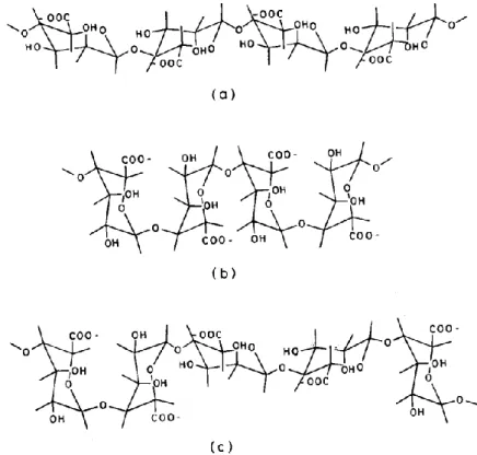
Figure 2.4. The structure of a) β-D-Mannuronic acid (M block), b) α-L-Guluronic acid (G block) and c) mixed block (Shilpa et al., 2003) ... 14
Figure 3.1. SA solutions ... 18
Figure 3.2. a) Gillmore needle and b) the mould for setting time ... 20
Figure 3.3. Injectability testing ... 21
Figure 3.4. a) 6 mm diameter and 12 mm height Teflon mould, b) scaffolds and c) measure of a scaffold ... 22
Figure 3.5. The mould for cell culture discs ... 27
Figure 4.1. XRD pattern of β-TCP calcined at 1000 ºC for 2 h ... 30
Figure 4.2. FESEM image and particle distributions of β-TCP sample calcined at 1000 ºC for 2 h ... 31
Figure 4.3. FTIR pattern of β-TCP calcined at 1000 ºC for 2 h ... 33
Figure 4.4. XRD patterns of pure DCP and SA/DCP cement composites ... 34
Figure 4.5. SEM images of a) DCP, b) 0.5% SA/DCP, c) 1.0% SA/DCP, d) 2.0% SA/DCP, e) 3.0% SA/DCP cement composites. Scale bar is 2µm ... 36
Figure 4.6. FTIR patterns of pure DCP and SA/DCP cement composites... 38
Figure 4.7. Thermograms of pure DCP and SA/DCP cement composites ... 39
Figure 4.8. a) Initial (IT) and final (FT) setting times, b) %injectability of pure DCP and SA/DCP cement composites and (c) digital image showing the injectability of 3.0% SA/DCP cement composites. Values are given as mean ± standard deviation (n =3); p*< 0.05 and p♣< 0.05 denote statistically significant difference between groups ... 41
Figure 4.9. Compressive strengths of pure DCP and SA/ DCP scaffolds, before and after immersion in SBF for 21 days. Values are given as mean ± standard deviation (n =3); p*< 0.05 and p♣< 0.05 denote statistically significant difference between groups. ... 44
xiii
Page
Figure 4.10. Viability of DPSCs after 24 h of incubation with the extraction media of cement discs (n=4). Viability of cells cultured with cell culture media was taken as 100% viable. A significant difference is shown with “*” (p<0.05) and the non-significant difference is shown with “♣” p>0.05. .. ... 46 Figure 4.11. Fluorescence images of a) DCP, b) 0.5% SA/DCP, c) 1.0% SA/DCP,
d) 2.0% SA/DCP, e) 3.0% SA/DCP cement composites and f) control. Cytoskeleton was stained with Alexa Flour 488-phalloidin (green) and cell nuclei stained with DAPI (blue). Scale bars are 100µm. Magnification 20x ... 47 Appendix A.1. Calibration curve of Ca2+ ions (mg/mL). ... 59
xiv
LIST OF TABLES
Page
Table 2.1. Main CPs forms (Bohner, 2010) ... 6
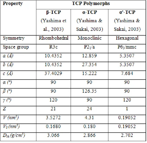
Table 2.2. Structural data of TCP polymorphs ... 10
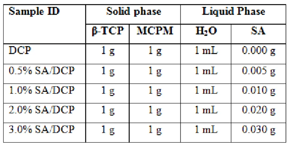
Table 3.1. Preparation of cements ... 18
Table 3.2. Order and amounts of reagents for preparing 500 mL of SBF (Tadashi & Takadama, 2006) ... 23
Table 3.3. Amount of the reagents used in PBS preparation ... 26
Table 4.1. Characteristic peaks of β-TCP in the FTIR spectrum ... 32
Table 4.2. Lattice parameters of pure DCP and SA/DCP cement composites ... 35
Table 4.3. Characteristic peaks of cements in the FTIR spectrum ... 37
Table 4.4. Cumulative weight loss, density, and porosity of DCP and SA/DCP scaffolds immersed in SBF for 1, 3, 7, 14 and 21 days and release of Ca2+ ions. ... 43
xv
SYMBOLS and ABBREVIATIONS
SYMBOLS
° : Degree
°C : The degree Celsius µL : Microliter
3D :Three Dimensional
Å : Angstrom
a-axis : lattice parameter b-axis : lattice parameter c-axis : lattice parameter
C : Carbon Ca : Calcium Cl : Chlorine cm : Centimeter Dth : Theoretical density g : Gram h : Hour H : Hydrogen
Ia : Monoclinic space group
K : Potassium kg : Kilogram kN : Kilonewton L : Liter Mg : Magnesium Min : Minute mL : milliliter MPa : Megapascal N : Nitrogen
xvi
Na : Sodium
nm : Nanometer
O : Oxygen
P : Phosphorus
P1 : Triclinic space group P21/a : Monoclinic space group P63/mmc : Hexagonal space group ppm : Parts per million
R3c : Rhombohedral space group rpm :Revolutions per minute
S : Sulfur
sec : Second
V : Cell volume
V0 : Volume per formula unit
v/v : Volume/volume
W : Watt
w/v : Weight/volume
Z : Number of formula units per cell α : Alpha, cell angle
β : Beta, cell angle
γ : Gamma, cell angle
2θ : The diffraction angle
ρ : Density
xvii
ABBREVIATIONS
ASTM : American Society for Testing and Materials
BSA : Bovine Serum Albumin
CPs : Calcium Phosphates
DAPI : 4′,6-diamidino-2-phenylindole
DCP : Dicalcium Phosphate
DCPA : Dicalcium Phosphate Anhydrous
DCPD : Dicalcium Phosphate Dihydrate
DMEM : Dulbecco's Modified Eagle's Medium
DPSCs : Dental Pulp Stem Cells
FBS : Fetal Bovine Serum
FDA : American Food and Drug Administration
FESEM : Field Emission Scanning Electron Microscopy
FT : Final Time
FTIR : Fourier Transform Infrared Spectroscopy
HA : Hydroxyapatite
ICP-OES : Inductively Coupled Plasma-Optical Emission Spectrometry
IT : Initial Time
JCPDS : Joint Committee on Powder Diffraction and Standards
MCPA : Mono Calcium Phosphate Anhydrous
MCPM : Mono Calcium Phosphate Monohydrate
MMA : Methyl Methacrylate
MTT : 3-(4,5-dimethylthiazole-2-yl)-2,5-diphenyltetrazolium bromide
PBS : Phosphate Buffered Saline
PMMA : Poly (Methyl Methacrylate)
SA : Sodium Alginate
SBF : Simulated Body Fluid
SEM : Scanning Electron Microscopy
xviii
TGA : Thermo Gravimetric Analysis
XRD : X-Ray Diffraction
α-TCP : α - Tricalcium Phosphate α′-TCP : α′ - Tricalcium Phosphate β-TCP : β - Tricalcium Phosphate
1
CHAPTER 1
INTRODUCTION
1.1. BONE STRUCTURE
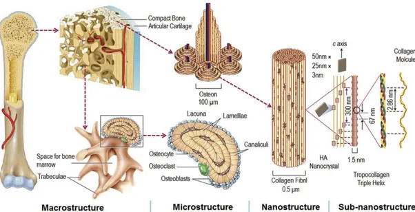
Bone is a mineralized connective tissue and protects numerous vital organs and soft tissues. Bone is classified based on its structure for two types (i) cortical (compact) bone and (ii) trabecular (cancellous) bone. Compact bone makes the outer surfaces of the bones, and it is denser than trabecular bone with 5 to 10% porosity (Zhang et al., 2007). Trabecular bone with a 50 to 90% porosity is very porous and softer than compact bone (Zhang et al., 2007). Bone is mainly composed of a collagen and calcium phosphates (CPs) composites that gives shape to the skeleton of the body. Bone has a honeycomb-like matrix inside that helps to supply the bone rigidity. Bone possess three types of cells: (i) Osteoblasts: These are derived from mesenchymal stem cells, making new bone matrix known as osteoid via osteogenesis process (Martin, 2003). (ii) Osteocytes: They are inactive osteoblasts, establishing connections between osteocytes and osteoblasts. (iii) Osteoclasts: They are derived from the hematopoietic lineage and responsible for osteolysis process (Hattersley et al., 1991). These cells dissolve the mineral of bone and destroy the organic matrix (Martin, 2003). Hierarchical organization of bone from the macro to the sub-nanoscale was illustrated in Figure 1.1.
2
Figure 1.1. Hierarchical organization of bone from the macro to the sub-nanoscale (Wang et al., 2016b).
1.2. CEMENTS AS BONE REPAIR MATERIALS
Bone cements are widely used for implant fixation in several trauma orthopedic surgeries (Vaishya et al., 2013). Their preparation is based on chemical reaction that takes place between the powder phase and the liquid phase, which results in paste formation as an intermediate stage. After this stage, it self-sets once implanted in the body. This implies that the material is flexible, which guarantees ideal fit on the implanted site providing high bone–material contact, even in strong geometric deformities. However, the way that the bone cements experience a setting response once embedded and framing a hard structure ensure a specific degree of mechanical support. This mechanical support could change based on cement type used. Latest developments in orthopedic surgery focus on use of minimally invasive surgical techniques (Ginebra & Montufar, 2019). However, there is no ideal cement in the medical market; nowadays all bone cements have some limitations. Bone cements divided into two main classes, acrylic bone cements and dicalcium phosphate (DCP) cements.
3
1.2.1. Acrylic Bone Cements
Poly (methyl methacrylate) (PMMA) used asacrylic bone cement, is one of the most common bone cement material used in orthopedic surgeries, dental applications, vertebroplasty and kyphoplasty. However, it has some limitations. PMMA polymerization is an exothermic reaction. The temperature rises about to 60-120 °C causing serious thermal damage surrounding the bone tissue (McMahon et al., 2012). Additionally, there is also concern about severe toxicity because of methyl acrylate monomer (MMA) (Pikis et al., 2015). The liquid monomer (MMA) can cause asthmatic reactions or lacrimation (Lewis, 1997). Furthermore, it was shown that monomers left decreased the growth rate of osteoblasts compared to other common materials, such as CPs and hydroxyapatite (HA) (Ricker et al., 2008).
1.2.2. Dicalcium Phosphate Cements
Dicalcium Phosphate (DCP) Cements have been suggested as an alternative option for PMMA. It has several advantages over PMMA like its good biocompatibility, capability of self-hardening at room temperature and causing no exothermic effect during the hardening process. Due to biocompatibility and ability to degrade inside the body that encourages bone growth, it is used in bone replacement in oral surgery and craniofacial applications and osteonecrotic sites in the body (Ambard & Mueninghoff, 2006). However, it has poor mechanical properties along with a low degradation rate (Julien et al., 2007). Some polymers such as alginate, chitin, chitosan, cellulose, collagen, gelatine and synthetic polymers have been used to improve properties like porosity, injectability, degradation rate and mechanical properties (Perez et al., 2012).
1.3. PROBLEM STATEMENT
DCP cements widely used for various applications of bone regeneration such as craniofacial and maxillofacial reconstruction (skull base defect repair, cranioplasty, augmentation genioplasty, cranial recontouring, on-lay grafting, cranial flap augmentation) (Gómez et al., 2005). However, it has poor mechanical properties
4
along with a low degradation rate (Julien et al., 2007).
1.4. OBJECTIVES OF THE THESIS
Generally, the main objectives of this research project are to investigate and improve the microstructural, mechanical, and biological properties of DCP cements using Sodium alginate (SA) as an additive.
Here the aim of this thesis can be summarized as,
- To synthesize β-Tricalcium phosphate (β-TCP) based biomaterials using microwave-assisted precipitation method and to characterize the above-mentioned β-TCP material utilizing XRD, FTIR, SEM and ICP-OES techniques.
- To synthesize DCP cements from the corresponding β-TCP precursors incorporated with different compositions of alginate and to characterize the obtained cements utilizing XRD, FTIR, FESEM, TGA and ICP-OES techniques.
- To evaluate the impact of alginate substitution up to the setting time, injectability and physicochemical properties of DCP cements.
- To study the effect of alginate substitution on the in vitro resorption and degradation of the DCP cements in simulated body fluid (SBF).
- To study the effect of alginate substitution on the mechanical properties of the DCP cements in SBF.
- To study the effect of alginate substitution on the in vitro osteoinduction, osteoconduction and osseointegration properties of the DCP and Alginate - DCP cements cultured with Dental pulp stem cells (DPSCs).
1.5. SIGNIFICANCE OF THE THESIS
Physicochemical properties of DCP cements play an important role in predicting in vivo performance of these materials. Cements injectability is important for invasive surgical techniques for filling bone defects; however, due to the liquid-solid stage
5
division, DCP cements without additives usually have the weak infusing ability. Sodium alginate (SA) can be used to improve injectability, physicochemical and mechanical properties of DCP cements. Controlling the resorption parameters of DCP cements, it may be easily feasible to control the flow of bone stimulating ions. In this thesis, it was aimed to investigate the effect of SA addition of different amounts on microstructural, mechanical, and biological properties of DCP cements. The synthesis of materials proposed in this thesis is a modest effort to produce biomedical materials, which will significantly reduce their cost and make them available to ordinary citizens at an affordable rate.
6
CHAPTER 2
LITERATURE REVIEW
2.1. CALCIUM PHOSPHATES
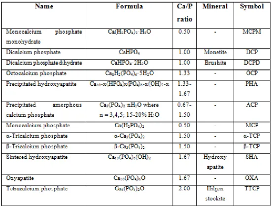
CPs highly resemble with the mineral part of the bone and tooth. It is classified based on Ca/P ratios (Table 2.1) and frequently used as bone graft material.
7
2.1.1. Monocalcium Phosphate Monohydrate
Monocalcium phosphate monohydrate (MCPM, Ca(H2PO4)2·H2O) is a very good water-soluble material resulted in low pH value. MCPM has a stable phase at a pH below 2.5 (Ginebra, 2008). Due to its high solubility and acidity nature, MCPM is not preferred to be used in biomedical applications. Drying the MCPM at 100 °C leads to evaporation of water molecule forming anhydrous form (MCPA). The density of MCPA is higher than that of MCPM (Farahani et al., 2012). However, MCPM is used as a source of DCP cements and the last used in dental, drug delivery and biomedical applications (Kouassi et al., 2003; Taha et al., 2017; Shu et al., 2019).
2.1.2. Dicalcium Phosphate
DCP materials are a kind of CPs with a Ca/P ratio of 1.0 (Kossler & Fuchs, 2009), commonly used in orthopedic and dental applications. Furthermore, they are used as a food additive and as a polishing agent in toothpaste (Dorozhkin & Epple, 2002; Qadir et al., 2014). Dicalcium phosphate dihydrate (DCPD, CaHPO4·2H2O) cements and dicalcium phosphate anhydrous (DCPA, CaHPO4) are two forms of DCP cements widely used as bone fillers. DCPD transforms into DCPA at temperatures above ∼80 °C
2.1.2.1. Dicalcium Phosphate Anhydrous
Dicalcium Phosphate Anhydrous (DCPA) is known as monetite, it is used to enhance skeletal repair (Desai et al., 2007), also used in orthopedic applications (Koju et al., 2018), drug delivery applications (Taha et al., 2017), dental applications (El Briak et al., 2008) and craniofacial bone augmentation (Tamimi et al., 2009a). DCPA cements was synthesized using different methods such as; hydrothermal method (Jinawath et al., 2001), precipitation from microemulsion (Kong et al., 2005), crystallization from solution (Sivakumar et al., 1998), electro and chemical deposition (Rohanizadeh & LeGeros, 2008) and microwave-assisted method (Kohn et al., 2002). DCPA crystallizes with a space group of Pl which is in triclinic crystal
8
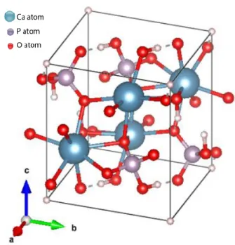
system and unit-cell parameters of a=6.910 Å, b=6.627 Å, c=6.998 Å , α=96.34º, β=103.82º and γ= 88.33º as seen in Figure 2.1 (Mathew & Takagi, 2001). DCPA is stable at a pH range of 2.5 - 4.2 (Ginebra, 2008). However, DCPA converts to HA when pH is changed to a physiological pH of 7.4. Furthermore, it is more stable than DCPD above 121 °C. Monetite’s crystal growth rate is slow, so DCPD forms easier than DCPA. The density of DCPA is 2.93 g/cm3 (Unosson et al., 2015). As with DCPD, the solubility of DCPA is pH dependent. Moreover, its solubility decreases with increasing the temperature.
Figure 2.1. The unit cell structure of DCPA.
2.1.2.2. Dicalcium Phosphate Dihydrate
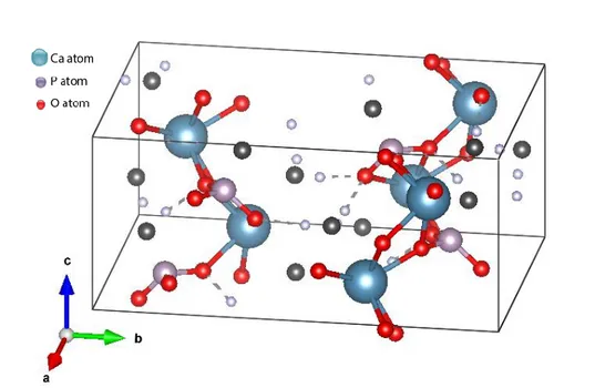
Dicalcium phosphate dihydrate (DCPD) is known as brushite, it is used for maxillofacial bone augmentation (Tamimi et al., 2009b), treatment of periodontal diseases (Tamimi et al., 2008), drug delivery applications (Taha et al., 2017), osteoplastic surgery (Fomin et al., 2017) and cranial defects (Ji & Ahn, 2010). DCPD crystallizes with a space group of Ia which is in monoclinic crystal system and unit cell parameters of a=5.812 Å, b=15.180 Å, c=6.239 Å and β=116.42° as seen in Figure 2.2 (Dumitraş et al., 2004). The density of DCPD is 2.33 g/cm3 (Unosson et al., 2015). Heat-treatment of DCPD leads to loss of water molecule,
9
forming an anhydrous form of DCPA (Klammert et al., 2009).
Figure 2.2. The unit cell structure of DCPD.
2.1.3. Tricalcium Phosphate
Tricalcium phosphate (TCP, Ca3(PO4)2) is well known biomaterial and commonly used in the biomedical area. It is used in particular to treat bone defects, repairing devices and as coatings on metal alloy prostheses. TCP introduces three polymorphs with stage change temperatures. β-TCP (rhombohedral) transforms to α-TCP (monoclinic) at 1125 °C and α-TCP converts to α′-TCP (hexagonal) at 1430 °C (Moreno et al., 2019) according to Kreidler and Hummel's phase diagram (Kreidler & Hummel, 1967) as shown in Table 2.2.
10
Table 2.2. Structural data of TCP polymorphs.
2.1.3.1. β - Tricalcium Phosphate
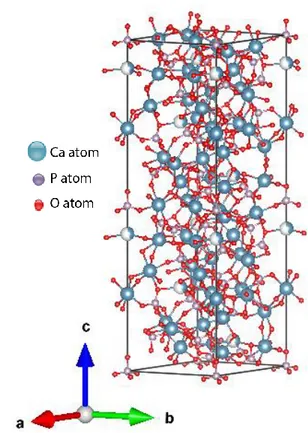
β - Tricalcium Phosphate (β-TCP, β-Ca3 (PO4)2) has seen much broader use as a biodegradable CPs or as a part of biphasic CPs in the form of dense or macroporous granules or scaffolds in clinical applications (Rahaman, 2014). Based on the crystal structure (Figure 2.3), β-TCP has R3c space group which in a rhombohedral structure with unit cell parameters of a=b=10.4352 Å and c=37.4029 Å with α=β=90°, γ=108.65°. It has a density of 3.07 g/cm3
(Mirhadi et al., 2011). β-TCP has complex structures consisting of tetrahedral phosphate centres connected to calcium ions with oxygen.
11
Figure 2.3. The unit cell structure of β-TCP.
Methods to Synthesize β-TCP Particles
Different methods have been used to synthesis pure phase of β-TCP, such as: electrochemical (Djošić et al., 2008), micro-emulsion (Dai et al., 2016), sonochemical (De Campos et al., 2007), hydrothermal (Gan et al., 2019), sol-gel (Sanosh et al., 2010) and microwave-assisted method (Taha et al., 2017). Wet chemical method (Fathi et al., 2015; Chang, 2016) has also been used for the production of HA nanoparticles. A brief detail of these methods is given below.
I. Wet precipitation method
Wet precipitation method is one of the most used techniques to prepare β-TCP powders. This method involves the mixing of Ca2+ and PO43- based precursors in a basic or neutral solution to prepare poorly crystalline β-TCP. Main advantages of using wet precipitation methods are; easy to process, no need for complex devices, having low reaction time and temperatures, low-cost raw materials, controllable
12
particle morphology and the mean size, high purity materials, scalability, reproducibility, possible for industrial application and synthesis conditions can be easily controlled (Ramesh et al., 2013; Krauklis et al., 2018; Rodríguez-Lugo et al., 2018).
II. Microwave-assisted wet precipitation method
Some benefits of using microwave are these superheating increases the reaction rate and enables faster synthesis, shape and size control by easily adjusting reaction parameters and because of better controlling of process parameters, greater reproducibility of chemical reactions than in conventional heating (Saleh et al., 2017). On the other hand, some of the limitations of using microwave have been observed such as lack of scalability, limited applicability for materials that absorb microwave and safety and health hazards about using of a microwave oven (Saleh et al., 2017).
III. Sol-gel method
A “Sol” is a scattering of colloidal amorphous or crystalline particles. A “Gel” is a 3D interconnected network shaped by sol particles. Sol-gel term refers to the processes of gels produced from colloidal suspensions. This process includes the production of inorganic matrices by the arrangement of colloidal suspension and shaping a bunch of gels. After drying it converts into xerogel shape (Aurobind et al., 2006). In sol-gel chemistry, hydrolysis and condensation reactions transform to molecular precursors.
The sol-gel method generally consists of the following basic steps: Hydrolysis of precursor, polycondensation, gelation, aging, drying and high-temperature process (Aurobind et al., 2006).
13 IV. Solid-state reaction method
Several forms of CPs particles can be synthesized using a solid-state method, the solid precursors are mixed and grinded in wet media to make the homogeneous mixture and reduce particle size. For wetting medium distilled water is used. At appropriate temperature solid-state reaction is carried out between the starting materials. This process is known as calcination. Calcination makes the starting precursors interact with them and results in a homogeneous phase. After calcination, the powder is shaped then it is densified with sintering. Sintering is a process of heating the material to increase its grain size and strength by bonding together the particles.
V. Hydrothermal method
In this method, from room temperature to high-temperature nanoparticles can be made. Depending on the vapour pressure of the principal components in the reaction, low pressure or high-pressure conditions are used to monitor the morphology of the materials to be prepared. The advantages of hydrothermal synthesis are at low-temperature materials with good crystallinity and homogeneous size and shape properties (Zhang & Vecchio, 2007).
2.2. ALGINATE AND ITS IMPORTANCE
Alginate is the foremost plenty of marine biopolymer within the world following to cellulose. The main alginate source is found in the cell walls and intercellular regions of marine brown algae such as Ascophyllum nodosum, Macrocystis pyrifera and Laminaria hyperborea (Shilpa et al., 2003). The alginate molecules provide flexibility and strength for the plant growth in the sea. Some bacteria synthesize alginate like Azotobacter and Pseudomonas species (Rani et al., 2019). Alginate exsited in different kinds such as: sodium alginate (SA), alginic acid, potassium, calcium and ammonium salts, an ester of alginic acid and propylene glycol alginate (Venkatesan et al., 2014) and SA consider the main type of alginate (McHugh, 2003).
14
Alginate is a linear and anionic polysaccharide and formed by the whole family of linear copolymers containing blocks of β-D-mannuronic acid (M) and (1,4) linked
α-L-guluronic acid (G) where these blocks are constituted by sequential G residues (GGGG), sequential M residues (MMMM) and also mixed G and M residues (GMGM) (Lee & Mooney, 2012). The structure of alginate blocks is given in Figure 2.4.
Figure 2.4. The structure of a) β-D-Mannuronic acid (M block), b) α-L-Guluronic
acid (G block) and c) mixed block (Shilpa et al., 2003).
Sodium Alginate (SA), a well-known polymer-based biomaterial, has been frequently used in the medical sector due to its biocompatibility and ease of gelation. SA in hydrogel form has been especially appealing in tissue engineering applications. Furthermore, these gels hold basic similarity to the extracellular networks in tissues (Lee & Mooney, 2012). Multivalent cations are regularly utilized to make crosslinks among carboxyl and hydroxyl moieties on neighbouring polysaccharide chains. Previous studies deduced that addition of SA improved mechanical properties of the composites. Zhang et al. (2013) found compressive modulus and the compressive yield strength of the nano-HA/collagen composites
15
increased as SA content was increased. Yu et al. (2019) revealed that SA can successfully improve compressive resistance and elasticity properties of κ-carrageenan composite gel. Dabiri et al. (2019) demonstrated that addition of 0.5% SA increased the elastic modulus and compressive strength of undoped brushite cement. Furthermore, numerous studies confirmed that adding SA to materials improves adhesion and proliferation of cells (Turco et al., 2009; Dabiri et al., 2019; Ni et al., 2019)
2.3. DENTAL PULP STEM CELLS (DPSCs)
Dental pulp stem cells (DPSCs) are obtained from the teeth recovered during a normal dental procedure. DPSCs can be isolated, cultured, and cryopreserved. DPSCs have an extremely proliferative ability and probability of differentiation into a variety of cells. Additionally, more than 80 passages can be passed with the capacity to protect differentiation (Laino et al., 2006). The osseo-specific markers of DPSCs are alkaline phosphatase (ALP), osteopontin and osteocalcin. In this thesis, DPSCs were used for cell culture tests.
16
CHAPTER 3
METHODOLOGIES
3.1. MATERIALS AND CHEMICALS
Calcium nitrate tetrahydrate (Ca(NO3)2·4H2O, Merck-Germany), di-Ammonium hydrogen phosphate ((NH4)2HPO4, Merck-Germany), Sodium chloride (NaCl, Merck-Germany), Sodium hydrogen carbonate (NaHCO3, Merck-Germany), Potassium chloride (KCl, Merck-Germany), Potassium Hydrogen Phosphate Trihydrate (K2HPO4. 3H2O, Merck-Germany), Magnesium Chloride Hexahydrate (MgCl2. 6H2O, Merck-Germany), Hydrogen chloride (1.0M–HCl, Merck-Germany), Calcium chloride (CaCl2, Merck-Germany), Sodium sulfate (Na2SO4), Tris (hydroxymethyl) aminomethane and Dimethyl sulfoxide (Merck-Germany) Monocalcium phosphate monohydrate (MCPM, Merck-Germany). Sodium Alginate (SA), o-cresolphthalein complex one, 8-hydroxyquinaline-5-sulfonic acid, 2-amino-2-methyl-1-propanol and Thiazolyl Blue tetrazolium bromide (MTT) were purchased from Sigma Aldrich, Germany. Dulbecco’s Modified Eagle Medium (DMEM), fetal bovine serum (FBS) and penicillin-streptomycin solution were purchased from Biowest, France.
3.2. SAMPLES PREPARATION
3.2.1. Synthesis of β-Tricalcium Phosphate
β-TCP material was prepared by microwave-assisted wet precipitation method (Motameni et al., 2020). About 42.51 g of Ca(NO3)2·4H2O was dissolved in 200 mL of distilled water, forming solution A. 15.85 g of (NH4)2HPO4 was dissolved in 200 mL distilled water, forming solution B. The solutions were kept under magnetic stirrer until the powders were completely dissolved in the distilled water. Solution B
17
was added dropwise under constant stirring to solution A. pH was kept at 7 using ammonium hydroxide. Then, the solution mixture was left under stirring for 30 min at room temperature and transferred to a microwave oven (SAMSUNG, MS23F301EAW). The mixture was heated at 800W for 5 min. The precipitated mixture was filtered and washed with distilled water. Then, dried in an oven set at 80 ºC (Eurotherm) for overnight. Finally, it was calcined at 1000 ºC Protherm PLF 140/5) for 2 h.
3.2.2. Preparation of Sodium Alginate / Dicalcium Phosphate Cement Composites
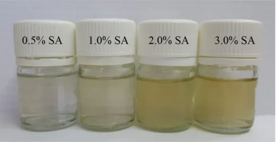
The pure phase of DCP cement was prepared by mixing 1 g of β-TCP and 1 g of Monocalcium phosphate monohydrate (MCPM) in a mortar. Then, about 1 mL of distilled water was thoroughly mixed with the powder mixture until a homogeneous paste was obtained. SA/DCP cement composites were prepared using the same method except for different amounts of SA were added to 1 mL of water. In Figure 3.1 there is a picture of SA solutions. Amounts of reactants used for DCP and SA/DCP cements preparation are listed in Table 3.1.
18
Figure 3.1. SA solutions.
3.3. CHARACTERIZATIONS
3.3.1. X-Ray Diffraction
The phase purity of the prepared materials was investigated using X-ray diffraction (XRD, Rigaku Ultima IV). The samples were scanned from 20º to 80º in 2θ angle and the lattice parameters were calculated using the integrated software package. The phase purity of the entire materials was detected via matching with standard patterns (Joint Committee on Powder Diffraction and Standards (JCPDS)).
3.3.2. Field Emission Scanning Electron Microscopy
Particle size and morphology were analysed using Field emission scanning electron microscopy (FESEM, Carl Zeiss Ultra Plus Gemini). Samples were pre-coated with a gold thin film to reduce the spark rate. Average particle size and particle size distribution were determined by measuring the size of 50 particles selected at random from FESEM images using image analysis software ImageJ (National Institutes of Health, USA).
19
3.3.3. Inductively Coupled Plasma - Optical Emission Spectrometry
Inductively coupled plasma - optical emission spectrometry (ICP-OES) is a technique used for the detection of chemical elements. This technique was used to detect chemical elements of β-TCP (Perkin Elmer Optima 4300DV). Approximately 500 mg of sample is decomposed overnight in a mixture of 10 mL of concentrated nitric acid (HNO3) and hydrochloric acid (HCl) at a ratio of 1:3. Then, the solution undergoes several dilutions prior to inject inside analyzing chamber of ICP-OES.
3.3.4. Fourier-Transform Infrared Spectroscopy
Presence of functional groups was confirmed by using Fourier-Transform Infrared Spectroscopy (FTIR, Bruker IFS 66/S) device. All spectra were recorded in transmission mode in the scanning range of 4000-400 cm-1.
3.3.5. Thermogravimetric Analysis
The thermogravimetric analysis (TGA) was carried out under a nitrogen atmosphere (Perkin Elmer Pyris 1) from room temperature to 950 ºC. Depending on the temperature, changes in the sample weights were monitored to study the thermal stability of the prepared materials.
3.4. SETTING TIME
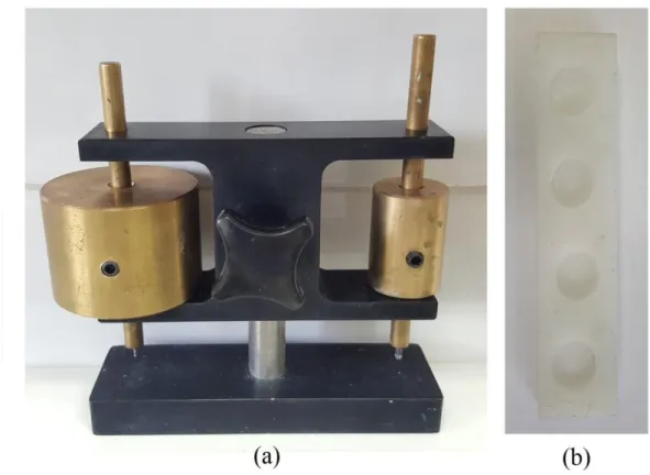
The initial time (IT) and final time (FT) of the cements were measured using a Gillmore needle (ASTM Standard C266-04, 2004). Briefly, about 1 g of the cement paste prepared in Table 3.1 poured in 16 mm diameter and 3 mm height Teflon mould. A needle with a diameter of 2.12 ± 0.05 mm and weight of 113.4 ± 0.5 g was placed on the surface of the cement paste and IT recorded when the needle does not leave any traces on the specimen. Similarly, a needle with a diameter of 1.06 ± 0.05 mm and weight of 453.6 ± 0.5 g was placed on the surface of the cement paste and FT when the needle does not leave any traces on the specimen. The experiments conducted in triplicate and measurements were recorded every 30 sec.
20
In Figure 3.2 there are pictures of the mould and the Gillmore needle.
Figure 3.2. a) Gillmore needle and b) the mould for setting time.
3.5. INJECTABILITY TEST
Injectability of the cement paste was investigated utilizing commercial 10 ml syringe sterile similar to the method reported in previous studies (Saleh et. al., 2016 ; Zhou et al., 2015). Briefly, after homogenization, the paste was poured into a commercial syringe with a slit tip 2 mm in diameter. About 5 kg of weight fixed vertically on the top of the plunger for 2 min. The amount of paste before and after injection was quantified and the injectability was determined by the following Equation (3.1). In Figure 3.3 there is a picture of injectability setup.
21
Figure 3.3. Injectability testing.
Inj% = (WF - WA) / (WF - WE) X 100 (3.1)
Where Inj (%) referred to the injectability (%), WF is the weight of the syringe packed with paste and WA is the weight of the syringe after injection and WE is the weight of the empty syringe. The tests were performed in triplicate.
3.6. IN VITRO BIODEGRADATION AND MECHANICAL ANALYSIS
3.6.1. Scaffold Shaping
The DCP and SA/DCP paste was poured into a split Teflon mould of 6 mm diameter and 12 mm height and extruded carefully in a disc shape. In Figure 3.4 there are pictures of mould and scaffolds.
22
Figure 3.4. a) 6 mm diameter and 12 mm height Teflon mould, b) scaffolds and c) measure of a scaffold.
3.6.2. Preparation of Simulated Body Fluid
In a plastic beaker 480 mL of distilled water was added and the beaker was covered with foil. The solution was mixed in a beaker with a magnetic stirrer and solution temperature was adjusted at 37 ºC. After the temperature reached to 37 ºC, reagents
23
were added and dissolved one by one in the order as seen in Table 3.2. When the first 9 reagents were added, the pH of the solution was adjusted to 7.4 by adding few drops of HCl (1.0 M). With 480 mL distilled water and 20 mL HCl, a total of 500 mL Simulated Body Fluid (SBF) were obtained.
Table 3.2. Order and amounts of reagents for preparing 500 mL of SBF (Tadashi & Takadama, 2006).
The biodegradation of the disc sample was evaluated by SBF. The discs were incubated for 1, 3, 7, 14 and 21 days into 15 mL sterile conical centrifuge tubes (one disc per tube) in shaking water bath at 37 ºC. At each period, the discs were gently removed then rinsed by distilled water followed by absolute ethanol and finally dried at room temperature.
3.6.3. Weight Loss, Density and Porosity
Weight loss, density, and porosity of cements after immersion in SBF for 0, 1, 3, 7, 14 and 21 days were measured. The percentages of weight loss were estimated using Equation 3.2 (Aydogdu et al., 2016).
24 Weight Loss(%) = (W0−Wf
W0 ) x100 (3.2)
Where W0 referred to the weight of scaffold before the immersion and Wf represents the weight of scaffold after a specific immersion period. The experiments were carried out in triplicates.
The apparent densities of discs of cements were measured by using the Archimedes principle (Equation 3.3).
p =A-BA p° (3.3)
Where p represents the apparent density, A is air-weight, B is water-weight and po is the water density (1 g/cm3).
The total porosity of the materials was calculated using Equation 3.4.
Total Porosity (%) = (Vtotal pore
Vtotal ) x 100 (3.4)
Where Φ is porosity, mdry is a dry weight of scaffold and mwet is water weight of scaffold.
3.6.4. Compressive Strength Tests
Firstly, samples were immersed in SBF at 37 °C for 0, 1, 3, 7, 14 and 21 days. After each immersion time point in SBF, the cement specimens (6 mm (diameter) x 12 mm (height)) were gently removed and dried at room temperature. The compression strength test was performed using Universal testing systems (Instron 5565A, USA) (ASTM Standard F451-16, 2016). It was applied with a capacity of 5 kN at a 2 mm/min crosshead speed. The tests were performed in triplicates.
25
3.6.5. In Vitro Ca2+ Ions Release
The release of Ca2+ ions from the cement formulations in SBF was determined at different incubation periods colorimetrically by a stable interaction with o-cresolphthalein complexone. Briefly, about 0.0024 g o-cresolphtalein complexone was dissolved in 10 mL distilled water and mixed thoroughly with a solution of 8-hydroxyquinoline-5-sulfonic acid (0.025 g dissolved in 10 mL distilled water). About 20 ml (0.5 M) of 2-amino-2-methyl-1-propanol was added to the solution mixture, and the colour of the solution turns a purple colour. At each immersion period, 100 µL of aliquot was collected from SBF and mixed with 900 µL (0.1M) HCl for 5 min. Then, 25 µL was taken from the last solution mixture and added to the 120 µL reagent in 96 well plates. After mixing their optical densities were measured at 560 nm by using a microplate spectrophotometer (SpectraMax iD3, USA) (Aydogdu et. al., 2016). The experiments were carried out in triplicates.
3.7. IN VITRO CELL CULTURE ANALYSIS
3.7.1. Preparation of Phosphate Buffered Saline
800 mL of distilled water was poured into a beaker to prepare 1 L of Phosphate Buffered Saline (PBS). The solution was mixed with a magnetic stirrer. All the reagent salts (Table 3.3) were dissolved in 800 mL of distilled water. By adding 1.0M-HCl or NaOH, the solution's pH was set to 7.4. Until the volume is 1 L, distilled water was added.
26
Table 3.3. Amount of the reagents used in the PBS preparation.
3.7.2. Cell Culture Tests
The prepared cements were cast in disc shape (6 mm (diameter) x 2 mm (height)) and the mould for producing these discs are shown in Figure 3.5. They were then sterilized by UV irradiation (254 nm) for 1 h for each side of the disc. The discs were rinsed with sterile phosphate-buffered saline (PBS, 0.1M, pH 7.4) for 1 h. This was followed by rinsing with Dulbecco's Modified Eagle Medium (DMEM) supplemented with 10% Fetal bovine serum (FBS) and 1% penicillin-streptomycin solution for 30 min and immersion in complete medium at 37 °C in carbon dioxide incubator for 7 days. After 7 days incubation in complete medium, the discs were gently removed, and the extracts of discs were collected and centrifuged at 14000 rpm for 15 min. The supernatants were collected and stored for future use. Meanwhile, human dental pulp stem cells (DPSCs) from the third molar (isolated and characterized as a stem cell by Rad et. al. (2018)) were sub-cultured for 3 passages. All experiments were performed in four replicates.
27
Figure 3.5. The mould for cell culture discs.
3.7.2.1. Cell Viability with MTT
In vitro cytotoxicity effects of the cement’s extracts were evaluated via the indirect method by measuring the formation of formazan from MTT (3-[4,5-dimethylthiazole-2-yl]-2,5-diphenyltetrazolium bromide) spectrophotometric.
DPSCs cultured in DMEM supplemented with 10% FBS and 1% penicillin-streptomycin solution. Then DPSCs were seeded in 96-well plates at a density of 2500 cells/well and incubated at 37 °C in a carbon dioxide incubator for overnight. The cell culture media was replaced with cements extracts media (prepared in section 2.7) and the DPSCs incubated with the extract’s media for 24 h. After a 24 h incubation period, the extractions medium was discarded, the cells and wells of tissue culture plate were rinsed with sterile PBS. After the rinsing process, about 100 µl of 0.5 mg/mL 1X MTT solution was added to each well, the well plates were covered with aluminium foil and incubated at 37 °C for 4 h. Then, the MTT solution in the wells was discarded and the formazan crystals formed inside cells. The formazan crystals were solubilized in 100 μl of dimethyl sulfoxide by gentle shaking
28
for 20 min on an orbital shaker in the dark. The supernatants were collected and transferred to a fresh 96-well plate. The absorbance was measured at 570 nm using a microplate reader (SpectraMax iD3, USA). The DPSCs were incubated in untreated cell culture media used as a control group.
To calculate the percentage of cell viability, for background control (negative controls) optical densities are averaged. This value is subtracted from all values. For control (healthy cells with 100% viability) average was calculated. Each value divided by control and multiplied by 100 ((value / control) * 100). This value is the percentage of cell viability. Then, the same group's values were averaged and the standard deviation was calculated.
3.7.2.2. Cell Fixation
100 mL PBS was put in a beaker and heated to 60 ºC with magnetic stirrer. Then 4 g paraformaldehyde powders were added to the heated PBS and waited for dissolving. Beaker was covered with foil to protect from the light.
DPSCs were cultured and grown in DMEM. Cells were trypsinized and collected by centrifugation at 2000 rpm for 5 min at room temperature. Then cells were counted by using a hemocytometer. 5x10⁴ DPSCs were seeded on 48 well plates, 500 µl DMEM low glucose with 10% FBS and 1% penicillin-streptomycin was put on wells and the cells were incubated at 37 ºC and 5% CO2 for 24 h.
After 24 h mediums in wells were removed and washed with 500 µl PBS in dark. 500 µl paraformaldehyde solution was added to each well and waited 20 min at room temperature. After 20 min paraformaldehyde solution was removed and 500 µl PBS was added to each well. After 5 min PBS was removed and 500 µl PBS was added again.
29
3.7.2.3. Cell Morphology Examination
The DPSCs were seeded on cover glasses at a cell density of 5000 cells/glass and cultured in complete medium for 24 h. After a 24 h incubation period, the cell culture media was replaced with extraction media and incubated for 24 h. At the end of the incubation period, the cells were fixed with 4% (w/v) paraformaldehyde solution for 15 min at 37 °C. Afterwards, the paraformaldehyde solution was discarded and cells were washed with PBS. Then, the cell membranes were permeabilized with 1% (v/v) Triton X-100 in PBS for 5 min at room temperature. After washing with PBS, cells were incubated in 1% (w/v) of bovine serum albumin (BSA) solution in PBS for 1 h at 37 °C. Then, to stain actin filaments, cells were incubated with Alexa flour 488 phalloidin in 0.1% (w/v) of BSA in PBS for 1 h at 37 °C. After washing with 0.1% (w/v) of BSA in PBS, to stain cell nuclei, cells were incubated with DAPI (4′,6-diamidino-2-phenylindole) in 0.1% (w/v) of BSA in PBS for 10 min at room temperature. After washing with PBS, cell morphology was assessed with Zeiss Axio Scope A1 (Zeiss, Germany) under fluorescence of excitation at 488 nm for Alexa flour (green) and 350 nm for DAPI (blue).
3.7.2.4. Statistical Analysis
All experiments were performed with n=3, except for MTT analysis. MTT analysis was replicated with n=4. All data are demonstrated as mean ± standard deviation. Results were analysed using a one-way analysis of variance (ANOVA) and Tukey's multiple comparison test.
30
CHAPTER 4
RESULTS AND DISCUSSION
4.1. CHARACTERIZATION OF β-TCP
The XRD pattern of β-TCP powder is illustrated in Figure 4.1. All diffraction peaks of the prepared β-TCP were in good match with the standard phase of β-TCP (whitlockite phase, JCPDS 09–0169). No other CPs phases were observed. Particularly, no peak was detected at 32.196º, indicating that the synthesized powder existed as single-phase and no HA phase (JCPDS 09–432) was present.
20 25 30 35 40 45 50 55 60 0 500 1000 1500 2000 2500 (42 8) (2 2 1 8) (2 3 1 4) (50 8) (3 0 1 8) (2 0 20 ) (05 4) (4 16 ) (12 5) (1 1 1 2) (3 06 ) (30 0) (20 2) (024) (1 0 10 ) (21 4) (12 2) (0 2 10 ) (22 0) (12 8) (4 04 ) (0 0 18 ) (2 2 1 2) (0 1 2 0) (2 38 ) (4 0 10 ) (04 8) (3 0 1 2) (1 0 16 ) (1 2 11 ) In te n si ty (a .u .) 2Degree) (2 1 10 )
31
SEM micrograph of β-TCP powder is shown in Figure 4.2. β-TCP particles appeared in globular shape with rounded edges, the particles coalesced due to heat-treatment at high temperature. However, similar observations were reported in previous studies (Mirhadi et al., 2011; Grigoraviciute-Puroniene et al., 2017; Taha et al., 2017). Particle distribution was plotted based on diameters of 50 particles and the results are ascribed in Figure 4.2. Average particle size was about 1000 nm (1 µm). Mirhadi et al., (2011) synthesized β-TCP particles using a wet chemical process, followed by calcination at 700 ºC for 2 h. The average size of the obtained particles was about 150 nm. Grigoraviciute-Puroniene et al., (2017) synthesized β-TCP particles using wet precipitation method and they calcined at 800 °C for 5 h. The group reported that the average size of the obtained particles was about 150 nm. However, in this thesis, the average size of the prepared particles was bigger than those reported in previous studies. This could be attributed to high particle growth rate during heat treatment. Ca/P ratio was found 1.39, which was slightly lower than the desired value (1.5).
Figure 4.2. FESEM image and particle distributions of β-TCP sample calcined at 1000 ºC for 2 h.
FTIR spectra of TCP powder are shown in Figure 4.3 and characteristic peaks of β-TCP in the FTIR spectrum were shown in Table 4.1. The bands at 431 and 496 cm−1 belong to the ʋ2 mode of PO43-. The bands at 542 cm-1, 589 cm-1 and 604 cm−1 were assigned to the vibration mode of PO43- (ʋ4). The bands located in between 943 cm-1
600 700 800 900 1000 1100 1200 1300 1400 0 2 4 6 8 10 12 14 16 Particle size(nm) N u m b er o f Pa rti cl es
32
and 1214 cm−1 were attributed to the ʋ1 (symmetric stretching) and ʋ3 (asymmetric stretching) mode of PO43- group. Locations and shapes of the peaks were well matched with FTIR results of previous studies (Raynaud et al., 2002; Chen et al., 2008). Stretching and bending bands corresponding to hydroxyl (-OH) groups were not detected at 630 cm-1 and 3570 cm-1 confirming synthesis of single-phase β-TCP. The peak at 727 cm−1 showed the presence of P2O74− ions which belongs to one of the β-TCP precursors [β-calcium pyrophosphate (β-Ca2P2O7)]. This peak becomes sharper which confirms the conversion of HPO42- to β-TCP after calcination at 1000 °C (Abdel-Fattah et al., 2008).
33 1500 1400 1300 1200 1100 1000 900 800 700 600 500 400 40 50 60 70 80 90 100 110 120 P2O7 4- ions PO43- (1) PO43- (3) PO43- (4) Wavenumber(cm-1) T ra n sm itta n ce (% ) PO43- (2)
Figure 4.3. FTIR pattern of β-TCP calcined at 1000 ºC for 2 h.
4.2. CHARACTERIZATION OF PURE DCP AND SA/DCP
XRD patterns of pure DCP and SA/DCP self-hardened at room temperature are illustrated in Figure 4.4. Diffraction peaks of DCP were in good match with a standard phase of monetite (JCPDS 71-1760). No peak was detected at 20.95º, indicating that prepared DCP existed as single-phase (Monetite), and brushite phase (JCDPS 72-1240) was absent. With incorporation of SA into DCP, no change in peaks intensity and shape was observed. Furthermore, a small increase in lattice parameters (a, b and c) was detected (Table 4.2). This expansion in lattice parameter could be due to the ionic exchange that took place between Na+ ions of SA and a part of Ca2+ of DCP, whereas Ca2+ ions are used for crosslinking of SA.
34 20 25 30 35 40 45 50 55 60
Inten
s
it
y
(
a
.u.)
2 Theta (deg)
0.5% SA/DCP
Monetite JCDPS 71-1760
2.0% SA/DCP
1.0% SA/DCP
3.0% SA/DCP
DCP
35
Table 4.2. Lattice parameters of pure DCP and SA/DCP cement composites.
Sample ID Lattice Parameters
a(Å) b(Å) c(Å) V(Å)3 Monetite 6.916 6.619 6.946 307.000 DCP 6.853 6.559 6.883 298.714 0.5% SA/DCP 6.889 6.594 6.919 303.445 1.0% SA/DCP 6.887 6.594 6.956 304.810 2.0% SA/DCP 6.860 6.566 6.890 299.611 3.0% SA/DCP 6.873 6.578 6.903 301.324
Micrograph images of pure DCP and SA/DCP set at room temperature are displayed in Figure 4.5. FESEM examinations showed that pure DCP particles had a large irregular plate-like structure with an average width ranging from micro to nanometer dimensions. This fluctuation in plate dimensions could be assigned to different degrees of agglomerations. However, a similar observation was reported by other researchers (Tas, 2009; Sridhar, 2010). Addition of SA resulted in the inhibition of growth of different forms of crystals with no clear changes in particle shape.
36
Figure 4.5. SEM images of a) DCP, b) 0.5% SA/DCP, c) 1.0% SA/DCP, d) 2.0% SA/DCP, e) 3.0% SA/DCP cement composites. Scale bar is 2µm.
FTIR spectra of pure DCP and SA/DCP are shown in Figure 4.6 and characteristic peaks of cements in the FTIR spectrum are shown in Table 4.3. The representative peaks existed in narrower shape, indicating structural distortion due to the existence of excess SA. Two peaks were located at 523 cm-1 and 558 cm-1 were ascribed to the stretching mode of P-O-H of the HPO42-. A single peak was detected at 887 cm-1 was assigned to the bending mode of P-O. The broadband was determined within the 975 cm-1 - 1128 cm-1 range was attributed to the stretching mode of P-O. Another peak
37
was observed at 1350 cm-1 was attributed to the bending mode of P-O-H. Furthermore, the broadband was observed at 1642 cm-1 which was ascribed to H-O-H bending and rotation of the residual free water (Mhla & Koutsoukos, 2017).
38 2000 1800 1600 1400 1200 1000 800 600 400
T
ra
n
s
m
itta
n
c
e
(%
)
P-O bending P-O-H stretchingWavenumber (cm
-1)
P-O stretchingDCP
H-O-H bending0.5%SA/DCP
1.0%SA/DCP
2.0%SA/DCP
3.0%SA/DCP
Figure 4.6. FTIR patterns of pure DCP and SA/DCP cement composites.
It was difficult to be detected using XRD, SEM and FTIR techniques because of low SA content of the cements. TGA analysis was conducted to study thermal properties and composition of the cements (Figure 4.7). TGA curves of the crystals consist of four regions (a) 30 °C - 100 °C, assigning to the removal of adsorbed water from the surface of samples, (b) 100 °C - 250 °C, corresponding to the removal of lattice
39
water (Liao et al., 1999), (c) 250 °C - 400 °C, in this stage of heating two CaHPO4 molecules combine and result in the decomposition that happens with the removal of water molecules, contributing to calcium pyrophosphate formation (Ca2P2O7) as shown in Equation 4.1 (Suryawanshi & Chaudhari, 2014).
2CaHPO4 Ca2P2O7 + H2O (4.1)
Furthermore, in the case of SA/DCP samples this stage was shifted a bit to the higher degree about 450 °C could be attributed degradation and decomposition of SA matrix to carbonate materials like CaCO3 (Dabiri et al., 2016) and (d) 450 °C – 950 °C neglected weight loss was detected, showing that all samples showed thermal stability to a temperature ≥ 450 °C. Total weight losses of pure DCP, 0.5% SA/DCP, 1.0% SA/DCP, 2.0% SA/DCP and 3.0% SA/DCP were 19.6%, 21.42%, 28.64%, 31.77% and 35.38%, respectively. 3.0% SA/DCP showed low thermal stability due to the presence of highest SA amount among others.
200 400 600 800 60 65 70 75 80 85 90 95 100 (b) (c) (d) 3.0% SA/DCP 1.0% SA/DCP 2.0% SA/DCP 0.5% SA/DCP W e ig h t (% ) Temperature (°C) DCP (a)
40
4.3. SETTING TIME AND INJECTABILITY MEASUREMENTS
The initial (IT) and final (FT) setting times of pure DCP and SA/DCP were evaluated employing a Gillmore needle at room temperature and the results are shown in Figure 4.8 (a). IT and FT of pure DCP were about 0.6±0.1 min and 7±1 min, respectively, which were close to the values reported in the previous studies (Huan & Chang, 2009; Taha et al., 2017; Luo et al., 2018). The addition of SA caused a significant rise in the setting time of DCP. The setting time of pure DCP raised to more than 7 times when 3.0% of SA was added to the liquid phase. DCP crystals formation by calcium and phosphate ions in water was diffusion controlled. The diffusion rate decreased, as the medium viscosity increased. SA significantly boosted the viscosity by forming viscous sol in aqueous media. Calcium alginate gel formation can play a major inhibitory role in DCP setting. SA sol in the presence of calcium ion is thought to form water-insoluble calcium alginate gel (Ishikawa et al., 1997) as shown in Equation (4.2).
2Na-Alg + Ca2+ CaAlg2 + 2Na+ (4.2)
The injectability measurements of the prepared cement compositions were performed at room temperature and the results are shown in Figure 4.8 (b). Injectability of pure DCP was about 82.97±1.55%. The injectability percentage significantly boosted with increasing SA content of DCP and it was about 95.38±2.49% for 3.0% SA/DCP. This behaviour could be related to the viscosity nature of the prepared cements. Ishikawa et al. reported that the addition of SA increased viscosity nature of DCP (Ishikawa et al., 1997). SA/DCP were found to be easy to handle (Figure 4.8 (c)) and they could be used to fill irregular gaps in a bone.
41
Figure 4.8. a) Initial (IT) and final (FT) setting times, b) %injectability of pure DCP and SA/DCP cement composites and (c) digital image showing the injectability of 3.0% SA/DCP cement composites. Values are given as mean ± standard deviation (n =3); p*< 0.05 and p♣< 0.05 denote statistically significant difference between groups.
4.4. IN VITRO DISSOLUTION ANALYSIS
Results of cumulative weight loss, density, porosity and Ca2+ ion leaching from pure DCP and SA/DCP discs immersed in SBF for 21 days are summarized in Table 4.4. After 1-3 days of incubation in SBF, no significant difference was observed between groups. However, it can be seen the cumulative weight loss of pure DCP discs was higher than that of SA/DCP. Initially, SA as a hydrogel absorbed some amount of water which was then followed by a low weight loss. The weight loss in 3.0% SA/DCP was significantly higher than the other groups between days 7 and 14 and after 21 days of incubation, the weight loss in 2.0% SA/DCP and 3.0% SA/DCP groups was significantly higher than observed in other groups for all days. This could be attributed to the dissolution of SA in SBF (Shahriari et al., 2016).
The density of the cement discs was determined. During 21 days of incubation only at day 3 the density of DCP discs was significantly higher than 3.0% SA/DCP discs.