ORIGINAL ARTICLE
Evaluation of different root canal filling techniques
in severely curved canals by micro-computed
tomography
Betul Aycan Alim
a,*, Y
ıldız Garip Berker
ba
Department of Endodontics, Bezmialem Vakıf University, Faculty of Dentistry, Fatih, Istanbul, Turkey
b
Department of Endodontics, Istanbul Kent University, Faculty of Dentistry, Taksim, Istanbul, Turkey
Received 19 June 2019; revised 21 August 2019; accepted 27 August 2019 Available online 4 September 2019
KEYWORDS
Continuous-wave obtura-tion;
Core carrier technique; Curved root canal; Micro-computed tomography
Abstract Aim: To fill the severely curved root canals with different filling techniques and to com-pare these techniques using micro-computed tomography (micro-CT).
Materials and methods: Sixty extracted mandibular first molars (degree of root canal curvature >25°) were selected. All samples were divided into four groups and filled with one of the following techniques: lateral condensation, single-cone, continuous-wave obturation, and core carrier. After they were scanned by micro-CT, the total area, filled area and void area of the root canal were cal-culated. The Mann–Whitney U and Kruskal–Wallis tests were used for statistical analysis (p < 0.05).
Results: It was shown that significantly more filling material was used in the lateral condensation and core carrier technique groups at 2 and 5 mm than in the single-cone and continuous-wave obtu-ration technique groups (p < 0.05). It was observed no statistically significant difference at 8 mm (p > 0.05).
Conclusion: No technique could completely fill in root canal. Regarding the coronal part, all techniques can be used for more effective filling as long as a good condensation is achieved. The use of a plugger with an optimal size according to the localisation of root canal curvature and the choice of a heat-resistant root canal sealer affects the success of the treatment in the thermoplas-tic techniques.
Ó 2019 The Authors. Production and hosting by Elsevier B.V. on behalf of King Saud University. This is an open access article under the CC BY-NC-ND license (http://creativecommons.org/licenses/by-nc-nd/4.0/).
1. Introduction
The removal of the pulpal tissue and necrotic materials, the disinfection procedure, the biomechanic preparation, and the complete filling of the canal are important for the success of endodontic treatment. The root canal morphology is also crit-ical for endodontic success (Vertucci, 1984; Sundqvist et al.,
* Corresponding author.
E-mail address:[email protected](B.A. Alim). Peer review under responsibility of King Saud University.
Production and hosting by Elsevier
King Saud University
Saudi Dental Journal
www.ksu.edu.sa
www.sciencedirect.com
https://doi.org/10.1016/j.sdentj.2019.08.009
1013-9052Ó 2019 The Authors. Production and hosting by Elsevier B.V. on behalf of King Saud University. This is an open access article under the CC BY-NC-ND license (http://creativecommons.org/licenses/by-nc-nd/4.0/).
1998; De Moor and Martens, 1999; Schafer et al., 2012; Gok et al., 2017). The root canal filling is affected by the extent of the root canal curvature (Leung and Gulabivala, 1994; Neuhaus et al., 2016). The most common method to describe the curvature was reported by Schneider in 1971. The degree of curvature divided the classification into three groups: straight (5° or less), moderate (10–20°) and severe (25–70°) (Schneider, 1971). The root canal morphology can be analysed non-invasively and correctly using micro-CT. The mineral con-centration of anatomical structures is also measured by micro-CT, with a resolution of 5–30lm and an accuracy of greater than 1% (Swain and Xue, 2009). The micro-CT and stereomi-croscopic images of the accessory canals of the maxillary and mandibular roots showed a statistically significant correlation (Acar et al., 2015).
Several root canal filling techniques are used during endodontic treatment, with lateral condensation as the most commonly used technique (De Moor and Martens, 1999; Cailleteau and Mullaney, 1997; Al-Dewani et al., 2000; Gilhooly et al., 2001). Although it is easy to control the work-ing length durwork-ing condensation, it can not fill irregularities in root canal as well as warm filling technique (Gilhooly et al., 2001; Wu and Wesselink, 2001). However, warm techniques have some disadvantages. The gutta-percha expands when heated, and during cooling it contracts. Therefore it may result in voids inside the root canal filling (Schmalz and Ho¨rsted-Bindslev, 2010; Peng et al., 2007).
The present study aimed to fill the curved root canals with the lateral condensation, single-cone, continuous-wave obtura-tion and thermoplasticised gutta-percha core carrier and to examine the images of the apical (2 mm), medial (5 mm) and coronal (8 mm) parts of the root canals using micro-CT for the comparison of these techniques. The null hypotheses tested were as follows:
1. All root canal filling techniques include voids in severely curved canals.
2. Continuous-wave obturation and core carrier techniques are more successful than others in curved canals.
2. Materials and methods
2.1. Calculation of root canal curvature
The study was approved by the Marmara University Health Sciences Ethics Committee (protocol no. 22-2016). Sixty extracted mandibular first molars without resorption, crack, fracture or restoration on the root surface were selected. All tissue remnants and debris on the root surface were removed with ultrasonic tips, and all of the crowns were removed. Radi-ological images were obtained with the paralleling technique in the buccolingual and mesiodistal plane. Radiographic images were transferred to the CorelDRAW X 8 (CorelDRAW Graphics Suite X 8-Corporate License, 2016, USA) program. According to Schneider’s method (Schneider, 1971), the degree of curvature was calculated with the same software. The 60 mandibular first molars with mesial root canal curvatures greater than 25° were included.
2.2. Preparation and obturation of the root canals
The working length (WL) was detected with #10 K file (Mani, Japan) at 1 mm back from the apical constriction. The root canals were instrumented with Protaper Next (Dentsply Maillefer) #X1–#X2 files using an X-Smart Plus Endomotor (Dentsply) and hand files (Mani, Japan). All samples were divided into four groups: group 1: lateral condensation tech-nique (LC), group 2: single-cone techtech-nique (SC), group 3: continuous-wave obturation (CW) (Elements Free; Sybro-nEndo, Orange, CA, USA) and group 4: core carrier technique (GC) (GuttaCore; Dentsply Tulsa Dental Specialties, Tulsa, OK, USA). Unlike in other groups, apical shaping was com-pleted with #40 K and H hand files (Mani, Japan) in the LC. During canal preparation, 2.5% 2 mL NaOCl was used between instruments. As a final irrigation, 17% EDTA was used. Afterwards, all samples were filled using four different filling techniques with the AH Plus sealer were used (Dentsply DeTrey, Konstanz, Germany).
Group 1: Lateral condensation technique: After a size 40/0.02 taper master cone (Meta Biomed, Korea) was covered with sealer, it was inserted into the root canal. The procedure was completed with a size finger 25 spreader and size 20/0.02 taper accessory gutta-percha cones.
Group 2: Single-cone technique: A size 25/0.06 taper gutta-percha cone (Dentsply Maillefer) was used for the filling. The cone was covered with sealer, and the obturation was completed after the insertion of the cone into the root canal.
Group 3: Continuous-wave obturation technique: A size 25/0.06 taper gutta-percha cone (Dentsply Maillefer) was cov-ered with sealer and placed in the root canal. The gutta-percha cone was down-packed with the 0.06 taper Buchanan Hand Plugger of the Elements Free (SybronEndo, Orange, CA, USA) according to the manufacturer’s recommendation. After this step, the root canal was filled with gutta-percha heated at 200°C using the Backfill part. Gutta-percha was compacted with a stainless steel plugger with size 1–3 (Dentsply Maillefer). Group 4: Core carrier technique: A size #25 GuttaCore obturator was selected and inserted into a Thermaprep 2 (Dentsply Maillefer) oven according to the manufacturer’s rec-ommendation. Upon the oven’s signal, the obturator was removed from the oven and placed into the canal for 5 s. The part of the cartridge that stood out of the root canal was removed with the help of a tungsten carbide bur (Meisinger).
2.3. Imaging of the root canals
After the closure of the cavities with Cavit-G (3M Espe), sec-tions were obtained at 2, 5 and 8 mm parts from the apex with the Micro-CT (SCANCO MEDICAL,lCT 50) to compare the success of these obturation techniques. The X-ray tube was operated at voxel size 20lm, FOV/diameter 20.5 mm, 70 kVp, 114lA and integration time 600 ms. The images were transferred to the analysis software ImageJ (ImageJ 1.42 m, NIH, USA). The filled area, void area and total area of the root canal were calculated in pixels by x600 magnification. In addition, 4 samples from each group were scanned in 3D (Fig. 1).
2.4. Statistical analysis
Percentage values were obtained with the proportion of the filled area to the total root canal area. All statistical analyses were conducted with SPSS v21.0 (Statistical Package for Social Sciences) for Windows software. The Kruskal–Wallis and Mann–Whitney U tests were used to determine the group that was causing the difference. P values less than 0.05 were consid-ered statistically significant.
3. Results
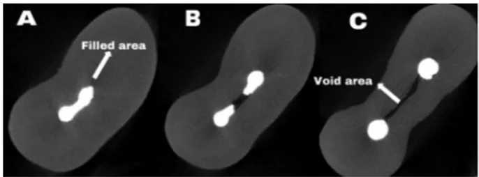
The filled area, void area and total area of the root canal were calculated for each sample (Fig. 2). The percentage of filled area of the root canals was 88.51% in the LC at 2 mm and it was significantly higher than in the SC (78.44%) (p < 0.05). Regarding the filling rate of the root canals at 5 mm, the filling success in the LC (92.32%) was significantly higher than that in the SC (85.09%) and CW (85.35%) tech-nique (p = 0.037). It was found no statistically significant dif-ference among the groups in the filling success at 8 mm (p = 0.207). The minimum filling material was found in the SC group (Table 1).
The intergroup comparison of each group revealed no sta-tistically significant difference in most gutta-percha areas of the root canals at the 2, 5 and 8 mm in the LC (p > 0.05) and the percentage of filled area was more than 88.51% for all sections. There was no statistically significant difference in the obtained sections in most filling material in the root canals in the CW (p > 0.05). The most gutta-percha areas at 2 mm were significantly lower than other sections in the SC (p < 0.05). It was observed no statistically significant differ-ence in the sections in the GC group with respect to most gutta-percha areas (p > 0.05).
4. Discussion
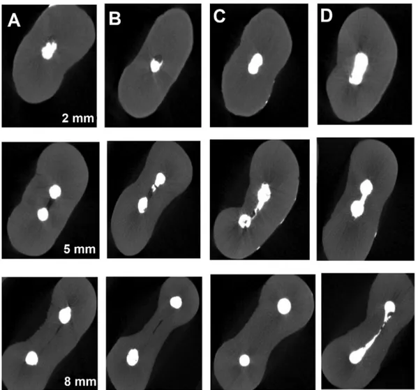
The extent of the root curvature affects the homogeneity and adaptation of the root canal fillings (Leung and Gulabivala, 1994; Neuhaus et al., 2016). The homogeneous filling of the curved root canals may be more difficult than that of the straight canals (Jafarzadeh and Abbott, 2007). Therefore, in this study, mandibular first molars with mesial root canal cur-vatures greater than 25° were preferred. The severely curved root canals were filled with the four different techniques and to examine the images of the 2, 5, and 8 mm of the roots using micro-CT in the present study. According to the results, LC was the most successful technique among the filling techniques at 2 and 5 mm (Fig. 3). In a similar study,Da Silva et al. (2002) compared the lateral condensation technique with thermoplas-ticised gutta-percha core carrier used in the curved canals using a digital radiography system. They did not find any voids in the core carrier group but observed small voids in several canals filled with the lateral condensation technique. Abarca et al. (2001)evaluated the mandibular first molars with curved mesial canals using the dye penetration test and found no sta-tistically significant difference between the thermoplasticised gutta-percha core carrier and lateral condensation techniques in terms of leakage. First, in the present study, it was believed that the result was affected by the obtainment of the sections only at certain levels and the usage of severely curved canals instead of straight canals. Second, the experimenter had more experience in the LC technique than other filling techniques. Therefore, this could have made the LC technique was more successful.
Some previous studies reported that the results of the single-cone technique were less successful than those of other filling techniques (McKissock et al., 2011; Obeidat and Abdallah, 2014). We noticed that the findings in the literature were in concordance with our own. As a result of the usage of the standard round-shaped gutta-percha cones, factors such as the lack of a full adaptation with the canal walls and filling failure of the isthmus and lateral canals contribute to the low success rate of this technique.
The obturation technique applied with continuous heat enables the plugging of the voids in the lateral canals, sulci and intracanal pockets (DuLac et al., 1998). Furthermore, this technique provides better sealing against coronal microbial penetration than the lateral condensation technique (Jacobson and Baumgartner, 2002). McKissock et al. (2011) compared the continuous-wave obturation and single-cone techniques regarding leakage and found that the number of voids in the root canals was significantly higher in the single-cone technique. In our study, the SC was the most unsuccessful technique among all groups. Moreover, the least mean per-centages were found for gutta-percha in all sections in this group. This result was not surprised for the present study. Gok et al. (2017) found more void areas in the warm gutta-percha obturation techniques than in the cold filling techniques in the C1 root canal configuration at 2 mm. It was similar to results of the present study. We believe that the curved canals have a compelling structure like the C1 root canal configura-tion and it is difficult to fill in the apical part of the canal cur-vature. In our study, the comparison of the filling rates in the sections obtained from the root canals filled with the CW did not reveal any statistically significant difference (p > 0.05).
Fig. 1 The 3D images of different mesial root canals.
Fig. 2 The filled area, void area and total area of the same sample: A. 2 mm, B. 5 mm, C. 8 mm.
The maximum filling material was found at 8 mm and the min-imum filling material at 5 mm in the CW group that surprised us a lot. The reason for the greater voids in the medial third can be explained by the location of the root curves on that level. It was assumed that the gutta-percha should be cut at a more coronal level because the Buchanan heat plugger can-not be inserted into the apical part of the canal curvature, which we consider as the reason for the voids in the apical part of the root curvature. It affects the success of the root canal treatment and the plugger with an optimal size according to the canal diameter in the CW technique.
It was observed no statistically significant difference in the sections in the GC (p > 0.05). The examination of the images taken from all three sections showed that the most successful filling was achieved in the coronal part and the least successful filling in the apical part. Our findings indicated that the Gutta-Core was the most successful technique after the lateral con-densation technique. We believe that the most important advantages of this technique are that the gutta-percha car-tridge can easily be moved until the apical part of the curved root canal and that the filling can be conducted with a single movement.
Table 1 The proportion of the filled area to total canal area at 2, 5 and 8 mm. (LC, lateral condensation; SC, single cone; CW, Elements Free; GC, GuttaCore; SD, standard deviation) Different superscript letters indicate a significant difference between groups.
LC SC CW GC p
Mean (%) SD Mean (%) SD Mean (%) SD Mean (%) SD Mean 2 mm 88.51a 8.31 78.44b 9.01 86.11a 11.01 86.69a 13.40 0.033*
5 mm 92.32a 8.37 85.09b 6.89 85.35b 11.83 89.29a 8.94 0.037*
8 mm 93.17a 5.85 88.39a 9.57 93.43a 7.20 92.44a 6.52 0.207
*
p< 0.05.
Fig. 3 The Micro-CT images of different root canal filling techniques (A, lateral condensation; B, single-cone; C, Elements Free; D, GuttaCore).
Compared with other filling techniques, it was found that the LC caused the development of dentinal defects (Shemesh et al., 2009, 2010). However, it was concluded that root filling procedures with GC, LC, and warm vertical compaction (WVC) techniques did not induce the development of new dentinal micro-cracks (De-Deus et al., 2017). In the present study, the development of dentinal micro-cracks after obtura-tion was not be evaluated.
The filling sealer considered the gold standard is AH Plus (Dentsply, DeTrey GmbH, Konstanz, Germany) (Silva et al., 2017). It is biocompatible (Leyhausen et al., 1999), having good physical properties that confer its long-term dimensional stability (Leonardo et al., 1999). Camilleri found that high temperature affected the chemical structure and setting time of AH Plus sealer. Therefore, it was not recommended for warm filling techniques (Camilleri, 2015). AH Plus sealer was used with different obturation techniques in the present study. One of the reasons why the CW was found to be less successful than LC technique might be due to changes in chemical struc-ture of AH Plus sealer with high temperastruc-ture. The choice of sealer according to the obturation technique should be considered.
There were several limitations in this study. First, the root canal curvature was not determined at which distance of the root canal. Although the degree of curvature was calculated, localization of the root canal curvature was not considered. The curvature may have been at middle part in some samples and at apical part in others. It may have been caused the results to be different. Future researchs might perform about effect of localization of the root canal curvature on the success of the filling techniques. Second, it was used only one size plug-ger to ensure standardization in continuous-wave obturation technique. Different size pluggers might use according to local-ization of the root canal curvature. Third, all samples were scanned as 2D except four samples due to high cost and it was received only horizontally sectional imagines. 3D scanning could provide more detailed examination for this study.
5. Conclusion
Within the limitation of this study, the most successful tech-niques at 2 and 5 mm were LC and GC, respectively. Regard-ing the coronal part, all techniques can be used for effective filling as long as a good condensation is achieved. Clinicians should use a plugger with an optimal size according to the canal diameter and the localisation of root canal curvature for the success of the root canal filling in the warm filling techniques.
Declaration of Competing Interest
Betul Aycan ALIM declares that she has no conflict of interest for ‘‘Evaluation of Different Root Canal Filling Techniques in Severely Curved Canals by Micro-computed Tomography”.
Yıldız GARIP BERKER declares that she has no conflict of interest for ‘‘Evaluation of Different Root Canal Filling Techniques in Severely Curved Canals by Micro-computed Tomography”.
Acknowledgements
None.
Funding
Marmara University Commission of Scientific Research Pro-jects, Istanbul, Turkey (Grant no: SAG-C-DUP-131016-0450).
References
Abarca, A.M., Bustos, A., Navia, M., 2001. A comparison of apical sealing and extrusion between Thermafil and lateral condensation techniques. J. Endod. 27, 670–672.
Acar, B., Kamburog˘lu, K., Tatar, _I., Arıkan, V., C¸elik, H.H., Yu¨ksel, S., O¨zen, T., 2015. Comparison of micro-computerized tomogra-phy and cone beam computerized tomogratomogra-phy in the detection of accessory canals in primary molars. Imaging Sci. Dent. 45, 205– 211.
Al-Dewani, N., Hayes, S.J., Dummer, P.M.H., 2000. Comparison of laterally condensed and low-temperature thermoplasticized gutta-percha root fillings. J. Endod. 26, 733–738.
Cailleteau, J.G., Mullaney, T.P., 1997. Prevalence of teaching apical patency and various instrumentation and obturation techniques in United States dental schools. J. Endod. 23, 394–396.
Camilleri, J., 2015. Sealers and warm gutta-percha obturation techniques. J. Endod. 41, 72–78.
Da Silva, D., Endal, U., Reynaud, A., Portenier, I., Orstavik, D., Haapasalo, M., 2002. A comparative study of lateral condensation, softened gutta-percha, and a modified master cone heat-softened backfilling technique. Int. Endod. J. 35, 1005–1011.
De-Deus, G., Belladonna, F.G., Silva, E.J.N.L., Souza, E.M., Carvalhal, J.C.A., Perez, R., Lopes, R.T., Versiani, M.A., 2017. Micro-CT assessment of dentinal micro-cracks after root canal filling procedures. Int. Endod. J. 50 (9), 895–901.
De Moor, R.J.G., Martens, L.C., 1999. Apical microleakage after lateral condensation, hybrid gutta-percha condensation and Soft-Core obturation: an in vitro evaluation. Endod. Dent. Traumatol. 15, 239–243.
DuLac, K.A., Nielsen, C.J., Tomazic, T.J., Ferrillo Jr., P.J., Hatton, J. F., 1998. Comparison of the obturation of lateral canals by six techniques. Endod. Prac. 1 (7–10), 13–16.
Gilhooly, R.M.P., Hayes, S.J., Bryant, S.T., Dummer, P.M.H., Wales, C., 2001. Comparison of lateral condensation and thermomechan-ically compacted warma-phase gutta-percha with a single cone for obturating curved root canals. Oral Surg. Oral Med. Oral Pathol. Oral Radiol. Endod. 1, 89–94.
Gok, T., Capar, I., Akcay, I., Kelesß, A., 2017. Evaluation of different techniques for filling simulated C-shaped canals of 3-dimensional printed resin teeth. J. Endod. 43, 1559–1564.
Jacobson, H., Baumgartner, J., 2002. Gutta-percha obturation of lateral grooves and depressions. J. Endod. 28, 269–271.
Jafarzadeh, H., Abbott, P.V., 2007. Dilaceration: review of an endodontic challenge. J. Endod. 33, 1025–1030.
McKissock, A.J., Mines, P., Sweet, M.B., Klyn, S.L., 2011. Ten-month in vitro leakage study of a single-cone obturation system. US Army Med. Dep. J., 42–47
Neuhaus, K.W., Schick, A., Lussi, A., 2016. Apical filling character-istics of carrier-based techniques vs. single cone technique in curved root canals. Clin. Oral. Invest. 20, 1631–1637.
Leonardo, M.R., da Silva, L.A., Almeida, W.A., Utrilla, L.S., 1999. Tissue response to an epoxy resin-based root canal sealer. Endod. Dent. Traumatol. 15, 28–32.
Leung, S.F., Gulabivala, K., 1994. An in-vitro evaluation of the influence of canal curvature on the sealing ability of Thermafil. Int. Endod. J. 27, 190–196.
Leyhausen, G., Heil, J., Reifferscheid, G., Waldmann, P., Geurtsen, W., 1999. Genotoxicity and cytotoxicity of the epoxy resin-based root canal sealer AH Plus. J. Endod. 25, 109–113.
Obeidat, R.S., Abdallah, H., 2014. Radiographic evaluation of the quality of root canal obturation of single-matched cone Gutta-percha root canal filling versus hot lateral technique. Saudi Endod. J. 4, 58–63.
Peng, L., Ye, L., Hong, T., Xuedong, Z., 2007. Outcome of root canal obturation by warm gutta-percha versus cold lateral condensation: a meta-analysis. J. Endod. 33, 106–109.
Schafer, E., Nelius, B., Burklein, S., 2012. A comparative evaluation of gutta-percha filled areas in curved root canals obturated with different techniques. Clin. Oral. Invest. 16, 225–230.
Schmalz, G., Ho¨rsted-Bindslev, P., 2010. Root canal filling materials. In: Bergenholtz, G., Horsted-Bindslev, P., Reit, C. (Eds.), Text-book of endodontology. Wiley-Blackwell Pub., Chichster, West Sussex, pp. 193–216.
Schneider, S.W., 1971. A comparison of canal preparations in straight and curved root canals. Oral Surg. Oral Med. Oral Pathol. 32, 271– 275.
Shemesh, H., Bier, C.A., Wu, M.K., Tanomaru-Filho, M., Wesselink, P.R., 2009. The effects of canal preparation and filling on the incidence of dentinal defects. Int. Endod. J. 42, 208–213.
Shemesh, H., Wesselink, P.R., Wu, M.K., 2010. Incidence of dentinal defects after root canal filling procedures. Int. Endod. J. 43, 995– 1000.
Silva, E.J., Perez, R., Valentim, R.M., Belladonna, F.G., De-Deus, G. A., Lima, I.C., Neves, A.A., 2017. Dissolution, dislocation and dimensional changes of endodontic sealers after a solubility challenge: a micro-CT approach. Int. Endod. J. 50, 407–414.
Sundqvist, G., Figdor, D., Persson, S., Sjogren, U., 1998. Microbio-logic analysis of teeth with failed endodontic treatment and the outcome of conservative re-treatment. Oral Surg. Oral Med. Oral Pathol. Oral Radiol. Endod. 85 (1), 86–93. 3.
Swain, M.V., Xue, J., 2009. State of the art of micro-CT applications in dental research. Int. J. Oral Sci. 1, 177–880.
Wu, M.K., Wesselink, P.R., 2001. A primary observation on the preparation and obturation of oval canals. Int. Endod. J. 34, 137– 141.
Vertucci, F.J., 1984. Root canal anatomy of the human permanent teeth. Oral Surg. Oral Med. Oral Pathol. Oral Radiol. Endod. 58, 589–599.