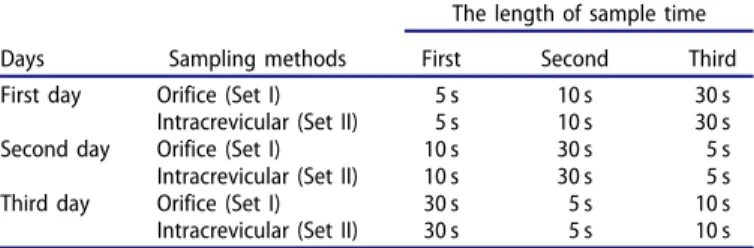
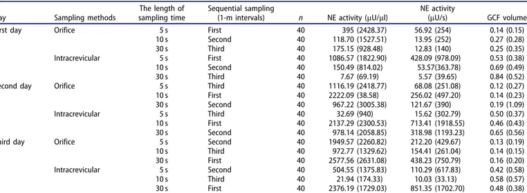
Methodological evaluation of gingival crevicular fluid volume and neutrophil elastase levels: sequential sampling, length of sampling time and two different sampling methods
Tam metin
Şekil

Benzer Belgeler
• 1961'de Avrupa, 1966 yı lında da Anadolu yaka sından kaldırılan tram vaylar, dün Tünel-Tak- sim arasında yeniden sefer yapmaya başladı.. •
Benim yetiştiğim kimseler ise Tahirülmevlevi, Darüşşafakalı mual lim Kâzım bey, Tahir Ağa Tekkesi Şeyhi Behçet Efendi, Ebussuut Efendi Zade Ali Emiri
Bu bildirim sonrasında çocuğun cinsel, fiziksel ve duygusal istismara uğramasında ebeveynlerin rol ve sorumlulukları olması halinde ya da çocuğun yaşadığı
İç Mekân Tasarımında Görselleştirme Yöntemleri “Lupa CR29 Projesi Üzerinden Örneklenmesi” 22 tasarım aşamaları bu makale kapsamında örnek olarak
Direct communicate with patient not family member Self-introduction of every hospital staff (doctor & nurse) & clear explanation before medical treatments (MUST)
Arzusunu, Hindistan hükümeti namına bize mihmandarlık eden ve sonradan CENTO Umumî Kâtipliği ile birkaç sene Ankara'da çalışmış olan Bin başı Osman Ali Bey’e
Aşağıda Sırbistan’ın 1909 ile 1911 yılları arasında Selanik limanı üzerinden gerçekleştirdiği tahıl ihracatını görmekteyiz... Sırbistan yavaş
Differences in subunit expression, posttranscriptional changes, organization of postsynaptic densities, contribution of numerous signal transduction proteins, different
