HACETTEPE UNIVERSITY INSTITUTE OF HEALTH SCIENCES
THE ACUTE FACILITATION EFFECTS OF GENERAL AND
LOCAL, MUSCULAR-ARTICULAR INTERVENTIONS IN
PHYSICAL THERAPY ON SHOULDER PROPRIOCEPTION
Dmitry CHAN (PT, MSc.)
Program of Physical Therapy and Rehabilitation Philosophy of Doctoral (PhD) Thesis
ANKARA 2015
HACETTEPE UNIVERSITY INSTITUTE OF HEALTH SCIENCES
THE ACUTE FACILITATION EFFECTS OF GENERAL AND
LOCAL, MUSCULAR-ARTICULAR INTERVENTIONS IN
PHYSICAL THERAPY ON SHOULDER PROPRIOCEPTION
Dmitry CHAN (PT, MSc.)
Program of Physical Therapy and Rehabilitation Philosophy of Doctoral (PhD) Thesis
ADVISOR OF THE THESIS Prof. Dr. Filiz CAN (PT, PhD, Full Prof.)
ANKARA 2015
ACKNOWLEDGEMENT
In accomplishing this dissertation I would like to thank, first of all my advisor Prof. Dr. Filiz CAN for her time and guidance throughout years.
I would like to thank my beloved wife Sonya Asgharifar for her emotional support, encouragement and patience. Without you I would not be able to walk this lengthily path.
I want to thank the members of jury Prof. Dr. Ayşe Karaduman, Prof. Dr.Ulunay Kanatlı, Prof. Dr. Zafer Erden, Assist. Prof. Ulaş Emin Erdem.
My special thanks go to my father Tran Quang Khoi for his help in designing original Proprioception Testing Device v2.0 and his wisdom though out the process.
Sincere gratitude to my friends and colleagues: Prof. Dr. Gül Şener, Mehdi Asgharifar, Esra Ateş Numanoğlu, Atila Güleç, Rustem Izzyatov, Ayla Fil, and many others who in various ways made this work easier and better.
Last but not least I want to thank everybody who participants in the study, without your participation and commitment this thesis would not be possible.
ÖZET
Dmitry Chan, Fizyoterapide Lokal ve Genel Kas-Eklem Uygulamalarının, Omuz Propriosepsiyonu Üzerine Olan Akut Fasilitasyon Etkileri, Doktora Tezi, Fizyoterapi ve Rehabilitasyon Programı, Ankara, 2015. Önceki çalışmalarda, bir
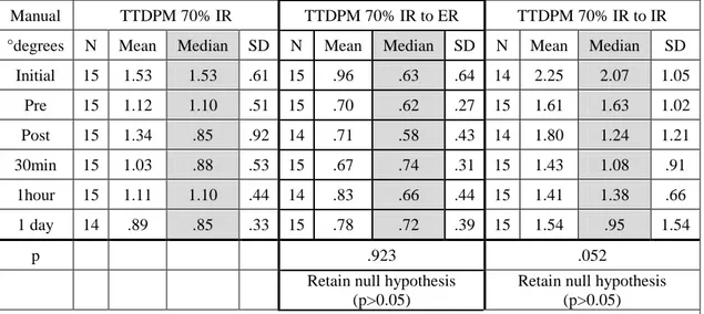
kaç haftalık eğitim programı olarak uygulanan çeşitli egzersiz yaklaşımlarının, propriosepsiyonu geliştirmede etkili olduğu bildirilmiş; ancak egzersize dayalı yaklaşımların etkileri, genellikle kuramsal olarak verilmiştir. Bu nedenle bu çalışma, sağlıklı bireylerde tek seanslık farklı fizyoterapi ve egzersiz uygulamalarının, omuz propriosepsiyonu üzerine olan akut etkilerini incelemek amacıyla yapılmıştır. Çalışmada, propiosepsiyon üzerine olan akut etkilerin, hem büyüklüğü, hem de süresi ölçülmüştür. Yaşları 19-37 arasında değişen, 55 kadın, 50 erkek, toplam 105 sedanter sağlıklı birey çalışmaya alınmış ve randomizasyon yolu ile 6 uygulama ve bir kontrol grubuna (n=15) ayrılmıştır. Gruplar, Aktif Egzersiz Grubu (belirlenen bir hızda aktif hareketler), Pasif Egzersiz Grubu (pasif hareketler), Manual Terapi Grubu (omuza manual terapi uygulaması), Stabilizasyon Grubu (omuza stabilizasyon egzersizleri), Pliometrik Egzersiz Grubu (omuza pliometrik egzersizler) ve Genel Isınma Grubu (koşarak yapılan ısınma) olarak ayrıldıktan sonra tek seanslık fizyoterapi uygulaması yapılmıştır. Omuz propriosepsiyonu, tüm bireylerde kinestezi (hareket hissi) ve eklem pozisyon hissi olarak, “2.0” versiyon orjinal propriosepsiyon aleti kullanılarak ölçülmüştür. Dominant omuzda ROM’un % 50’si içinde, %70 iç rotasyon ve % 90 dış rotasyonda yapılan ölçümler, oturma pozisyonunda, ilk gün, uygulama öncesi, uygulama sonrası, uygulamadan 30 dakika, 1 saat ve 1 gün sonrasında tekrarlanmıştır. Kontrol grubundaki bireylerin propriosepsiyon değerlerindeki aşırı değişkenlik nedeniyle 105 bireyin tümünün propriosepsiyon değerleri ölçülmüş ve alınan bu baz değerler, her bir grup için karşılaştırma referansı olarak kullanılmıştır. Tek seanslık uygulamadan sonra Aktif Hareket Grubunda baz değerlere göre kinestetik duyuda belirgin bir azalma olmuştur (p<0.05). Hem Pasif Hareket, hem de Manuel Terapi grubundaki bireyler, uygulamadan sonra (çoğunlukla ROM sınırları içinde olmak üzere) uygulama öncesine göre ve baz değerlere göre propriosepsiyonda anlamlı gelişmeler göstermişlerdir (p<0.05). Buna karşılık Stabilizasyon Grubu ve Pliyometrik grup için, tek bir uygulama seansının propriosepsiyonu değiştirmeye yetmediği gözlenmiştir (p>0.05). Genel Isınma grubunda ise, bir sonuca varamayan bulgular ortaya çıkmıştır (p>0.05). Ek olarak tüm bireylerde, hareketin yönünün, pasif hareket algılama eşiğinin testi sırasında propriosepsiyon ölçümlerini etkilediği bulunmuştur (p<0.05). Ayrıca proprisepsiyon ölçümünde kullanılan kinestezinin ( hareket hissi), eklem pozisyon hissine göre daha hassas bir ölçüm yöntemi olduğu sonucuna varılmıştır. Sonuç olarak, sağlıklı bireylerde tek seanslık bir fziyoterapi uygulaması, propriosepsiyonu anlamlı derecede etkilemek için yeterli olmayabilir. Pasif egzersiz ve manuel terapi uygulamaları ile bazı gelişmeler görülebilir. Aktif egzersiz uygulamaları sırasındaki kortikal adaptasyon ve öğrenmenin, mekanoreseptörlerin lokal stimulasyonundan daha önemli olabilir. Tekrarlayıcı aktif hareketler ile ortaya çıkabilecek kas yorgunluğu sonucunda propriosepsiyonda görülebilecek azalma, klinik uygulamalar sırasında daima gözönünde bulundurulmalıdır.
Anahtar Kelimeler: Propriosepsiyon, eklem pozisyon hissi, kinestezi, omuz,
ABSTRACT
Dmitry Chan, The Acute Facilitation Effects of General and Local, Muscular-Articular Interventions in Physical Therapy on Shoulder Proprioception, Doctoral Thesis in the Program of Physical Therapy and Rehabilitation, Ankara, 2015. Various exercise interventions applied as a course lasting for several
weeks have been reported to be effective on improving proprioception. The underlying causes are only hypothesized about. This study aimed to investigate the acute effects of one time intervention derived from different aspects of exercises on shoulder proprioception of healthy subjects. Both the magnitude and the duration of effect were aimed to measured. 55 female and 50 male, a total of 105 sedentary healthy subjects aged between 19 and 37 years old, were randomly divided into 6 intervention and a control group (n=15). The groups were: Active Movement Group (active movement at predetermined speed), Passive Movement Group (passive movements), Manual Therapy Group (joint play of glenohumeral joint), Stabilization Group (shoulder stabilization exercises), Plyometric Exercise Group (shoulder plyometrics) and General Warm Up Group (warm up through jogging). Each group received single intervention. Shoulder proprioception has been assessed as Joint position sense, and kinesthesia using original proprioception testing device version 2.0. Dominant shoulder at 70% internal rotation, 90% external rotation and 50% of ROM were measured in sitting position on first day, pre, post, 30 minute, 1 hour and 1 day post interventions. Increased variability of proprioception in control group necessitated use of baseline proprioception values of all 105 subjects as comparison reference. In active movement group there was a decrease in kinesthetic sense compared to baseline after intervention (p<0.05). Subjects in both passive movement and manual therapy group showed improvements in proprioception mainly at the extremes of ROM, either within group or against baseline (p<0.05). Single session of intervention was not sufficient to change proprioception in stabilization and plyometric groups (p>0.05). Inconclusive results were seen in warm up group (p>0.05). Additionally the direction of movement during testing of threshold to passive movement detection was found to affect proprioception measurements (p<0.05). Furthermore, kinesthesia testing might be more sensitive test for proprioception then joint position sense test. In conclusion, one time intervention might not be sufficient to significantly affect proprioception in healthy subjects. Although some improvements may be seen with passive exercises and manual therapy, cortical adaptation and learning rather than local stimulation of mechanoreceptors might be more critical to increase proprioception with the other exercises. Clinicians should be aware of possible decrease in proprioceptive acuity with repetitive active movements due to muscle fatigue.
Key words: proprioception, joint position sense, kinesthesia, shoulder, exercise, warm-up, plyometric
TABLE OF CONTENTS
page
APPROVAL PAGE iii
ACKNOWLEDGEMENT iv
ÖZET v
ABSTRACT vi
TABLE OF CONTENTS vii
SYMBOLS AND ABBREVIATIONS xi
LIST OF FIGURES xiv
LIST OF TABLES xvi
1 INTRODUCTION 1
2 GENRAL KNOWLEDGE 9
2.1 Shoulder joint anatomy and physiology 9
2.1.1 Bones 9
2.1.2 Joint capsule and ligaments 12
2.1.3 Muscles 13
2.2 Stability of glenohumeral joint 19
2.3 Proprioception and sensorimotor system 22
2.3.1 Homeostasis - human organism against external environment 22
2.3.2 Sensorimotor system 23
2.3.3 Definition of proprioception 23
2.3.4 Proprioceptors 24
2.3.5 Proprioceptive input 29
2.3.7 Methods to assess sensorimotor system 32
3 MATERIALS AND METHODS 36
3.1 Study design 36 3.2 Subjects 36 3.3 Procedures 39 3.4 Equipment 41 3.5 Proprioception testing 48 3.6 Interventions 51
3.7 Data reduction, transformation and statistical analysis 58
4 RESULTS 60
4.1 Demographics and general characteristics of subjects 60
4.2 Initial proprioception values prior to any intervention 60
4.3 Comparison of proprioception at various moments of assessment
within the groups 63
4.3.1 Analysis of proprioception within the "Active Movement
Group" 64
4.3.2 Analysis of proprioception within the "Control Group" 68
4.3.3 Analysis of proprioception within the "Manual Therapy
Group" 73
4.3.4 Analysis of proprioception within the "Passive Movement
Group" 77
4.3.5 Analysis of proprioception within the "Plyometric Exercise
Group" 79
4.3.6 Analysis of proprioception within "Stabilization Exercise
Group" 82
4.3.7 Analysis of proprioception within "Warm Up Exercise
4.4 Between group comparison at specific moments of assessment pre and
post interventions 87
4.4.1 Comparison of groups at initial assessment 87
4.4.2 Comparison of groups at Pre assessment 89
4.4.3 Comparison of groups at Post assessment 94
4.4.4 Comparison of groups at 30 minute assessment 96
4.4.5 Comparison of groups at 1 hour assessment 99
4.4.6 Comparison of groups at 1 day assessment 102
4.5 Comparison of proprioception of each group at various moments of
assessment against 105 subject baseline 105
4.6 Additional analysis role of joint angle on proprioception 116
4.7 Additional analysis gender differences 123
5 DISCUSSION 127
5.1 Study design 127
5.2 Subjects and general assessment 128
5.3 Proprioception testing method and device 130
5.4 Control group 138
5.5 Active movement intervention and the role of musculotendonous
structures 142
5.6 Passive movement and manual therapy effects of joint
mechanoreceptors 148
5.7 Stability exercises and proprioception 155
5.8 Plyometric exercises 160
5.9 General warm up effects 165
5.10 Comparison of intervention groups 168
6 CONCLUSION 173
6.1 Study limitations 175
6.2 Directions for future research 175
6.3 Clinical relevance 176
REFERENCES 178
SYMBOLS AND ABBREVIATIONS
cm centimeter
kg kilogram
mm millimeter
n/N number
oPTD Original Proprioception testing device
p statistical significance value
sec second
sig significance
v version
AC Acromioclavicular
BMI Body Mass Index
C Cervical
CNS Central Nervous System
EMG Electromyography
ER external rotation
GH Glenohumeral
GTO Golgi tendon organs
HR heart rate
IGHL Inferior Glenohumeral Ligament
IR internal rotation
JPS Joint position sense
L left
LHB Long head of biceps
M male
Max maximum
MGHL Middle Glenohumeral Ligament
Min minimum
N. Nervous
PNF Proprioceptive Neuoromuscular Facilitation
PNS Peripheral Nervous System
PTD Proprioception testing device
R right
SC Sternoclavicular
SD standard deviation
SGHL Superior Glenohumeral Ligament
T Thoracic
TTDPM Threshold to detection passive movement
TTDPMD Threshold to detection passive movement direction
ULTT Upper Limb Tension test
VO2 volume of oxygen Z statistical coefficient α Alpha Ø diameter ° degree γ Gamma % percent ± plus minus
LIST OF FIGURES
Figure page
2.1. Scapular position 10
2.2. Motions of scapular 11
2.3. Ligaments of glenohumeral joint 13
2.4. Rotator cuff muscles 17
2.5. Scapulohumeral rhythm 18
2.6. Muscle spindles 28
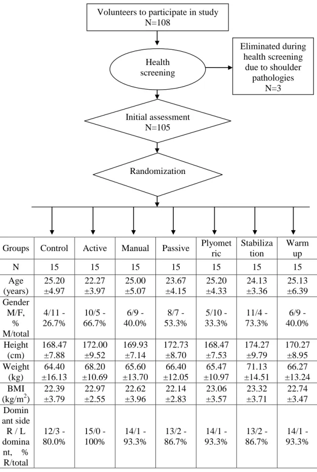
3.1. Chart elimination and distribution of subjects participated in study 38
3.2. iGaging AngleCube Digital Level 42
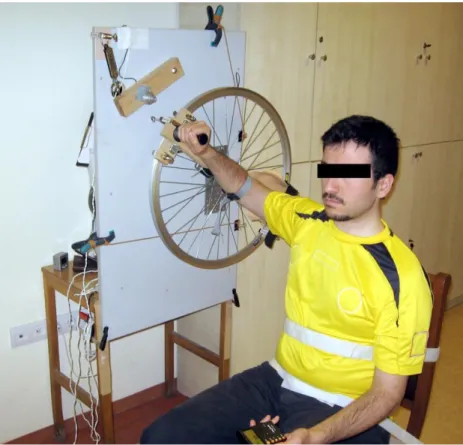
3.3. oPTD v2.0 43
3.4. JPS testing with oPTD v2.0 44
3.5. Kinesthesia testing with oPTD v2.0 45
3.6. Metronome and digital counter 46
3.7. Treadmill and heart rate monitor 47
3.8. Medicine balls and theratubes® used for plyometric exercises 48
3.9. Adaptation of oPTD v2.0 for active movements through ROM at preset
speed guided by metronome 51
3.10. Manual Therapy Intervention 53
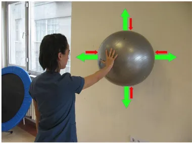
3.11. Shoulder stabilization against perturbations at 90° shoulder flexion
against wall 54
3.12. Shoulder stabilization against perturbations at 90° abduction against
wall 55
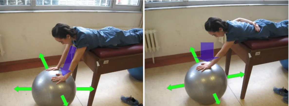
3.13. Two hand and single handed shoulder stabilization supporting body
3.14. Plyometric internal and external rotations with theratube 57
3.15. Plyometric ball overhead throw 57
4.1. Boxplot of TTDPM at initial assessment of 105 subjects accounting for
direction of movement 122
4.2. Bar plot gender differences of JPS at initial and pre evaluation of 105
subjects 124
4.3. Bar plot gender differences of TTDPM at initial and pre evaluation of
LIST OF TABLES
Table page
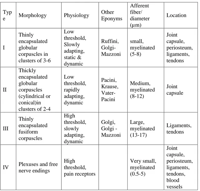
2.1. Joint mechanoreceptors adapted from Wyke and Hogervost 26
4.1. Physical characteristics of subjects 60
4.2. JPS of all subjects at initial assessment 61
4.3. TTDPM of all subjects at initial assessment disregarding direction of
movement 61
4.4. TTDPM at 90% of ER of all subjects at initial assessment accounting
for direction of movement 62
4.5. TTDPM at 50% of ROM of all subjects at initial assessment accounting
for direction of movement 62
4.6. TTDPM at 70% of IR of all subjects at initial assessment accounting for
direction of movement 63
4.7. Within the "Active Movement Group" analysis of JPS 64
4.8. Within the "Active Movement Group" analysis of TTDPM disregarding
direction of movement 64
4.9. Post Hoc analysis, comparison of TTDPM at 50% of ROM in "Active
Movement Group" between specific assessment moments 65
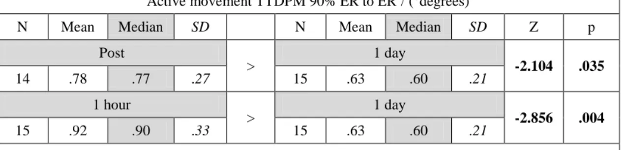
4.10. Within the "Active Movement Group" analysis of TTDPM at 90% of
ER to ER and IR 66
4.11. Post Hoc analysis, comparison of TTDPM at 90% of ER to ER in
"Active Movement Group" between specific assessment moments 66
4.12. Within the "Active Movement Group" analysis of TTDPM at 50% of
ROM to ER and IR 67
4.13. Within the "Active Movement Group" analysis of TTDPM at 70% of
IR to ER and IR 67
4.15. Post Hoc analysis, comparison of JPS at 50% of ROM in "Control
Group" between specific assessment moments 68
4.16. Within the "Control Group" analysis of TTDPM disregarding direction
of movement 69
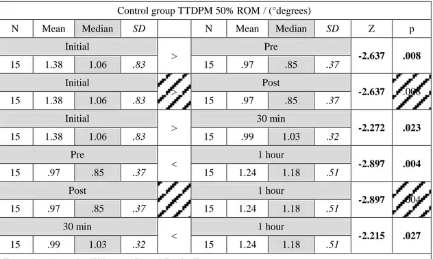
4.17. Post Hoc analysis, comparison of TTDPM at 50% of ROM in "Control
Group" between specific assessment moments 70
4.18. Within the "Control Group" analysis of TTDPM at 90% of ER to ER
and IR 70
4.19. Within the "Control Group" analysis of TTDPM at 50% of ROM to
ER and IR 71
4.20. Within the "Control Group" analysis of TTDPM at 70% of IR to ER
and IR 71
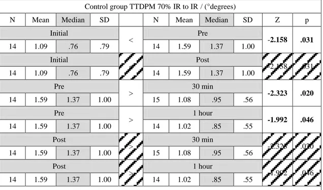
4.21. Post Hoc analysis, comparison of TTDPM at 70% of IR to IR in the
"Control Group" between specific assessment moments 72
4.22. Within the "Manual Therapy Group" analysis of JPS 73
4.23. Within the "Manual Therapy Group" analysis of TTDPM disregarding
direction of movement 73
4.24. Post Hoc analysis, comparison of TTDPM at 90% of ER in the
"Manual Therapy Group" between specific assessment moments 74
4.25. Post Hoc analysis, comparison of TTDPM at 70% of IR in the
"Manual Therapy Group" between specific assessment moments 74
4.26. Within the "Manual Therapy Group" analysis of TTDPM at 90% of
ER to ER and IR 75
4.27. Post Hoc analysis, comparison of TTDPM at 90% of ROM to IR in the
"Manual Therapy Group" between specific assessment moments 75
4.28. Analysis of TTDPM within the "Manual Therapy Group" at 50% of
4.29. Within the "Manual Therapy Group" analysis of TTDPM at 70% of IR
to ER and IR 76
4.30. Within the "Passive Movement Group" analysis of JPS 77
4.31. Within the "Passive Movement Group" analysis of TTDPM
disregarding direction of movement 77
4.32. Analysis of TTDPM within the "Passive Movement Group" at 90% of
ER to ER and IR 78
4.33. Within the "Passive Movement Group" analysis of TTDPM at 50% of
ROM to ER and IR 78
4.34. Within the "Passive Movement Group" analysis of TTDPM at 70% of
IR to ER and IR 79
4.35. Within the "Plyometric Exercise Group" analysis of JPS 79
4.36. Within the "Plyometric Exercise Group" analysis of TTDPM
disregarding direction of movement 80
4.37. Within the "Plyometric Exercise Group" analysis of TTDPM at 90%
of ER to ER and IR 80
4.38. Within the "Plyometric Exercise Group" analysis of TTDPM at 50%
of ROM to ER and IR 81
4.39. Within the "Plyometric Exercise Group" analysis of TTDPM at 70%
of IR to ER and IR 81
4.40. Within the "Stabilization Exercise Group" analysis of JPS 82
4.41. Within the "Stabilization Exercise Group" analysis of TTDPM
disregarding direction of movement 82
4.42. Within the "Stabilization Exercise Group" analysis of TTDPM at 90%
of ER to ER and IR 83
4.43. Within the "Stabilization Exercise Group" analysis of TTDPM at 50%
4.44. Within the "Stabilization Exercise Group" analysis of TTDPM at 70%
of IR to ER and IR 84
4.45. Within the "Warm Up Exercise Group" analysis of JPS 84
4.46. Within the "Warm Up Exercise Group" analysis of TTDPM
disregarding direction of movement 85
4.47. Within the "Warm Up Exercise Group" analysis of TTDPM at 90% of
ER to ER and IR 85
4.48. Within the "Warm Up Exercise Group" analysis of TTDPM at 50% of
ROM to ER and IR 86
4.49. Within the "Warm Up Exercise Group" analysis of TTDPM at 70% of
IR to ER and IR 86
4.50 Post Hoc comparison of TTDPM at 90% of ER between the groups at
Initial assessment 87
4.51. Post Hoc comparison of TTDPM at 50% of ROM between the groups
at Initial assessment 88
4.52. Post Hoc comparison of TTDPM at 50% of ROM to ER between the
groups at Initial assessment 88
4.53. Comparison of JPS between the groups at Pre assessment 89
4.54. Post Hoc comparison of JPS at 70% of IR between the groups at Pre
assessment 90
4.55. Comparison of TTDPM between the groups at Pre assessment 90
4.56. Post Hoc comparison of TTDPM at 50% of ROM between the groups
at Pre assessment 91
4.57. Comparison of TTDPM at 90% of ER to ER and IR between the
groups at Pre assessment 92
4.58. Comparison of TTDPM at 50% of ROM to ER and IR between the
4.59. Post Hoc comparison of TTDPM at 50% of ROM to ER between the
groups at Pre assessment 93
4.60. Comparison of TTDPM at 70% of IR to ER and IR between the
groups at Pre assessment 93
4.61. Comparison of JPS between the groups at Post assessment 94
4.62. Comparison of TTDPM between the groups at Post assessment 94
4.63. Comparison of TTDPM at 90% of ER to ER and IR between the
groups at Post assessment 95
4.64. Comparison of TTDPM at 50% of ROM to ER and IR between the
groups at Post assessment 95
4.65. Comparison of TTDPM at 70% of IR to ER and IR between the
groups at Post assessment 96
4.66. Comparison of JPS between the groups at 30 minute assessment 96
4.67 Post Hoc comparison of JPS at 70% of IR between the groups at 30
minute assessment 97
4.68. Comparison of TTDPM between the groups at 30 minute assessment 97
4.69. Comparison of TTDPM at 90% of ER to ER and IR between the
groups at 30 minute assessment 98
4.70. Comparison of TTDPM at 50% of ROM to ER and IR between the
groups at 30 minute assessment 98
4.71. Comparison of TTDPM at 70% of IR to ER and IR between the
groups at 30 minute assessment 99
4.72. Comparison of JPS between the groups at 1 hour assessment 99
4.73. Comparison of TTDPM between the groups at 1 hour assessment 100
4.74. Comparison of TTDPM at 90% of ER to ER and IR between the
groups at 1 hour assessment 100
4.75. Post Hoc comparison of TTDPM at 90% of ER to ER between the
4.76. Comparison of TTDPM at 50% of ROM to ER and IR between the
groups at 1 hour assessment 101
4.77. Comparison of TTDPM at 70% of IR to ER and IR between the
groups at 1 hour assessment 102
4.78. Comparison of JPS between the groups at 1 day assessment 102
4.79. Comparison of TTDPM between the groups at 1 day assessment 103
4.80. Comparison of TTDPM at 90% of ER to ER and IR between the
groups at 1 day assessment 103
4.81. Comparison of TTDPM at 50% of ROM to ER and IR between the
groups at 1 day assessment 104
4.82. Comparison of TTDPM at 70% of IR to ER and IR between the
groups at 1 day assessment 104
4.83. Comparison of JPS of all 105 subjects at Initial and Pre assessments 105
4.84. Comparison of TTDPM of all 105 subjects at Initial and Pre
assessments 106
4.85. Comparison of proprioception of "Active Movement Group" at
various assessment points against baseline proprioception 107
4.86. Comparison of proprioception of "Control Group" at various
assessment points against baseline proprioception 108
4.87. Comparison of proprioception of "Manual Therapy Group" at various
assessment points against baseline proprioception 109
4.88. Comparison of proprioception of "Passive Movement Group" at
various assessment points against baseline proprioception 110
4.89. Post Hoc comparison of proprioception of "Passive Movement Group"
at specific points of assessment against baseline proprioception 111
4.90. Comparison of proprioception of "Plyometric Exercise Group" at
4.91. Comparison of proprioception of "Stabilization Exercise Group" at
various assessment points against baseline proprioception 113
4.92. Post Hoc comparison of proprioception of "Stabilization Exercise Group" at specific points of assessment against baseline
proprioception 114
4.93. Comparison of proprioception of "Warm Up Exercise Group" at
various assessment points against baseline proprioception 115
4.94. Post Hoc comparison of proprioception of "Warm Up Exercise Group"
at specific points of assessment against baseline proprioception 116
4.95. Comparison of JPS between various angles of all 105 subjects at
Initial and Pre assessments 117
4.96. Comparison of TTDPM disregarding direction of movement between
various angles of all 105 subjects at Initial and Pre assessments 118
4.97. Comparison of TTDPM at 90% of ER accounting for direction of movement between various angles of all 105 subjects at Initial and
Pre assessments 119
4.98. Comparison of TTDPM at 50% of ROM accounting for direction of movement between various angles of all 105 subjects at Initial and
Pre assessments 120
4.99. Comparison of TTDPM at 70% of IR accounting for direction of movement between various angles of all 105 subjects at Initial and
Pre assessments 121
4.100. Comparison of JPS between genders at Initial and Pre assessments 123
4.101. Comparison of TTDPM between genders at Initial and Pre
1 INTRODUCTION
A human movement is an intricate balance between external forces acting on the body and internal forced working within body to oppose external forces and to propagate voluntary or involuntary movements by a human. The responding units are muscles that span between skeletal bones and act through the joints causing movement of body parts and skeleton in total. The orchestration of movements is coordinated by various levels of the Central Nervous system (CNS), and is delivered to muscles by means of electrical signals via Peripheral Nervous System (PNS). Each joint in a human body can be viewed to be in two different states. One is immobile or static and the other one is moving or dynamic. Most joints are meant to be moved, therefore are inherently unstable in one or more directions. Therefore, in order for a joint to be immobile it should be stabilized. Muscles, ligaments and the shape of the joints act to provide stability of the joint. Similarly during motion, an internal or external force must be exerted over a body segment in order to move. An indiscriminate force will likely disrupt the joint movement rather than it will cause a functionally meaningful motion. Therefore, a dynamic stability of the joint is essential for coordinated motion. Once again muscles, ligaments and shape of joints function to provide the dynamic stability. The system responsible for providing functional stability of the joint, static or dynamic stability, is called sensori-motor system. Sensori-motor system works parallel and in the background of human locomotion or even while holding a static position(1).
Sensori-motor system can be divided into 3 distinctive components. The afferent system or proprioception that is responsible of sensing the position or movement of body segments as well as forces through the joints. The central integration or processing of signals coming from proprioceptive system, and the efferent output system or motor control(1-3).
While sensori-motor system functions are adequate and capable to cope with demands of daily living, its' functions can be disrupted during pathological conditions such as injury, pain, etc. Similarly state of fatigue negatively affects functional stability of the joint(1, 3-14). Vice versa a disrupted joint stabilization
system might cause an injury or disruption. For an example, poor perception of movements in the ankle joint can predispose a person to have an inversion trauma of an ankle(15).
As it becomes evident, it can be beneficial to improve functioning of sensori-motor system to allow it to cope with wide range of activity demands. Three components of the system can be influenced. While in theory these three components are well defined, however, it is next to impossible to separate them and influence one by one in practice. In a human body sensori-motor system works as a whole. Recently many researchers have been focused on proprioception- the afferent component of sensori-motor system. How well does the human body is able to percept the forces, position or perturbations on the body segment or a joint might be critical in shaping the response to the stimuli.
From a physical therapy and athletic training point of view, exercise training is the most common tool of training and conditioning of human body. Many authors agree that proprioception can be affected by exercises(2, 3, 16-26). Several studies in the literature have been devoted to prove the effectiveness of one or another exercise based programs on proprioception. Various exercise programs have been tested lately, and some were found to be effective the others were not. Each time the researchers propose various theories to argue the obtained results. Some of the theories are in coherency with the others, while others are not. Therefore, it might be beneficial to deconstruct various exercises into basic characteristics and to test them against the proposed hypotheses. A good starting block would be to find out acute effects of single session physical therapy intervention or exercise on proprioception.
From a perspective of proprioception receptors, the afferent input can arise mainly either from joint receptors or from muscle receptors. The role of both has been already identified. Almost any exercise involves both the joint and the muscle. Joint receptors such as Pacinian corpuscle and Ruffini end organs as well as free nerve endings, do play role in proprioception(27). David Suprak found that at higher elevation angles of shoulder where there a greater capsular stretch the JPS, proprioception is more acute then in lower angles(28, 29). Furthermore, J. Munn's
narrative review pointed out that ligament laxity associated with ankle instability negatively affected JPS in ankle(30). However, a hypothesised decreased viscosity of joint capsule and ligaments introduced via cold pack over joint for 20 minutes did not show to decreased joint proprioception in two studies(31, 32). Finally and most valuable the study by Ju focused on positive effects of repetitive passive joint motion on proprioception. Ju and colleagues found that at high angular velocity of 90 and 150 degrees per second, both JPS and kinaesthesia improved. However, this effect was not observed at low speed of 2 degrees per second. Such low speed might have not been sufficient to cause sufficient capsular and ligament stretch(33, 34). Some body of evidence exists suggesting that during mid ranges of motion of the joint the capsule and ligaments are not being stretch sufficient to cause discharge of joint mechanoreceptors(35, 36).
There is still lack of enough information in the literature regarding the effect of manual therapy on proprioception, although manual therapy has been using commonly for shoulder problems in the clinical practice. One interesting aspect of capsular stimulation without angular displacement via manual therapy joint play techniques was poorly studied up to date(37). However, knowing the proven effect of manual therapy on proprioception would be valuable in planning of physical therapy and rehabilitation program for the physical therapists. This may also clarify which mechanism would be more critical for stimulating of the joint receptors. Furthermore, it might enlighten to differentiate the effect of local stimulation of the mechanoreceptors.
The role of mechanoreceptors located in muscles and tendons during mid ranges of motion have been studied and revealed possible lack of stimulation of joint receptors in mid ranges. Muscle of mammals and humans have complex system of muscle spindles that are able to sense not only the stretch of muscle tissue, but also the rate of the stretch. Additionally, Golgi tendon organs located parallel to muscle tissue give feedback on the amount of stretch going through the muscle tissue(1, 3, 20, 30, 38-43). Stimulation of muscle itself was observed to have both positive and negative effects on proprioception. Nonetheless, the effects on proprioception in both cases are evident. Over stimulation of muscle to a point of fatigue was found to
decrease proprioception(7, 8, 34, 44, 45). Lee Hung-Maan found that at 50% of maximal voluntary peak torque decline due to fatigue proprioception decreases(46). However, loading a muscle less than its fatigue level proves beneficial to proprioception. A study by David Suprak found that giving load to an arm in lifting task improves proprioception(47). Furthermore, in the earlier mentioned article, David Suprak argues that a greater tension associated with increased elevation angels is responsible for greater accuracy of JPS at extremes of range of motion(29). Otmar Bock applied vibration of 50Hz frequency to muscle tendon and observed perceived sense of motion due to interference with muscle spindles afferent output(48). Similarly degeneration of muscle and tendon in chronic supraspinatus impingement degraded force sensation and proprioception in several studies (5, 11).
Clinically many protocols aimed at improving proprioception focus on strength building(17, 18, 27, 49-51).Muscle bulk might be responsible for increased joint stability and proprioceptive afferent (52-54). Other valuable exercises are balancing and stability exercises. The proposed mechanism of benefit is the level of muscle co-contraction associated with these exercises. In order to provide active stability to a joint a simultaneous activation of agonists and antagonists is happening(2, 17, 27, 49-51). While this theory largely derives from clinical experience, there is still lack of scientific evidence aside from wobble board exercises supporting it. Wobble board exercises were found beneficial for increased muscle onset of peroneal muscles of a foot and subsequent functional stability(55). Another possible explanation is the cortical adaptation and learning effect evolved from increased afferent input from muscle spindles, as well as joint and articular receptors.
Some exercises cannot be classified as mainly affecting joints or muscles. The best examples are plyometric exercises. While originally plyometric exercises were aimed at explosive power training method, lately a beneficial effect was found to proprioception. K.Swanik studied effect of plyometrics on shoulder proprioception of female swimmers, and found great benefits of it(25).In accordance with the study of Swanik, we have observed similar benefits of plyometrics on JPS and kinesthesia on sedentary subjects in our previous study(26). According to these two studies, the
proposed mechanism of action is the increased stimulation of muscle spindles under high load of eccentric and concentric contraction, as well as stimulation of joint receptors at the ends of range of motion through which a joint is brought at each plyometric exercise(25, 26).
Finally, a warm up effect associated with performance level should not be disregarded as a method to influence proprioception. Temperature increase of a body has been proven to cause many beneficial effects to human body, such as increase in metabolic activity of muscle cells, increase in nerve conduction velocity and overall improvements in muscle performance(56-60). There are two possible ways of raising body temperature, passive and active one. Passively, direct application of hot pack might increase skin, joint and muscle temperature. In turn, this application can aid in increased viscosity of a joint(57, 61, 62). However, actively exercising can have a similar increase in area temperature via repetitive active motion of the joint. The demands of muscle activity will cause a greater peripheral circulation and increased blood flow, which eventually will increase the temperature of muscle and joint itself. Much literature agrees that warm up exercises are beneficial for muscle activity and contract ability(43, 57, 63-66). Concentric warm ups showed to decrease soreness from eccentric activity(67).A leading cause of muscle soreness is a buildup of lactic acids in the muscles as waist products of its activity. These are the same lactic acids which are responsible for muscle fatigue and degradation of proprioception. Following this logic an active warm up exercises might be beneficial to proprioception. Subasi's study on health young subjects confirms this theory(68). Although majority of researchers focus of direct activity of joint in interest from warm up perspective, unfortunately, this method for this proposal is invalid due to possible overlap of effects with active or passive joint motions. Therefore, it might be valuable to approach a joint warm up as a part of whole body warm up achieved through aerobic activities such as running. These activities are also effective in raising body temperature. Not surprisingly, there is no study in the literature to this date on benefits of whole body warm-up exercises on proprioception of a joint not primarily involved in the activities.
In the literature, there is not enough study which assess both active and passive joint position sense using objective measurement devices for shoulder joint. Some authors have used measurement of passive joint position sense, whereas the others have claimed measurement of active joint position sense will give more accurate data for assessment of proprioception. However, both active and passive joint position senses may be more valuable parameters for measuring proprioception and would be helpful for discussion. Furthermore, there is still a debate on effect of various type of exercises like plyometrics, stabilization exercises, active or passive range of motion exercises for shoulder proprioception. It is also unclear the results of some physical therapy interventions such as manual joint mobilization, vibration or whole body warm up exercises which have never been studied to investigate the effectiveness of them on shoulder proprioception.
From a numerous proposed mechanisms that physical therapy interventions like manual therapy and various types of exercises might have an effect on proprioception, the aim of this study was to deconstruct a shoulder exercises and other physical therapy interventions like manual joint mobilization into various components and test each one in particular. In other words what affect, if any, does each component of exercise has on proprioception. It has been aimed to focus on proprioceptive system rather than learning component or central integration of the central nerve system. Therefore, the interventions were limited to acute effects of single time stimulus rather than a training protocol. Finally, this study based on not only the magnitude, but also the duration of the effect on proprioception is important to judge which component of exercise or intervention has the greatest effect. In addition to that comparing each intervention will be a sensible analysis to perform based on gathered data. This kind of detailed approach has not been attempted yet in any of the studies, and will help to gain deeper understanding of the mechanism in effect.
Based on outlined goals of the study, several research hypotheses were established.
H1- There will be difference in shoulder joint proprioception between intervention group and a control group immediately after interventions
H2- There will be difference in shoulder proprioception within intervention group between initial assessment and at a specific time interval after interventions
H3- There will be difference in shoulder proprioception between intervention groups immediately after interventions
H4- There will be difference in shoulder proprioception between intervention groups at specific time intervals after interventions
In order to test our hypotheses, multi group randomized control trial with variation of repeated measures study was designed. Glenohumeral joint of dominant arm was studied. The choice of investigating glenohumeral joint was made by considering functionality of shoulder joint which allows motion in great variety of planes and requires both capsular ligamental structures as well as muscles to stabilize the joint.
Six intervention groups and one control group comprised the study. The intervention groups were: active movement, manual joint mobilization, passive movement, plyometric exercises, stabilization, and general warm up. Each group consisted of 15 healthy sedentary subjects, who were recruited from university students, administration and academic population. Total number of participants was 105. The study was conducted at Physical Therapy and Rehabilitation Department, Institute of Health Sciences of Hacettepe University. The study typically lasted 3 days for each participant and took place between June 2014 and January 2015.
The results of this study will be beneficial to lay foundation for the effects of various components of exercises and other physical therapy interventions as manual therapy for shoulder joint on proprioception. Based on the results greater
understanding could be reached about the physiology of proprioception and effects of exercises on it. Furthermore, clinician could benefit from knowing the magnitude and duration of particular exercises on shoulder proprioception and subsequently adjust the treatment program for shoulder based on treatment requirements.
2 GENRAL KNOWLEDGE
2.1 Shoulder joint anatomy and physiology 2.1.1 Bones
Shoulder girdle is a major part of human body, the main role of which is to put arm into functional positions for hand manipulations. The shoulder girdle is composed of three bones, numerous of muscles which make the arm move as well as moving the shoulder girdle itself in relation to thorax and several ligament structures that help to restrict the movements. Human typically needs to reach various objects in a great field in front, therefore shoulder girdle should allow great range of motion.
The three bones of shoulder girdle are clavicle, scapulae and humerus. The clavicle is a long slightly S-shaped bone having two articulations on each end. Proximal clavicle articulates with sternum, 1st and 2nd rib, making the only true joint between the shoulder girdle and the rest of the body(69-71). Sternoclavicular joint allows 40-45 degrees of elevation and 5° depression. 15° of anterior and posterior glide, and finally 30° to 50° of axial rotation. Distal end of clavicle meets with acromion of scapular bone forming acromioclavicular joint (35, 52, 69, 72). The role of the clavicle is to provide attachment of upper extremity to thorax as well as giving base for muscle attachment. Additional role of clavicle is to protect subclavial vascular and nervous structures from compression of hanging arm.
Scapular is thin, triangular bone. It serves as a major attachment site for muscles. Numerous prominences are notable on the scapular. Spine of scapular divides the bone into supraspinatus and infraspinatus fossas where the similar named muscle originate. The spine of scapular itself serves as attachment side for trapezius muscle acting as an effective lever arm for the muscle. From superior edge of scapular, laterally and anteriorly the acromion protrudes. The deltoid muscle broadly originates from acromion. Furthermore acromion articulates with clavicle. Acromion hangs over the head of humerus creating a space in between called subacromial space. Numerous rotator cuff tendons and biceps long head tendon pass in the
subacromial space. The space is being protected by subacromial bursa. However, pathological conditions commonly arise if the subacromial space is compromised.
The coracoid process comes of the scapular upper anterior neck. It passes anterior and hooks to the lateral position. It functions as an origin of short head of biceps muscle and an insertion of pectorals minor muscle.
At the lateral corner of scapulae the final process called glenoid. The glenoid is a concave articulation surface of glenohumeral joint between scapulae and head of humerus. The shape of glenoid resembles a pear being narrower superiorly and wider at the base. The glenoid is tilted 3-5° superiorly and has 6-7.4°of retro tilt in relation to the sagital plane of scapulae(54, 71-73).
The position of scapulae and the motion with relation to the thorax is critical. 30° protraction forms an angle between scapulae plane and frontal plane. 3° of external rotation and 20° of anterior tilt is also present(35). While there is no true articulation between the scapulae and thorax, none the less by the actions of muscles between scapulae and thorax several motions of are possible: elevation, depression, adduction, abduction, upward and downward rotation. In addition to this, anterior and posterior tilts are present(35, 52, 74).
Figure 2.2. Motions of scapular(75)
The function of scapulae is to provide stable base of support for rotation of the humerus. In addition to that, the movement of the scapular bone ensures the proper orientation of glenoid fossa allowing the scapulohumeral muscles to retain optimal length-tension relationship. Furthermore scapular bone provides a platform for muscles attachments of muscles that move upper extremity as well as muscles that stabilize shoulder girdle on the thorax. Finally, movement of the scapulae on the thorax gives additional range of motion in shoulder without overstressing glenohumeral joint(35, 70-72).
Humerus is a long bone of upper arm. The proximal part of humerus has several prominent sites, humeral head, neck, greater and lesser tuberosities and intertubercular groove(71). The cartilage surface of greater head of humerous articulates with glenoid fossa having 135° medial inclination and 25±5° retroversion(35, 69-72). The lesser tuberosity lies anteriorly on the neck of the humerus, where as the greater tuberosity faces laterally. The grove in between is called intertubercular groove and causes passage of long head of biceps. Subscapularis muscle inserts at lesser tuberosity, while the greater tuberosity functions as insertion for supraspinatus, infraspinatus and teres minor muscles. This group of four muscles is called rotator cuff due to joint tendon that cuffs around humeral head. Furthermore, latisimus dorsi and pectoralis muscles insert at medial
and lateral lips of humeral shaft respectively. Finally deltoid muscle attaches at the deltoid tuberosity(71).
2.1.2 Joint capsule and ligaments
Glenohumeral (GH) joint is the main articulation of shoulder girdle with the greatest range of motion. This is a synovial articulation comprised on one side of ball shaped head of humeruos and the pear shaped concave glenoid fossa of scapulae. The humeral head has diameter ranging from 37 to 55 mm, covered with cartilage tissue. On the concave side glenoid fossa is much smaller than humeral head with longitudinal measurement of about 41 mm or just 75% of head of humerous. Transversally only 25mm or 60% of humeral head is covered by glenoid fossae. Furthermore the convexity of humeral head is greater than concavity of glenoid fossae. This makes the glenohumeral joint rather unstable ball and socket joint(53, 72, 74).
In order to increase depth and the contact area of the joint fibrocartilage structure called glenoid labrum surrounds the glenoid fossa creating a rim. The glenoid labrum blends into articular capsule of the joint(35, 53, 54, 72, 74).
The glenohumaral capsule running from the glenoid labrum encircles surgical neck of humerous. The capsule is lax and allows traction of about 1 cm, however it performs valuable function of creating negative pressure inside the joint, which ensures joint integrity and provides additional stability(35, 53, 70, 72).
Number of intrinsic to the capsule ligaments of glenohumeral joint provide stability to the inherently unstable joint. The superior glenohumeral (GH) ligament runs over the long head of biceps whereas middle GH ligament lies under sub subscapularis tendon. The inferior GH ligament has 3 distinctive portions: anterior, posterior and inferior bands that have specific functions. The inferior band of GH ligament is being loose in resting position. The anterior band is becoming taut during abduction and external rotation, whereas posterior band of GH ligament is becoming taut in adduction and internal rotation. Inferior band of inferior GH ligament prevents inferior subluxation of humerus(35, 53, 54, 69, 70, 72).
Figure 2.3. Ligaments of glenohumeral joint(76)
2.1.3 Muscles
Muscles perform the actions, which allow us doing all ranges of activities in our daily life. Aside from making the functional movements, muscles have a wide range of other activities, which are not appreciated. This is particular true for shoulder joint where muscles act as stabilizers of the joint, restrictors of the movement, shock absorbers, primary and secondary movers. Before considering interactions of shoulder muscles the review of each group of muscles of shoulder girdle should be done. Based on origin, insertion and isolated function muscles can be divided into ccapulothoracic muscles and glenohumeral muscles.
Scapulothoracic muscles:
Scapulothoracic muscle ensure position and movement of scapulae on the thorax. Trapezius muscle originates from spinal process C7-T12 and inserts along distal 1/3 of clavicle, acromion and spine of scapulae. The muscle has three distinct portions the descending part, transverse and ascending parts. If all portions of muscles fire together the retraction of the scapulae will occur. However, the coordinated contraction of upper and lower fibers create upward rotation of scapulae
which is critical for scapulohumeral rhythm between the motion of scapulae and humerous is shoulder elevation(70, 72).
Rhomboid muscle consists of minor and major rhomboids also functions as scapular retractor and assists in scapular elevation. Levator scapulae acts to elevate superior angle of scapular bone. However, coupled with action of serratus anterior causes upward rotation of the scapulae. The serratus anterior is a broad muscle made of three slips originating from first till ninth rib. The serratus anterior muscle inserts along medial border of the scapulae from superior to inferior angles. Serratus anterior protracts scapula(70, 72). Furthermore during the elevation and in particular flexion of the arm the muscle keeps the medial border close to the thorax essentially limiting the winging of the scapulae. This action is particularly important for providing proper position of glenoid fossae and humerous(52, 70, 72). Pectoralis minor originates from anterior surface of second to fifth ribs and inserts into medial side of coracoid process. The function of this muscle is protraction of scapulae from the retraction position. In addition to that the muscle can cause downward rotation of scapular from upward rotated position(70).
Glenohumeral muscles:
Glenohumeral muscles, based on its name, act over glenogumeral joint to cause motion of the upper arm. Deltoid muscle is the largest prime mover of GH joint. It originates broadly from anterior lateral 1/3 of clavicle, acromion and spine of scapulae. All three branches have common insertion at deltoid tubercule of humerous. Anterior and middle deltoid causes elevation of humerus in scapular plane with a minor involvement of posterior deltoid at above 90° angle. Abduction movement in coronal plane performed by middle and posterior deltoid. Finally the flexion in sagital plane is a product of anterior and middle deltoid with assistance of clavicular portion of pectoralis major muscle. 60% of force produced for abduction is done by deltoid muscle(53, 70, 72).
A group of muscles called rotator cuff has a distinctive structure and a functional role. Structurally, the tendons of these muscles create a cuff that envelope
humeral head, anteriorly, superiorly and posteriorly. The main action of these muscles is to provide stability to glenohumeral joint in different positions as well as causing primary movements in the joint itself.
Supraspinatus muscle originates in supraspinatus fossae inserting on the greater tuberosity of humerus. Insertion of supraspinatus is blended together and shared with two other rotator cuff muscles; infraspinatus and teres minor. Supraspinatus is active in elevation of the humerus with maximum strength exerted at 30° of elevation. With the positioning of the muscle and its tendon covering humeral head superiorly, the muscle creates force which is directly pointed into glenoid fossa, therefore compressing glenoid and humeral head together. This action is specifically important for stabilization of glenohumeral joint(35, 71).
Infraspinatus muscle with origin in infraspinatus fossae of scapular inserts just below the insertion of supraspinatus muscle at the greater tuberosity of humerous. Infraspinatus causes strong external rotation accounting to 60% of all external rotation force. Due to its orientation, the muscle together with teres minor, infraspinatus pulls humerus downwards. This pull is opposing the action of deltoid muscle, which pulls upwards to compress humerus and acromion together. The specific importance of downward pull is during initial elevation of the arm, where deltoid lever arm is yet short and large vector of deltoid muscle force is projected upwards. Infraspinatus has a major role of providing an opposing action and effectively stabilizing GH joint at various positions. Infraspinatus stabilize the joint against posterior subluxation in internal rotation by surrounding humeral head and acting as passive block. In contrast the muscle pulls to stop anterior subluxation in abduction and external rotation(35, 71, 72). Similarly to infraspinatus, teres minor is a strong external rotator causing up to 45% of force. It also pulls humerus downward and provides stability against anterior subluxation(71).
Subscapularis muscle is the anterior muscle of rotator cuff group. It originates from subclapularis fossae, which covers anterior surface of scapulae. The muscle inserts at the lesser tuberosity and just below it with 60/40% ratio. Subscapularis has a function of internal rotation as well as preventing of anterior subluxation of
humerus by providing passive block. The lower fibers of the muscle act together with infraspinatus and teres minor to resist upward pull of deltoid. With coactivity of other rotator cuff muscles subscapularis provides centering force and compression of humerus into glenoid(35, 71, 72).
Teres minor, coracobrachialis, pectoralis major, latisimus dorsi and biceps are not rotator cuff muscles but these muscles cause movements in glenogumeral joint. Teres minor causes internal rotation, adduction and extension of the arm being active only against resistance. Coracobrachialis a small muscle from anterior running from choracoid process to antromedial surface of mid portion of humerus. It acts to flex and adduct arm(71).
Multi joint muscle the span across several joint cause movements in each joint simultaneously, Pectoralis major has three distinctive portions. Clavicular, medial and inferior portions originate from medial half of clavicle, mandibulum and anterior sternum, second - sixth ribs and external obligue fascia. The muscle inserts at and just below the lateral lip and bicipital groove. The actions of pectoralis major depend on the staring position of the arm. Clavicular portion can assist flexion of the arm, whereas lower portion would pull hand into extension till neutral position. Pectoralis major is a large adductor muscle. Latisimus dorsi – the large muscle originating from broad aponeurosis from dorsal spines of T7-L5, part of sacrum and crest of ilium wraps around teres major and inserts into medial floor of bicipital groove. It acts to retract and internally rotate, particularly from abducted position to adducted and extended arm and cause indirect scapular depression via pool on humerus. Finally biceps brachii, while being mainly elbow flexor, if no sufficient force is generated by supraspinatus, through the pull on the long head, causes downward and centering force to humeral head on to glenoid(71).
Figure 2.4. Rotator cuff muscles(77)
During flexion motion the arm is being brought in from of the body, within the visual field for hand manipulations. Therefore, flexion is arguably the most important movement of shoulder girdle. The movement of flexion is a complex interaction of movements in glenohumeral and scapulothoracic joints and is described by scapulohumeral rhythm. The exact ratio between the movements has been studied extensively. For the whole movement the ratio is considered to be 2:1 between GH and ST joints. However, the ratio changes depending of the angle of flexion. During first 25-30° of flexion a ratio 4:1 or 7:1 was noted with movement largely happening in glenohumeral joint. Following that, 5:4 or even 1:1 movements occur in glenohumeral and scapulotharacic joint. Furthermore scapulohumeral rhythm is affected by a speed of motion. During faster movements a motion in glenohumeral joint predominates in the beginning of flexion(35, 52, 53, 73, 74, 78, 79).
Figure 2.5. Scapulohumeral rhythm(80)
In addition to that lateral rotation and anteroposterior tilt occurs with the scapulae. During the first 90° of flexion, 6° of anterior tilt happens. Following that 16° of posterior tilt is takes place up to the full arm flexion. This gives a total of 10° posterior tilt at the maximum of arm flexion. Finally, external rotation is required during arm elevation in any plane anterior to a scapular plane. this is required to avoid the impingement of tuberosity against acromion. The other reason is that upon the rotation, inferior glenohumeral ligament becomes lax allowing more free movement. A humerus externally rotates a total of 35° in full arm flexion(35, 53).
In order to cause smooth scapulohumeral rhythm motion the action of numerous of muscles working as force couples is crucial. Lower trapezius, upper trapezius and serratus anterior muscles form the force couple responsible for upward rotation of the scapular during arm elevation. Acting over different portions of scapular simultaneous rotation of the scapular is ensured over approximate center of the bone(35, 52, 69, 72). Similarly rhomboid, teres major and latisimus dorsi muscle are force couple which is responsible for lowering the arm to the side of the body(72).
In addition to that, another force couple is responsible for elevation of the arm in glenohumeral joint. Anterior and middle portions of the deltoid muscle are mainly responsible for arm elevation. Nevertheless, teres minor, supraspinatus and infraspinatus are also active as force couples for deltoid muscle. Also, rotator cuff
muscles are greatly responsible for centering of humeral head in glenoid, against shear pull of deltoid muscle at low angles of elevation(35, 52, 53, 69, 72).
2.2 Stability of glenohumeral joint
Glenohumeral joint stability is an delicate and complex task since the joint is inherently unstable . There are many factors that play a role, such as articular constraints, passive and active structures as well as its interrelationship that play a role in glenohumeral joint stability.
Articular structures. Glenoid covers only 24-30% of humeral head surface, which gives inherent instability to the joint. The ratio of glenoid length to humeral head length is 0.86 in sagital plane and 0.58 in transverse plane(35, 53, 54, 72). The apparent 7° retroversion of glenoid to the body of scapula contributes to stabilization against anterior dislocations. In addition, the shoulders with greater glenoid depth were found to be more stable(35, 53, 54, 69, 72).
Glenoid labrum which is made out of three layers of collagen is effectively deepens the glenoid and increases the contact area with humeral head. Up to 20% more translation force is needed to dislocate a shoulder with a healthy labrum(35, 50, 54, 72). In addition to that is was found that shoulders with neutral or up facing glenoid were more stable than the ones with down facing glenoid. Inferior stability of humerus was increased by bulk effect of rotator cuff muscles in upward facing glenoid(35, 69, 72).
Finally, intra-articular pressure has a contribution to shoulder stability. There is a negative pressure at all times found in the intra-articular space of shoulder joint. In case this pressure is compromised, the subluxation of shoulder joints occurs more readily (35, 69, 72).
Ligamental structures. Capsular-ligaments of shoulder are superior, middle and inferior portions of glenohumeral ligament, as well as extra-capsular
coracohumeral ligament. Coracohumeral ligament was observed to have an inferior stabilization role while arm is in external rotation, but not in neutral or internal rotations(35, 53, 54, 69). Through a number of studies, superior glenohumeral ligament (SGHL) was confirmed to be an important inferior stabilizer. In accord to Dempster’s global concept of stability, during inferior glide of humerus, the superior structures – SGHL are getting pulled on limiting the translation. The greatest tension on SGHL is exhibited at arm in adduction and external rotation(35, 53, 54, 69).
Middle glenohumeral ligament (MGHL) becomes most taut in external rotation and abduction. This structure provides greater stabilization against anterior dislocations in abduction and external rotation. However, there is low contribution to anterior stability from MGHL in neutral or internal rotation positions since the ligament is lax at these positions(35, 50, 69).
Careful examination of humeral capsule presents inferior glenohumeral ligament (IGHL) with 2 specific bands. Anterior band is present as a part of IGHL in all subjects, whereas posterior band is found in only 62.8% of cases. IGHL is found to be a primary anterior and inferior stabilizer in arm in external rotation and abduction. As a part of IGHL anterior band is most tight while arm is in external rotation and abduction, where as posterior band is tighter in abduction and internal rotation. IGHL is the most important static stabilizer of shoulder in anterior and inferior direction, and is plays its role while the hand is in abduction or flexion(35, 50, 53, 69, 73, 81).
Dynamic stabilizers. Muscles of shoulder have several ways to increase the stability of glenohumeral joint. Passive muscle tension from the bulk effect of the muscle, contraction that causes compression of articular surfaces, joint motions that cause secondary pulls on ligament restrains and the barrier effect of a contracted muscle. The most recognized active stabilizers of shoulder joint are rotator cuff muscle, which include supraspinatus, infraspinatus, teres minor and subscapularis, as well as long head of biceps muscle(35, 50, 52-54, 69, 72, 73, 78).
The subscapularis muscle, of rotator cuff group, is one of the most important stabilizer in anterior direction in wide variety of angles of hand elevation. The body of the muscle makes a barrier against anterior dislocation. It was also found that during arm elevation, posterior muscle – infraspinatus, teres minor and supraspinatus are being activated as well as subscapularis. Joined contraction of these muscles provides compression force of glenoid and humeral head that further increases joint stability. Furthermore, it was found that there is no even need of balanced activity of anterior and posterior cuff muscle for humeral head centering action(35, 53, 54, 69, 72, 73). Similarly to subscapularis, teres minor and infraspinatus also provide barrier against posterior dislocation(35, 53, 54, 69, 72, 73). Supraspinatus is known to be an inferior stabilizer of the shoulder joint, however this muscle also moves humeral head into external rotation and flexion. This action tightens the inferior glenohumeral ligament. Therefore, action of supraspinatus muscle gives also secondary stability to the joint via ligament structures(69, 72). In all cases rotator cuff muscles has shown to provide stability in both end and middle ranges of motion(72).
Long head of biceps (LHB) muscle was thought to provide joint stability by depressing humeral head and centering it in glenoid. However, further investigation shown that LHB to be electromyographically active only in unstable shoulders, while in stable ones there is no EMG activity of this muscle(35, 53, 69, 72).
There is interrelationship between passive and active stabilizers of the shoulder joint. The role of passive and active stabilizers of shoulder joint cannot be separated. The active structures provide greater stability at lower angles of movement, whereas passive structures play their role at the end ranges of motion. This is logical since at the lower angles of movement, the passive structures are not taut to provide greater stability(35, 36).
The position, movements, and load in the joint are detected by mechanoreceptors of passive as well as active structures. Through a reflex arch, the signal reaches active stabilizers, which in response contract and provide stability to the glenohumeral join. The critical process of recognition of position, motion or loading of a joint is called proprioception. In shoulder joint the proprioception is
crucial aspect joint stability, since the dynamic stabilizers have so important role in stabilization of this joint (2, 3, 13, 14, 19, 35, 50, 82).
2.3 Proprioception and sensorimotor system
2.3.1 Homeostasis - human organism against external environment
Human body is a very complex and delicate organism which is able to operate effectively only within a narrow range of environmental conditions. While external environment exerts pressure on a human organism, human body works to preserve its most optimal working condition, this process is widely called homeostasis. Homeostasis is defined as the dynamic process by which an organism maintains and controls internal environment despite perturbations from external forces(1). In order for a human body to maintain homeostasis it must sense the environment, effectively comprehend it and produce the most appropriate response. Two different types of response control were identified. The first one is called feed backward response. In this type of response there is a reaction by the body to an external stimulus. The second response type is called feed forward, and is characterized by an anticipatory action of the body before the stimulus is sensed by the body. While some responses by human organism are automatic or reflex based, the others are more complicated and largely depend on learning process where most optimal response is refined(1).
Many different systems are responsible for homeostasis of human organism, such as thermoregulatory system made of sweat glands and circulation system, immune system etc. However, the greatest and the most effective interaction the human body has with the surrounding environment is through the movement. Every day we get up, sit, stand carry things around and more, with each movement the position of our body and body parts with respect to surrounding environment changes. Consequently the action of gravity on body part changes constantly.