Corresponding Author Burak Kaya
Ankara Üniversitesi Tıp Fakültesi
Plastik, Rekonstrüktif ve Estetik Cerrahi Anabilim Dalı 06590 Cebeci, Ankara
Phone : 0 312 362 30 30 / 6175 Fax : 0 312 319 81 39 E-mail : [email protected] Received: Dec 10, 2004 • Accepted: Dec 21, 2004
M
edical photography dates back to only mid 19th century. Gordon Buck(1807-1877) inscribed his name to medical history as the pioneering surgeon who has incorporated a preoperative photography of his pati-ent into an article in 1845. Seven years later, an orthopedist Dr. Berhrend took the pre and postoperative photos of a patient he operated in 1852. The very first use of photography in plastic surgery was realized by James Balossa (1814-1868) of Budapest University, when he depicted a two-stage nasal reconstruction pro-cedure with seven photographs in his book published in 1863 (1).
Standardization of lighting, consistent camera-to-subject distances, proper patient positioning and the standardized positioning of the camera (vertical or horizontal view) are the essential features for standardized photographic docu-mentation in esthetic surgery (2).
For clinical photography, a camera, adequate lenses, a lighting system and an appropriate space to be used as a background are the required equipment.
1 Denizli Social Security Authority Hospital 2 Ankara University, School of Medicine,
Department of Plastic, Reconstructive and Aesthetic Surgery
Standardized photo-documentation acquired an increased significance in modern esthetic sur-gery. Archives set up with the photographs of the cases enable surgeons to better assess their performances and share experiences and data with the colleagues. Such archive materials, in-dispensable for the scientific presentations and publications are also valuable instruments in the training of medical students and residents as well as in forensic medicine. Photographs are also functional in the planning process of the treatments and help surgeons to better inform the pa-tients.
Systems of photography are rapidly developing parallel to technological improvements. However, it is imperative to employ the same system for the preoperative and postoperative photography. The aim of this article is to provide fundamental insights to achieve standardized preoperative and postoperative photographs for each group of procedure in esthetic surgery.
Key words: photography, documentation, surgery, plastic
Estetik cerrahide standart fotografik arşivleme çok önemli bir yer tutar. Olguların fotoğraflanması ve bunların arşivinin oluşturulması ile cerrahın klinik olarak kendi sonuçlarını değerlendirmesi, meslektaşları ile deneyimlerini paylaşması, bilimsel toplantı ve yayınlarda referans olarak kulla-nılması, öğrenci ve asistan eğitiminde, adli durumlarda kaynak olarak kullanılması ve hastaları bilgilendirme ve tedavi planının bunlar ışığında yapılması mümkün olabilmektedir.
Bu amaçla kullanılan sistemler teknolojik gelişim ile paralel olarak ilerlemektedir. Ancak standar-dizasyon için önemli olan ameliyat öncesi ve sonrası takiplerde aynı sistemin kullanılmasıdır. Bu makalenin amacı tüm fotoğraflama sistemlerinde, ameliyat öncesi ve sonrası takiplerde kulla-nılabilecek temel bilgilerin verilmesi ve estetik cerrahi uygulamalarında, her operasyon grubu için hasta pozisyonuna bir standardizasyon getirmektir.
Anahtar sözcükler: fotoğraf, dokümantasyon, plastic cerrahi
Corresponding Author Burak Kaya
Ankara Üniversitesi Tıp Fakültesi
Plastik, Rekonstrüktif ve Estetik Cerrahi Anabilim Dalı 06590 Cebeci, Ankara
Phone : 0 312 362 30 30 / 6175 Fax : 0 312 319 81 39 E-mail : [email protected] Received: Dec 10, 2004 • Accepted: Dec 21, 2004
The most important part of the system, without any doubt, is the lens. Two different types of lenses are sugges-ted. The first is the standard macro lens with 50 to 60 mm short focal length. They are primarily used to photograph the wider areas. The second type of macro lenses with a 90-105 mm focal length are recommended for facial views from shorter distances (3).
As for the lighting arrangements ideal type, apart from the day light, is the studio lighting. However, in daily practices, a flash system is more frequently used. They are especially functional when photography is needed in the operation room. In photography, it should be kept in mind that different, even undesired results can be obtained de-pending on the system of lighting used. Likewise, lighting arrangements such as the direction of the light source, its angle or density can have effects on the quality of the pho-tographs (4).
Another factor that accounts for the quality of the photography is the background. A sky blue background is more appropriate for both black and white or colour pho-tographs. Some clinical photographers prefer a black back-ground since it eliminates shadows but it is obviously not suitable for dark skinned subjects. However, this obstac-le can be surmounted with simpobstac-le lighting arrangements (3).
If a permanent separate room can be reserved it would be decrease the amount of time spent on photography. Si-milarly, it would enable to establish a studio type of
ligh-ting allowing better quality results. Moreover, this will help patients to better perceive the importance of clinical pho-tography.
Another important aspect of clinical photography is the preparation of patients. In facial medical photography, for hairs not to cover the face they should be pulled off and placed behind the ears. Ornaments such as earrings and necklaces or eyeglasses should be removed. Makeup is tole-rated if not excessive. Nevertheless, if the upcoming proce-dure is related with problem such as laser, dermabrasion, peeling, scar revisions or skin lesions, all makeup should also be removed (5).
The camera should be perpendicularly positioned in a manner to set lens at the same level as the area being pho-tographed. Otherwise, distorted images could be produced since the size of the subject within the frame will differ.
Finally, as a technique of obtaining standard patient position anatomic landmarks such as hair line, eyelid, um-bilicus, etc. can be used as a reliable frame to adjust the position of the subject.
Rhinoplasty
Necessary visual information in the events of rhinop-lasty patients are the anterior, profile (left and right), basal, oblique and cephalic views. To obtain consistent poses ana-tomic frame comprises the area between the top of the head and clavicle. When photographing the standard views the camera is positioned vertically at the eye level of the patient
Figure 1. Standard views for rhinoplasty
A. Anterior view, B. Right lateral view, C. Right oblique view, D. Left lateral view, E. Left oblique view, F. Basal view, G. Cephalic view.
A B C D
G E F
while he/she is looking straight ahead. For the oblique view the whole body of the patient (feet and shoulders) should rotate to place the nasion at a distal medial canthus. To have the lateral view the patient should be rotated until the philtral columns are aligned. A lateral view can be taken while the patient is smiling in order to asses the mobility of the nasal tip. For the basal view camera should be oriented horizontally while the tip of chin aligned with upper eye-lid crease. To evaluate the external nasal deformity and the dorsum of the nose, with a horizontally oriented camera cephalic view should be taken (6-8) (Figure 1).
Rhytidectomy
Anterior, lateral and oblique views of full-face are ta-ken with a vertically positioned camera like in rhinoplasty. Then, with a horizontally oriented camera, to evaluate buccal fat pads a close-up oblique view, to evaluate plats-ymal bands an anterior view of the neck while the patient gnashing his/her teeth and to evaluate submental fat and the skin of the area lateral view while patient is in reading position (neck is flexed) should be taken (9) (Figure 2).
Figure 2. Standard views for rhytidectomy
A. Anterior view, B. Right lateral view, C. Right oblique view, D. Left lateral view, E. Left oblique view, F. Right close-up view, G. Left close-up view, H. Anterior view of the neck while the patient is gnashing her teeth, I. Left lateral view in reading position, J. Right lateral view in reading position.
G
F H
I J
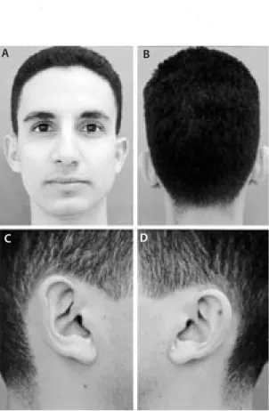
Figure 3. Standard views for otoplasty
A. Anterior view, B. Posterior view, C. Right close-up view, D. Left close-up view.
A B
Otoplasty
Firstly, just like in rhinoplasty, anterior view of the full-face should be taken by a camera positioned vertically, and back of the head should be then viewed. Long hair should be pulled off of the face and placed behind ears with the help of hair clips. Finally, an oblique view including the ears should be photographed (4) (Figure 3).
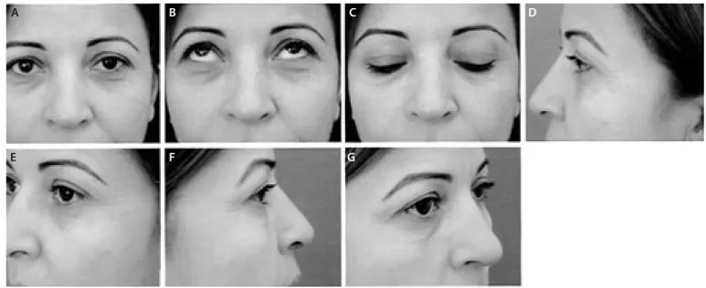
Blepharoplasty
The camera is positioned at the eye level of the patient having the hair line as the upper, tip of the nose as the lower border of the frame to photograph anterior views,
with the patient looking straight ahead, downwards and upwards. Then, the lateral and oblique views of the same frame are taken (9) (Figure 4).
Mammoplasty
Camera is oriented horizontally while the arms at side, anterior view is taken with a frame including the area bet-ween clavicles, shoulders and the inferior margin of the ribs. Then, lateral and oblique views are photographed for both sides (5) ( Figure 5).
Figure 4. Standard views for blepharoplasty
A. Anterior view while the patient is looking straight ahead, B. Anterior view while the patient is looking upwards, C. Anterior view while the patient is looking downwards, D. Left lateral view, E. Left oblique view, F. Right lateral view, G. Right oblique view.
Figure 5. Standard views for mammoplasty
A. Anterior view, B. Right lateral view, C. Right oblique view, D. Left lateral view, E. Left oblique view.
A B E F C D G B C D A E
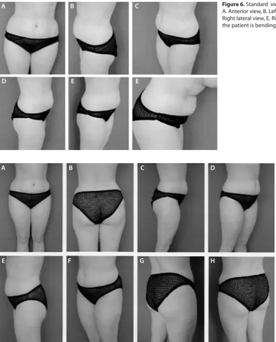
Abdominoplasty
Landmarks of standard photographs for abdominop-lasty are inframammary fold at he top and medial thigh at the bottom. With a camera oriented horizontally –arms kept upwards- anterior, lateral and oblique poses of both sides are taken as standard photographs. A close-up view of the abdominal area may also be helpful for assessing the quality of the abdominal skin. To evaluate the laxity of the abdominal musculature and lower abdominal adiposity, a lateral view of the patient, bending downwards gently, can be taken (10, 11) (Figure 6).
Body contour surgery
To obtain standard photographs for buttock and thigh –while keeping arms upwards- anterior, anterior medial-oblique, anterior lateral-medial-oblique, posterior, posterior obli-que right buttock, posterior obliobli-que left buttock views and lateral views of frame extending from subcostal margin to beneath the knees must be taken with a vertically orien-ted camera. In addition to these standard views a posterior view with gluteal muscles relaxed and tightened will help to better evaluate trochanteric lipodystrophy. The patient may also be viewed while she/he is lifting her/his excess fat (Figure 7).
Figure 7. Standard views for contour
surgery of the buttock and thigh A. Anterior view, B. Posterior view, C. Right lateral view, D. Right oblique view, E. Left lateral view, F. Left oblique view, G. Right posterior oblique view, H: Left posterior oblique view.
D E E
B C D A
References
1. Rogers BO. The first pre- and postoperative photographs of plastic and reconstructive surgery: contributions of Gurdon Buck (1807-1877). Aesthetic Plast Surg 1991;15:19-33.
2. Ellenbogen R, Jankauskas S, Collini FJ. Achieving standardized photographs in aesthetic surgery. Plast Reconst Surg 1990;86:955-961.
3. DiBernardo BE, Adams RL, Krause J, Fiorillo MA, Gheradini G. Photographic Standards in Plastic Surgery. Plast Reconst Surg 1998;102-2:559-568.
4. Yavuzer R, Smirnes S, Jackson IT at al. Guidelines for standard photography in plastic surgery. Ann Plast Surg 2001;46-3:293-300.
5. Jemec BI, Jemec GB. Photographic surgery: Standards in clinical photography. Aesthetic Plast Surg 1986;10:177-180.
6. Galdino GM, Da Silva D, Gunter JP. Digital photography for rhinoplasty. Plast Reconst Surg 2001;109-4:1421-1434.
7. Webber WB. Rhinoplasty: The importance of consistent
documentation and significant long-term follow-up. Plast Reconst Surg 1987;79:640-654.
8. Staffel JG. Photo documentation in rhinoplasty. Facial Plast Surg 1997;13:317-332.
9. Schwartz MS, Tardy ME. Standardized photodocumentation in facial plastic surgery. Facial Plast Surg 1990;7:1-12.
10. Zarem HA. Standards of photography. Plast Reconst Surg 1984; 74:137-146.
11. DiBernardo BE, Adams RL, Krause J, Fiorillo MA, Gheradini G. Photographic Standards in Plastic Surgery. Plast Reconst Surg 1998;102-2:559-568.
12. Galdino MG, Vogel JE, Vander Kolk CA. Standardizing digital photography: It’s not all in the eye of the beholder. Plast Reconst Surg 2000;108:1334-1344.
To photograph the lower extremity knees and the who-le feet must be placed in the frame. Then, anterior, lateral and oblique views are are taken with a vertically positioned camera. A lateral view of the patient, standing on tiptoe is also helpful to differentiate gastrocnemius musculature from adiposity of the culf.
To view the arms camera must be positioned horizon-tally. The upper arm kept perpendicular to axilla, the el-bow flexed at 90 degrees, the excess fat and skin can thus be evaluated by the help of gravity (11).
Discussion
Standardizing photographs in plastic surgery is very im-portant, even indispensable for objective analysis and
eva-luation of the archived results. Standardized photography is essential and necessary since it is thus possible to establish a standardized photographic documentation of the patients in terms of the procedure groups. With the arrival of the digital technology photo documentation in plastic surgery became easier, more effective and economical when com-pared to conventional photography. Nevertheless, it sho-uld be admitted that all digital cameras do not produce the same quality and accuracy as to their outcomes (12).
To have consistent and objective evaluations of the tre-ated patients through clinical photographs, they should be taken with the camera lens, lens setting, lighting, film and standard patient position.