Original Paper
Med Princ Pract 2019;28:418–424
Thyroid Nodules Are More Prevalent in
Subjects with Colon Polyps, Independent
of Insulin Resistance
Umut Mousa
aCuneyd Anil
bCanan Cicek Demir
aYusuf Bozkus
aKubra Ozturk
cNeslihan Bascil Tutuncu
aAlptekin Gursoy
baDepartment of Endocrinology and Metabolism, Baskent University Faculty of Medicine, Ankara, Turkey;
bDepartment of Endocrinology and Metabolism, Guven Hospital, Ankara, Turkey; cDepartment of Internal Medicine Baskent University Faculty of Medicine, Ankara, Turkey
Received: June 28, 2018 Accepted: March 12, 2019 Published online: March 13, 2019
Umut Mousa
Department of Endocrinology and Metabolism
Baskent University Faculty of Medicine, 5. Sokak No. 48 Cankaya © 2019 The Author(s)
Published by S. Karger AG, Basel
Significance of Study
• This study establishes a positive association between colorectal polyps and thyroid nodules. According to our analysis, this association seems to be independent from insulin resistance, which was previ-ously argued to be a possible central mechanism in both disorders.
DOI: 10.1159/000499527
Keywords
Thyroid volume · Nodule · Polyps
Abstract
Objective: Colorectal polyps and thyroid nodules are
com-mon disorders linked to hyperinsulinemia and metabolic syndrome (Mets). The direct association between these two diseases is not clear. We aimed to analyze the prevalence of thyroid nodules in subjects with and without colorectal pol-yps. The secondary aim was to establish the prevalence of Mets and its parameters in both disorders and to determine if insulin resistance and hyperinsulinemia are common underlying pathophysiological mechanisms. Subjects and
Methods: One hundred and five subjects with colorectal
pol-yps (71 males, 34 females) and 68 controls (28 males, 40
fe-males) were enrolled. The parameters of Mets together with TSH, insulin, low-density lipoprotein cholesterol, and ho-meostasis model for assessment of insulin resistance levels were calculated. We performed thyroid ultrasonography in all participants. Results: The prevalence of Mets was similar in the colorectal polyp and control groups (37.1 vs. 37.3%, p = 0.982). The prevalence of Mets was nonsignificantly high-er in subjects with a documented thyroid nodule compared to subjects without a thyroid nodule (43.0 vs. 32.6%, p = 0.205). The prevalence of thyroid nodules in subjects with colorectal polyps was significantly higher than in subjects without polyps (52.9 vs. 35.3%, p = 0.017). Compared to sub-jects with no colorectal polyps, we established a significant increase in the odds of having thyroid nodules (OR 2.05; 95% CI: 1.097–3.860, p = 0.017). The presence of colorectal polyps and age in the adjusted model were established to be
inde-pendent risk factors for having thyroid nodules (p = 0.025 and p = 0.007, respectively). Conclusion: These results may support the presence of other common mechanisms in the development of these two pathologies other than insulin re-sistance and hyperinsulinemia. © 2019 The Author(s)
Published by S. Karger AG, Basel
Introduction
Colorectal polyps and thyroid nodules are common disorders, which are linked independently to hyperinsu-linemia and the metabolic syndrome (Mets). Any direct association between these two entities is not clear.
Limited studies have reported the association between colorectal polyps and thyroid nodules. In a study by Du-ran et al. [1], thyroid nodules were demonstrated to be more prevalent in subjects with colonic polyps. In that study, the components of Mets and insulin levels were not included in the analyses. A study by Park et al. [2] dem-onstrated that thyroid nodules were associated with co-lonic polyps in subjects with BMI >25, but not in subjects with BMI <25.
Mets is defined as a cluster of abnormalities, which is associated with cardiovascular risk and is comprised of abdominal obesity, abnormal glucose tolerance, dyslipid-emia, insulin resistance (IR), and hypertension [3]. In many studies, Mets has been associated with some can-cers in adults [4]. The molecular mechanisms and patho-physiology of this syndrome have not been completely resolved. However, some of the carcinogenic effects are attributed to IR and hyperinsulinemia.
It is known that insulin uses the same signaling path-ways with the insulin-like growth factor-1 (IGF-1) tor. In peripheral tissues, insulin binds to the insulin recep-tor, thus leading to intracellular phosphorylation of pro-teins. Different tissues have different sensitivities to insulin depending on the number of receptors on the cell mem-brane. Increased insulin levels lead to an increase in levels of free IGF-1 and decreased production of IGF-1-binding proteins [5]. Insulin displays a mitogenic effect via increas-ing DNA synthesis and decreasincreas-ing apoptosis [6].
Obesity and IR increase the risk of colon cancer. Ad-enomatous colon polyps are considered to be precancer-ous lesions. The effects of increased insulin and IGF-1 levels on cellular proliferation have been accused to be a potential risk for the development of colon cancer [7, 8]. Some studies have shown an increased prevalence of colorectal adenomas in Mets [9–14].
Our group has previously demonstrated a relationship between IR and thyroid volume (TV) together with the formation of thyroid nodules [15]. This has been attrib-uted to high levels of insulin in the circulation. IGF-1-de-pendent mechanisms have also been partially accused [15, 16]. Thus, a probable association between thyroid nodules and colorectal polyps has gained our interest due to common physiological mechanisms.
In the present study, we aimed to analyze the preva-lence of thyroid nodules in subjects with and without colorectal polyps. The secondary aim was to establish the prevalence of Mets and its parameters in both disorders and ascertain whether IR and hyperinsulinemia are com-mon underlying pathophysiological mechanisms.
Subjects and Methods
Prior to the study, a statistical power calculation revealed that at least 160 cases would be sufficient with 85.72% reliability. We included 105 subjects (71 males, 34 females) who were diagnosed as having a colorectal polyp by total colonoscopy between January 2009 and April 2010. As the control group, we included 68 subjects (28 males, 40 females) who underwent total colonoscopy in the same time period with no visualized polyps.
Exclusion criteria were a previous history of any thyroid dis-ease, including subclinical or overt hypo-/hyperthyroidism, previ-ous use of levothyroxine; antithyroid drugs; any drug that can in-terfere with the thyroid function tests, previous use of oral anti-diabetic drugs, any previous history of radiotherapy or surgery to the head and neck, positive thyroid autoantibodies, kidney and hepatic insufficiency or abnormal laboratory results, hypercorti-solemia, pregnancy, lactation, and a previous history of a neuro-logical or psychiatric disease.
Criteria of the National Cholesterol Education Program (NCEP) Adult Treatment Panel III (ATP III) were used in evaluat-ing the presence of Mets in the participants. A patient havevaluat-ing at least three positive values out of five in the NCEP ATP III criteria was diagnosed as Mets [17]. Subjects with a blood pressure ≥130/85 mm Hg or those using antihypertensive drugs were recorded as being hypertensive. Subjects with a fasting blood glucose (FBG) ≥100 mg/dL or those with a glucose value ≥140 mg/dL at the 120th min of 75 g glucose load were recorded as having abnormal glucose tolerance. Subjects with an FBG ≥126 mg/dL or those with a glu-cose value ≥200 mg/dL at the 120th min of 75 g gluglu-cose load were recorded as being diabetic.
Body weight was measured to the nearest kilogram and height to the nearest 0.1 m simultaneously with the measurement of the waist circumference (WC), which took place with a tape at the um-bilical site. BMI was calculated by dividing the weight in kilograms to the height in meters squared. All anthropometric measurements were performed by the same investigator to avoid interobserver variability. We also measured the patients’ blood pressure with a sphygmomanometer after the patient had rested for at least 30 min.
A blood sample was collected from each patient for analysis of fasting plasma glucose (glucose oxidase method; Roche Diagnos-tics GmbH, Mannheim, Germany), insulin (chemiluminescent
as-say; Bio-DPC Diagnostic Products Corporation, Los Angeles, USA, via Immulite 2000), triglyceride, high-density lipoprotein (HDL) cholesterol (Hitachi Modular systems; Roche Diagnostics GmbH, Mannheim, Germany), TSH (immunochemiluminescent assay; Bio-DPC Diagnostic Products Corporation, Los Angeles, USA, via Immulite 2000). We performed Tg-ab and TPO-ab mea-surements by ICMA (Immulite 2000; Siemens). In addition, a 75-g oral glucose tolerance test was performed to obtain the glucose levels at the 120th min for each patient. The homeostasis model for assessment of IR (HOMA-IR) was calculated by the formula fast-ing plasma glucose (mg/dL) × fastfast-ing insulin (µU/mL) / 405 [18]. The same investigator performed thyroid ultrasonography (Logiq 5 Pro; GE Medical Systems, WI, USA) with a 10 MHz probe. We evaluated the presence of a thyroid nodule and calculated the TV in mL by the formula (depth × width × length × π) / 6 [19]. Thyroid ultrasonography was performed within 1 month of the initial colonoscopy.
Smoking and drinking habits together with the pathology of the colorectal polyp (adenomatous, hyperplastic, mixed) were record-ed. Subjects with detected thyroid nodules were offered fine-nee-dle aspiration cytology if indicated by the researcher.
Statistical Analysis
We used the SPSS for Windows v. 16.0 (Statistical Package for Social Sciences, Chicago, IL, USA) software program for the statis-tical analysis. We used the Shapiro-Wilk and Levene tests for con-firmation of the normality and homogeneity of variances of our data and the χ2 test to compare the ratios between the groups, the
independent samples t test to compare the means between the groups, and the Mann-Whitney U test for comparing the means of nonparametric values. We performed a logistic regression analysis to identify independent variables. A p value of <0.05 was defined as being statistically significant.
Results
We included 105 subjects (71 males, 34 females) who underwent total colonoscopy between January 2009 and April 2010 and in whom colorectal polyps were detected. As the control group, we included 68 subjects (28 males, 40 females) who underwent colonoscopy, which was re-ported as normal in the same time period. A total of 99 male (57.2%) and 74 female (42.8%) subjects was en-rolled.
Prevalence of Thyroid Nodules and Analyses of Risk Factors in the Study Group
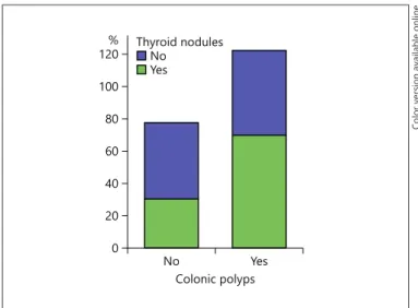
The prevalence of thyroid nodules in subjects with colorectal polyps was significantly higher than in subjects without polyps (55/105 [52.9%] vs. 24/68 [35.3%] p = 0.017) (Fig. 1). Compared to subjects with no colorectal polyps, in subjects with colorectal polyps, we established a significant increase in the odds of having thyroid nod-ules (OR 2.05; 95% CI: 1.097–3.860, p = 0.017).
The prevalence of thyroid nodules was similar in male and female subjects (45/99 [44.9%] vs. 35/74 [47.3%], p = 0.760).
A total of 112 subjects were ≥50 years of age, and 61 subjects were <50 years. The prevalence of thyroid nod-ules was significantly higher in subjects ≥ 50 years com-pared to those <50 years (62/112 [55.4%] vs. 17/61 [28.3%], p = 0.001). Compared to subjects <50 years, in subjects ≥50 years, we demonstrated a significant in-crease in the odds of having thyroid nodules (OR 3.13, 95% CI: 1.59–6.15, p = 0.001). However, the prevalence of colorectal polyps was similar in both age groups.
The prevalence of thyroid nodules was higher in sub-jects with abnormal glucose tolerance (i.e., diabetes or prediabetes) compared to those with normal glucose tol-erance (39/71 [54.9%] vs. 40/102 [39.6%], p = 0.048). Compared to subjects with normal glucose tolerance, in subjects with abnormal glucose tolerance, we established a significant increase in the odds for developing thyroid nodules (OR 1.85, 95% CI: 1.01–3.43, p = 0.048). Again, the prevalence of colorectal polyps was similar in subjects with abnormal glucose tolerance compared to normal glucose tolerance.
The prevalence of thyroid nodules in subjects with hy-pertension was significantly higher than in normotensive subjects (30/52 [57.7%] vs. 49/121 [40.8%], p = 0.047). Compared to normotensive subjects, in hypertensive subjects, we determined a significant increase in the odds for developing thyroid nodules (OR 1.9, 95% CI:1.02–
120 Thyroid nodules■ No ■ Yes % 100 80 60 40 20 0 No
Colonic polypsYes
Fig. 1. Bar charts displaying the presence of thyroid nodules in
subjects with and without colonic polyps.
3.80, p = 0.047). Subjects in the colorectal polyp group were prone to be more hypertensive compared to the con-trol group. This comparison did not reach statistical sig-nificance (37/105 [35.2%] vs. 15/68 [22.1%], p = 0.089).
The prevalence of thyroid nodules was similar in sub-jects with triglyceride levels ≥150 mg/dL compared to those <150 mg/dL (48/110 (44.0%) vs. 31/63 (50.8%), p = 0.529). Again, the presence of colorectal polyps was simi-lar in both triglyceride groups.
The presence of thyroid nodules and colorectal polyps was similar in subjects with HDL values, and WC was lower than the values determined by the NCEP ATP III criteria compared to those with normal values (i.e., HDL ≥40 mg/dL and WC <102 cm for male and HDL ≥50 mg/ dL and WC <88 cm for female subjects) (Table 1).
We performed a logistic regression analysis to define the independent variables for the presence of thyroid nodules. The regression models were adjusted for the presence of colorectal polyps, age group, abnormal
glu-cose tolerance, and hypertension. Presence of colorectal polyps and age were established to be independent risk factors for having thyroid nodules (p = 0.025 and p = 0.007, respectively).
The prevalence of thyroid nodules did not differ be-tween smokers and nonsmokers. Again, the prevalence of thyroid nodules was similar in all 3 types of colorectal polyps (adenomatous, hyperplastic, and mixed).
Prevalence of Mets in the Study Group
The presence of Mets was similar in the colorectal pol-yp and control groups (39/105 [37.1%] vs. 25/68 [37.3%], p = 0.982). A total of 39 subjects with colon polyps were Mets(+). Out of these 24 had documented thyroid nod-ules (61.5%). A total of 66 subjects with colon polyps were Mets(–). Out of these, 31 (48.5%) had thyroid nodules. The comparison did not reach statistical significance (p = 0.224). The sex distribution was detected to be similar in Mets(+) and Mets(–) subjects with colorectal polyps.
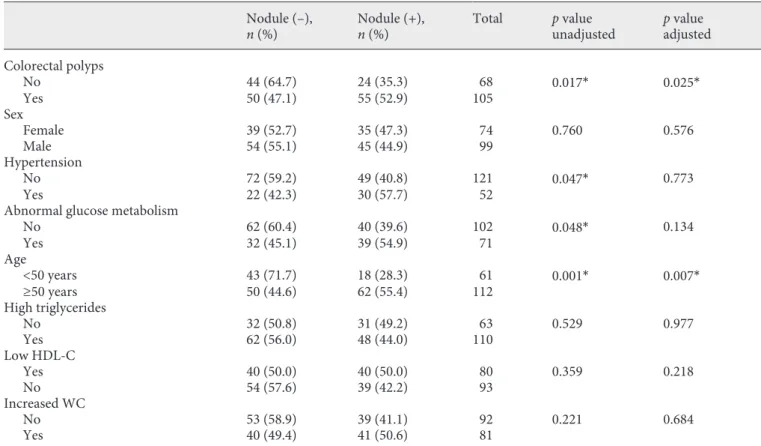
Table 1. Risk factors for thyroid nodules in unadjusted and adjusted models
Nodule (–),
n (%) Nodule (+),n (%) Total p valueunadjusted p valueadjusted
Colorectal polyps No 44 (64.7) 24 (35.3) 68 0.017* 0.025* Yes 50 (47.1) 55 (52.9) 105 Sex Female 39 (52.7) 35 (47.3) 74 0.760 0.576 Male 54 (55.1) 45 (44.9) 99 Hypertension No 72 (59.2) 49 (40.8) 121 0.047* 0.773 Yes 22 (42.3) 30 (57.7) 52
Abnormal glucose metabolism
No 62 (60.4) 40 (39.6) 102 0.048* 0.134 Yes 32 (45.1) 39 (54.9) 71 Age <50 years 43 (71.7) 18 (28.3) 61 0.001* 0.007* ≥50 years 50 (44.6) 62 (55.4) 112 High triglycerides No 32 (50.8) 31 (49.2) 63 0.529 0.977 Yes 62 (56.0) 48 (44.0) 110 Low HDL-C Yes 40 (50.0) 40 (50.0) 80 0.359 0.218 No 54 (57.6) 39 (42.2) 93 Increased WC No 53 (58.9) 39 (41.1) 92 0.221 0.684 Yes 40 (49.4) 41 (50.6) 81
Low HDL-C: <50 mg/dL for female and <40 mg/dL for male subjects; increased WC: >102 cm for male subjects and >88 cm for female subjects; high triglycerides: ≥150 mg/dL. * Statistically significant.
However, the age group was higher in Mets(+) subjects with colorectal polyps compared to Mets(–) subjects with colorectal polyps (44.6 vs. 19.4%, p = 0.016).
Out of the total 173 (46.2%) subjects, 79 were demon-strated to have thyroid nodules. Out of these, 44 (55.0%) had a uninodular goiter, and 35 (45.0%) had a multinod-ular goiter. The prevalence of Mets was higher in subjects with a documented thyroid nodule compared to subjects without a thyroid nodule. This analysis did not reach sta-tistical significance (34/79 [43.0%] vs. 30/94 [31.9%], p = 0.205). The prevalence of Mets did not differ between subjects with uninodular and multinodular goiter (21/44 [47.7%] vs. 13/35 [38.2%], p = 0.365).
Comparison of Means
A comparison of some anthropometric, laboratory, and thyroid parameters of the study groups are presented in Table 2. The mean BMI, FBG, insulin, HOMA IR, and triglyceride levels were similar in both the colorectal pol-yp and control groups. The TV was higher (16.38 ± 11.16 vs. 12.92 ± 7.33 mL, p = 0.027), the low-density lipopro-tein cholesterol (LDL-C) was higher (132.36 ± 34.57 vs. 121.25 ± 30.71 mg/dL, p = 0.032), and the TSH level was lower (1.40 ± 0.84 vs. 1.72 ± 0.97 mIU/L, p = 0.026) in the colorectal polyp group compared to the control group. The mean age, FBG, and TV were significantly higher in subjects having a documented thyroid nodule compared to those without a thyroid nodule (55.39 ± 9.18 vs. 51.75 ± 11.15 years, p = 0.032; 108.49 ± 28.06 vs. 100.42 ± 19.88 mg/dL, p = 0.029; 18.13 ± 13.26 vs. 12.36 ± 4.88 mL, p < 0.0001). The mean BMI, insulin, HOMA-IR, triglycer-ides, HDL-C, and LDL-C were similar in subjects with and without thyroid nodules.
Discussion
Colorectal polyps are known to be precursors of colon adenocarcinoma together with other known risk factors such as age, high-lipid and low-fiber diet, hyperinsu-linemia, and IGF-1 [9, 10]. Mets is an endocrinopathy in which IR plays the central role and links abdominal obe-sity, glucose tolerance defects, lipid anomalies, hyperten-sion, and coronary artery disease. Furthermore, Mets is known to be related to some cancers of adulthood. Many studies have demonstrated an increased prevalence of colorectal adenomas in subjects with Mets. Hyperinsu-linemia, IR, high-fat and low-fiber diet, changes in the microbiota, and increased IGF-1 levels have been accused in this relationship [9–11, 20, 21].
Recent studies have demonstrated increased TV and higher prevalence of thyroid nodules in subjects with Mets [15]. This has been attributed to high insulin levels and partially to IGF-1-related mechanisms.
According to our results, age and the presence of colorectal polyps were determined to be independent risk factors for thyroid nodules, and TV is larger in subjects with colorectal polyps. We failed to correlate our findings with Mets and its components in subjects with colorectal polyps. The prevalence of thyroid nodules was associated with two of the components of Mets (i.e., glucose metab-olism defect and hypertension), which were determined to be dependent risk factors.
Mets and hyperinsulinemia have been implicated as factors in the development of both thyroid nodules and colorectal polyps [11, 12, 15]. However, in our study, the prevalence of Mets and its components was similar in subjects with and without colorectal polyps. We also failed to demonstrate higher insulin levels and HOMA-IR scores in these subjects. This finding was not consistent with some previously reported studies. Morita et al. [11] demonstrated that Mets and its parameters were associ-ated with the development of colorectal polyps in a study including 756 subjects with colorectal polyps and 1,751 controls. Similarly, Kim et al. [12] established that the prevalence of Mets was significantly higher in subjects with colorectal adenomas compared to a control group. Lee et al. [14] demonstrated that the development risk of colorectal adenomas was higher even in subjects with only one component of Mets. The relatively low number of participants in the present study may be a reason for
Table 2. Comparison of some anthropometric parameters between
the groups
Colorectal polyp
group Control group p Age, years 54.48±10.01 51.87±10. 86 0.107 BMI 28.88±7. 66 27.99±4. 06 0.425 TV, mL 16.38±11.16 12.92±7.73 0.027* FBG, mg/dL 101.88±23.95 107.25±24.54 0.155 Insulin 9.54±4.64 10.52±8.10 0.315 HOMA-IR 2.43±1.33 2.84±2.88 0.204 LDL-C, mg/dL 132.36±34.57 121.25±30.71 0.032* Triglycerides, mg/dL 144.92±85.59 143.34±89.17 0.907 TSH 1.40±0.84 1.72±0.97 0.026*
Values are presented as mean ± standard deviation. * Statistically sig-nificant.
not documenting the increased prevalence of Mets in subjects with colorectal polyps.
Pathophysiological mechanisms other than hyperin-sulinemia have been demonstrated in the development of colorectal polyps. IGF-1 plays important roles in growth and cellular metabolism. IGF-1 also inhibits apoptosis. Normal and cancerous colorectal epithelium express IGF-1 receptors. Saubry et al. [10] previously demonstrated that IGF-1 levels were correlated with the development of colorectal adenomas. Another substance involved in the pathogenesis of colorectal polyps is glu-cose-dependent insulinotropic peptide (GIP), which stimulates the secretion of insulin from the pancreas and leads to lipogenesis. Sasaki et al. [22] analyzed the rela-tionship between GIP and the development of colorectal adenomas. Fasting GIP levels, FBG, triglycerides, insulin levels, and HOMA-IR were significantly higher in the disease group compared to the control group. In this study, multiple logistic regression revealed that the high-est quartile of fasting GIP levels was associated with a significantly higher risk of colorectal adenoma (OR, 2.1; 95% CI, 1.08–3.96; p = 0.01) in comparison with the low-est quartile.
It is known that increased epithelial proliferation oc-curs in subjects with acromegaly, which is correlated with IGF-1 and growth hormone levels. The pathogenesis of colonic lesions in acromegaly is not well understood. A study by Foltyn et al. [23] demonstrated that hyperinsu-linemia, IR, and colonic lesions were significantly higher in a cohort of 40 acromegalic subjects. In the same study, they demonstrated that in subjects with multiple colonic polyps, the number of hyperplastic and adenomatous polyps were correlated with HOMA-IR.
In the present study, the prevalence of Mets was non-significantly higher in subjects with thyroid nodules compared to those without thyroid nodules. The compo-nents of Mets that were associated with the prevalence of Mets were hypertension and abnormal glucose metabo-lism. However, in the adjusted models, these compo-nents were not defined to be independent. Age and the presence of colorectal polyps were defined as the inde-pendent variables associated with thyroid nodule preva-lence. Supporting this finding, the thyroid volume was significantly larger in subjects with colorectal polyps compared to the control group. Studies associating colorectal polyps and thyroid nodules are limited. Simi-lar to our findings, a study by Duran et al. [1] detected that thyroid nodules were more prevalent in subjects with colorectal polyps. In another study, Park et al. [2] demonstrated a significant correlation between
colorec-tal polyps and thyroid nodules only in subjects with a BMI ≥25. This relationship did not exist in subjects with a BMI ≤24.9.
It is known that the insulin/IGF-1 pathway modulates thyroid gene expression and acts as a cofactor for thyro-cyte proliferation and differentiation. It has been report-ed that in cell cultures, TSH has mitogenic and antiapop-totic effects with insulin. IGF-1 and insulin receptors have been demonstrated in thyrocyte cultures. In addi-tion, the high goiter prevalence in subjects with acromeg-aly shows the activity of IGF-1 in vivo as a co-mitogenic factor. Rezzonico et al. [24] reported that the TV was higher, and the risk of development of thyroid nodules was higher in subjects with IR.
The main limitation of our study was the relatively small number of participants enrolled. This may have had an impact on our findings regarding the prevalence of thyroid nodules in the different subgroups related to age, glucose intolerance, blood pressure, triglycerides, and smoking habits. A large prospective cohort would be more meaningful with regard to these subclassifications. Despite this limiting factor, our study was an interven-tional study in which the thyroid ultrasonography was performed concurrently with the total colonoscopy, and all anthropometric and ultrasonographical evaluations were performed by the same investigator.
Conclusion
Our findings support the presence of common mecha-nisms other that IR and hyperinsulinemia in the develop-ment of these two entities, such as IGF-1. Our findings need to be proven with high-scale prospective studies in-cluding IGF-1 assays.
Statement of Ethics
This study was approved by the Baskent University Research and Ethics Committees. All procedures performed in studies in-volving human participants were in accordance with the ethical standards of the institutional and/or national research committee and with the 1964 Helsinki Declaration and its later amendments or comparable ethical standards. All subjects provided informed written consent.
Disclosure Statement
All authors declare that they have no conflicts of interest to dis-close.
References
1 Duran C, Gokturk HS, Kulaksizoglu M, Bak-dik S, Unler GK, Erbayrak M, et al. Frequency of thyroid nodules among patients with co-lonic polyps. Gastroenterol Res Pract. 2012; 2012:178570.
2 Park CW, Kim EK, Yun YB, et al. The correla-tion between thyroid nodules and the inci-dence rate of colonic polyps based on body mass index. Korean J Fam Pract. 2015;5:354– 60.
3 Balkau B, Charles MA; European Group for the Study of Insulin Resistance (EGIR). Com-ment on the provisional report from the WHO consultation. Diabet Med. 1999 May; 16(5):442–3.
4 Braun S, Bitton-Worms K, LeRoith D. The link between the metabolic syndrome and cancer. Int J Biol Sci. 2011;7(7):1003–15. 5 Giovannucci E. Metabolic syndrome,
hyper-insulinemia, and colon cancer: a review. Am J Clin Nutr. 2007 Sep;86(3):s836–42.
6 Tan KT, Luo SC, Ho WZ, Lee YH. Insulin/ IGF-1 receptor signaling enhances biosyn-thetic activity and fat mobilization in the initial phase of starvation in adult male C. elegans. Cell Metab. 2011 Sep;14(3):390– 402.
7 Siddiqui A, Pena Sahdala HN, Nazario HE, Mahgoub A, Patel M, Cipher D, et al. Obesity is associated with an increased prevalence of advanced adenomatous colon polyps in a male veteran population. Dig Dis Sci. 2009 Jul;54(7):1560–4.
8 Bruce WR, Cirocco M, Giacca A, Kim YI, Marcon N, Minkin S. A pilot randomised controlled trial to reduce colorectal cancer risk markers associated with B-vitamin defi-ciency, insulin resistance and colonic in-flammation. Br J Cancer. 2005 Sep;93(6): 639–46.
9 Sato T, Takeda H, Sasaki Y, Kawata S. In-creased homeostasis model assessment-insu-lin resistance is a risk factor for colorectal ad-enoma in Japanese males. Tohoku J Exp Med. 2011 Apr;223(4):297–303.
10 Soubry A, Il’yasova D, Sedjo R, Wang F, Byers T, Rosen C, et al. Increase in circulating levels of IGF-1 and IGF-1/IGFBP-3 molar ratio over a decade is associated with colorectal adeno-matous polyps. Int J Cancer. 2012 Jul;131(2): 512–7.
11 Morita T, Tabata S, Mineshita M, Mizoue T, Moore MA, Kono S. The metabolic syndrome is associated with increased risk of colorec- tal adenoma development: the Self-Defense Forces health study. Asian Pac J Cancer Prev. 2005 Oct-Dec;6(4):485–9.
12 Kim JH, Lim YJ, Kim YH, Sung IK, Shim SG, Oh SO, et al. Is metabolic syndrome a risk fac-tor for colorectal adenoma? Cancer Epidemi-ol Biomarkers Prev. 2007 Aug;16(8):1543–6. 13 Liu CS, Hsu HS, Li CI, Jan CI, Li TC, Lin WY,
et al. Central obesity and atherogenic dyslip-idemia in metabolic syndrome are associated with increased risk for colorectal adenoma in a Chinese population. BMC Gastroenterol. 2010 May;10(1):51.
14 Lee GE, Park HS, Yun KE, Jun SH, Kim HK, Cho SI, et al. Association between BMI and metabolic syndrome and adenomatous co-lonic polyps in Korean men. Obesity (Silver Spring). 2008 Jun;16(6):1434–9.
15 Ayturk S, Gursoy A, Kut A, Anil C, Nar A, Tutuncu NB. Metabolic syndrome and its components are associated with increased thyroid volume and nodule prevalence in a mild-to-moderate iodine-deficient area. Eur J Endocrinol. 2009 Oct;161(4):599–605. 16 Minuto F, Barreca A, Del Monte P, Cariola G,
Torre GC, Giordano G. Immunoreactive in-sulin-like growth factor I (IGF-I) and IGF-I-binding protein content in human thyroid tis-sue. J Clin Endocrinol Metab. 1989 Mar; 68(3):621–6.
17 National Cholesterol Education Program (NCEP) Expert Panel on Detection, Evalua-tion, and Treatment of High Blood Choles-terol in Adults (Adult Treatment Panel III). Third Report of the National Cholesterol Ed-ucation Program (NCEP) Expert Panel on Detection, Evaluation, and Treatment of High Blood Cholesterol in Adults (Adult Treatment Panel III) final report. Circulation. 2002 Dec;106(25):3143–421.
18 Matthews DR, Hosker JP, Rudenski AS, Nay-lor BA, Treacher DF, Turner RC. Homeosta-sis model assessment: insulin reHomeosta-sistance and beta-cell function from fasting plasma glu-cose and insulin concentrations in man. Dia-betologia. 1985 Jul;28(7):412–9.
19 Brunn J, Block U, Ruf G, Bos I, Kunze WP, Scriba PC. [Volumetric analysis of thyroid lobes by real-time ultrasound (author’s transl)]. Dtsch Med Wochenschr. 1981 Oct; 106(41):1338–40. German.
20 Saetang J, Sangkhathat S. Diets link metabolic syndrome and colorectal cancer development (Review) [Review]. Oncol Rep. 2017 Mar; 37(3):1312–20.
21 Lim HS, Shin EJ, Yeom JW, Park YH, Kim SK. Association between Nutrient Intake and Metabolic Syndrome in Patients with Colorectal Cancer. Clin Nutr Res. 2017 Jan; 6(1):38–46.
22 Sasaki Y, Takeda H, Sato T, Orii T, Fujishima S, Nagino K, et al. Increased levels of serum glucose-dependent insulinotropic polypeptide as a novel risk factor for human colorectal ad-enoma. Metabolism. 2011 Sep;60(9):1253–8. 23 Foltyn W, Kos-Kudla B, Strzelczyk J, Matyja
V, Karpe J, Rudnik A, et al. Is there any rela-tion between hyperinsulinemia, insulin resis-tance and colorectal lesions in patients with acromegaly? Neuro Endocrinol Lett. 2008 Feb;29(1):107–12.
24 Rezzonico J, Rezzonico M, Pusiol E, Pitoia F, Niepomniszcze H. Introducing the thyroid gland as another victim of the insulin resistance syndrome. Thyroid. 2008 Apr;18(4):461–4.