30 (3) : 361-367, 1983
A:\ OUTBREAK OF A DISEASE OF FARMED EEL (ANGUII.LA ANGUILLA)
DUE TO AEROMONAS I-IYDROPHILA İl\' TeRKE'Y: HİSTOPATHOLOGICAL
AND BACTERIOLOGICAL STUDIES
Metin Timur*
Yurdumuz kültür yılan balıklarında (Anguilla anguilla) etkeni Aeromonas hyd-rophila olan hastalığın histopatolojik ve bakteriyolojik yönden incelenmesi.
Özet:
Bu çalışmada Eskişehir, Çifteler Sakaryabaşı Balık üretim ve Araştırma istasyonunda değişik rasyonlarla beslenen yılan balıklarında (An-guilla an(An-guilla), Aeromoııas hydrophila'nın neden olduğu hastalık, bakteriyo-lojik ve histopatol~jik yihıden incelenmiştir.Klinik ve histopatol~jik bulgularda yaygın deri ülseri, pullarda dökülme, uzun süreli olaylarda ise deri altındaki kas tabakasına kadar ilerlqen etkenin miyopati ve miyofajiye neden olduğu saptanmıştır.
Summary: The present study describes an outbreak of Aeromonas
hyd-rophila irifection among the eel (Anguilla anguilla) at Çifteler-Sakaryabaşı Fish Breeding and Research Station in Eskişehir- Turkq.
The main clinical and histopathological feature of the diseases was the development of extensive skin ulcere, mOlJeout of scales and in longer standing lesions the m)'opathy and myophagia of underiying musculature.
Introduction
Baeterial fish disease eaused by members of the gencra
Aeromo-nas and PseudomoAeromo-nas are very eommon among fishes. The great
epizoo-tics described in Europea (5). The disease is eommon in warmwater
pondfishes in the United States in rainbow trout (Salmo gairdneri) in
the Western States and in northem pike (Esox lucius) in Canada (12).
The disease was eharaeterized by ulceration of the ilkin and
ex-tcnding down to penetrate the underıying museulature (2, 3, 6, iO, 1)).
Materials and Methods
Live speeimens of eels (Angilla anguilla) with eharaeteristie
lesi-ons were obtained from the Çifteler-Sakarya baş! Fish Breeding and
362 METiN TiMUR
Rcsearch Station. Measured
ı
6,60+
O,
ı
7 cm., in length and 12,381-0,33 g., in weight. Affected live fish were brought to the Unit of
Fis-heries and Fish Disease in Ankara for histopathological and
bacteri-ological studies.
For patological investigation at
iO
per cent farmalin fixeel andparaffin embedded samples of skin, kidney, gills, livcr, spken from
affected fish were cut at .') microns and staineel with heamotoxylin
and eosin.
For bacteriological examination, the sample was taken
ascpti-cally from the lesion and from kidney, liver, heart, blood and SpieCIL
lt
was streakeel on the blood agar (addedı
O per cent sheep blood)and frıınculosis agar (8). Smears from these tissues \vere also made
and stained by Gram's method. Bıochemical examination of the
iso-lates were carried out according to the technique of Osbaldistün (7).
The sensitivity disc was used in the therapeutic use of <1ntibiotics.
Results
Gross Pathology
The disease was characterized by external lesions on both flanks
varying in severity from small areas of scale loss to large uleers which
extending down to penetrate the undcrlying musculature.
Haemorr-hage on the fins wc re also present. Internally numerous petechial
lıac-morrhages observed on the walls of intestinal tract.
Histopatholog)'
Gill: Gill flament were lost their morphologic features
Hypert-rophic epithclial cells of the seconder gill !amellar were occupieel with
bacteria (Fig.
i)
and loss of cellular outlines and phyknosis ofnccro-tic cells were presen t.
Liver: N ecrosis of hepa tic perenchymall cells was first eviden t.
Surrounding paranchymall cells of vena centralis wc re lost their cell
membrane and nuc1ei, and many bacteria were scen around them (Fig.
2) .
Kidney: Loss of cellular outıines and nuclei of neerotic kidney
tubules were present. The normally distributed reticuloendothelial
tissue was spread through the kidney tubules, but some have lost
the-ir nuclci. Man)' bactcria were seen either within the
Figure ı. :\e(TGıic gill "pil1ıclin! edls ",iılı ,I.!~rdın:)liil(, (ermwed) H.E. >: ::l75 Nckrotik solung:ı( qıiıhcli:ı! 1ı,len.kri \T Il.lirt/rc/)fiilıı (okla işaretli) .
..
"
J:.4'~~ ~
''fb .ii;
Figmc 2. Hepaıic parcnehyma! cclls ",iıh Lass of edlular outlines and nudear staining and somc hacıeria :ıround ılıe eclis (arrowed). H.E. x 375 Hücre zan kaybolmuş ve çekirdck-leri iyi boya almayan karaciğer panlJlşima hücrderi r;evrcsinde yer alan bakteriler (ok La
364 METiN TiMUR
,
.•
'
~.... i
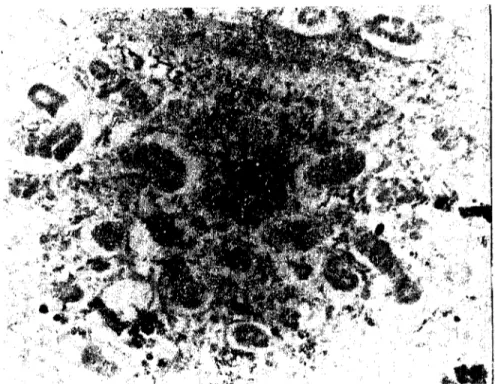
Figure 3. Melanin cdls (;\1) and bacteria (B) within the necrotie reticuloendothelial tis-sue and kidney tubules. H.E. X 250 Nekratik retiküloendotclial doku ve böbrek tübülleri
arasında yer alan melanİn hücreleri (l"I) ve bakterİler (B).
Spleen: The Iymphoid tissue of the white pulp was neerotie and
the baeteria were not mu ch spread throughout the neerotie eclIs.
Skin: The lesions were varied depending on the stage of the
in-feetion. The ed ge of the ulcer showed haemorrhages and leueoeytie
infiltration. In ehronie skin lesions the ulcer had extended to depth
into the myotomal muscle fibres. Museulatire showed myopathy and
some baeteria were seen around and within the muscle bundles
(my-ophagia). At this stage, lcueoeyte infiltration had invaded among the
eapillary blood vesseIs of the muscle fibres.
Bacteriology
Smcars from the spleen, liver, kidney and heart of the infceted
fish were showed Gram negativc, rod shape baeteria. From the
vis-eeral organs and blood of the İnfeeted fish, cireular wİth
ı
-2 mm.,in diameter, regular bordered, pİgmented and spreading eolonies
wc re isolated and eharaeterized as a straİn of
Aeromonas hydrophila
reaeti-ons. The results of the biochemical tests and the sensitivity test are
shown in (Tablc 1,2).
Tablc
ı.
Biochemical rcaclİons of A.Iı)'drolllıiln Tests Motiliıy Gram's rcacıion Hcmolvsis Lactos~ GIlIcose l'vlalıosc Galaclosc i Saccarose Arabinosc Laevıılosc Raffinosc lnositol ]-I,S Vögcs-Preskauer reaction Metlıvl-rcd .., lndol~ Cytochrome-Oxidasc Oxidation-Fcrmentatioıı (Oi F) Discussion Real'ıions-, ----
---I
i ~i ,!
i
i"'-
-j-+
+
FClinical signs observ'ed in the present outbreak were similar to
those described elsewhere for haemorrhagic septicemia except that
the severe necrosis and many bacteria were see n cither within the
necrotic eclIs or around them described by Reynolds et al (9) in
gold-fish
(Carassius _al/ratus)
were not scen in this study.Tablc 2. Reactioıı to antibiotics ı~nl~bio~~c_s _
Chloramphcnicol Nitrofunton Ch lortet racycıine
Rcactions Sensitiye Slight sensitiye Slight Sensitiye
The lesion varied depending on the stage of the infection.
His-topathological changes in the dcrmis and Stratum spongiosum
simi-lar to those described for
Pseudoınonas
infection.in eel (1) with uleer,loss of scales, mark ed hyperemia, minor haemorrgahes and
lcueocy-tie infiltration. In longer standing skin lesions the underlying
muscu-lature showed myopathy and myophagia.
Natural epizootics in fish population are not uncommen but
most of Gram negativc bacteria unablc to act as primary pathogen
st-366 METİN TİMUR
ress-free conditions.
Mortality
in the spav.;ning period has also bcen
reported
in browıı trout (Salmo trutta)
by Tharpe
and Roberts
(13).Aeromonas Iz)'droplzila has becn identified
as a true fish pathagen
(4). The major
pyogenic companent
of Gram
negati\"e bacteria
is
though to be a lipopolysaccharide
constitucnt
of the ecU waH,
rcfcr-red to as endoJoxin
(1 I). This bacteria
(~Ildotoxin is presumably
the
exogcnous pyogen present in Aeromoıws l!J'droplzila whieh initiatcs
the
fcbrilc response in fishes and other animals
(9).References
Andre, P.G., Conroy, D.A., McGregor, D., Roberts, Roj. and Yound, H. (I 972) :
Acııle haeıııonlwgic sel)liceıııi" iıı wl)liu Eurol)eaıı eel., (Aııgııilla wlg"risj,' .1 diııical aııd IWlllOlogica/ sludy. Vr.l. Teeord 90, 72(,-729.
2- Bach, R., Chen, P.K., and Chapman, G.B. (ı !J78j: Chaııgcs iııIhe sl)/eeıı o/Ihe Chmı-Ile! cal Iish, /c1"lurtls Iıımelalus ra{/ııe.ı,!ue iııdured by /ıı{aıioıı wııh AerOlllOlI"J 1~J'drol)hi/(/.
.J.
["islı Disease 1, 20'1-218.
3 Gunstrup, A.S.P. and Hansen, L.C. (ı97(,): ..lcrOlııoııas h)'droı)hi/a .mıııtII.wg ıii 1)/111-selig .fisked i"kuırier. Dansk vetcrinaria t"dsskrirı 'i9, (':;()-(i'iG.
4. Hastein, T., Lakob saltveit, S., R and Robcrts, R. L. (ı 978):,\tass ıııurl"lıl)' "iiioll.!; miııııows, PhoxilıllS 1)IIOXiııuJ iıı Lake Tc'eilw"lıı, .1I.'(l/wa)', dile Iİİ aıı aberıııııl sımiıı o/ Aero-ıııoııas J"lmoııhid" ..I.Fislı Diseases i, 241 249.
5 .. Levis, W. and Bender, M. (I9(iOj: [-{Wi)' ıııor/"liı.y dile o baclClilı1ıı ıf! ıhe geııııs ACT()-IIIOllaS.The I'rog. Fish Cull. 22(1), 11-14.
G, Miller, R.W. and Chapman, W.R. (I !J76):r:pis(ylıs "ııd Aeıoıııolı"S h,-dml)hi/a
iııfeJ-liolı.1 iııfishesfroııı Norıh Carolın" RCJfI'uoirs. Prof rislı Cul\. 3B, J(i5- 168.
7- Osbaldislon, G.W. (1973): L"boraıol)'lm/(:cdııres 11/cl/ııic,,1 cclcriııar)' b"c/criology. Univer-siıy Park Press :;0 .. 1 10.
8- Ramon, L.S., Aııen, D.A., Loc1<man, H., Loseph, S.W. and Dai1y, D.P. (1980):
ho/alioıı, enıııııemlıoıı "ııd characler/zaıion of Aero/llaııas .fimıı I)olll/Ied 1wlen cııcol/ıılered iıı
diviııg Ol)cralioııs. Appl. Em.iron. Microbiol. 39, 1010-1018.
9 Reynolds, W.W., Covert, L.B. and Caterling, M.E. (19781:Felm/e mj)ol/Jes ojgold-IiJh, Carııs.!iıı.! ml/'(llus lo Acmıııoı/().f lıy1bol)hillı mıd lo Escheridı;a eoli eııdolotiıı ..J. Fislı
Dise-ase, I, 271.274.
10- Ross, A.L. (19(;2): lso/ıılum o{ a {)igıııCllI Inodııciııg sl/(/;ii 0/;lcraıııoııas Iıque{aoeıı.ı ji-olll siluer .mlmoıı, OlıwdıyııcllııS kisuıdl. .I.Bacl. 84, :,90-591.
1ı-Snell, E.S. (1971): lôııdoloxiıı mıd l)aıhogellCJis olIeuer lıı: ,\ticrobial eııdoloxill.L Vol. 5
Baclerial endoloxillS (cd. 'by S.Kadis., G.\\'einbaunl and S ..I. AjJ.) pp 277 340. Acadc-mi" Press, New York.
12 Snieszko, S.F., Bullock, G.L. (1965): heshıvaler Iish diseases ccıuded by bacleri" be/oıı-giııg lo ıhe geııeı" Aeroıııoııııs aııd l'seudo/lloıws. U .5 ..'\. Fish and 'o\'ildlife Service. Fishery Jeafl,,! ;\io. 459.
ı
3- Thorbe, L.E., Roberts, R.K. (ı972): Aıı aewıııoııad el)ideıııic iıı ıhe browıı 11'0111(SalןIIo lrulla L.) . .I. Fish BioL. 4, 441 4:)1.Acknowledgements
This \York was carricd
out while i was o mcmber
of the
Fa-culty of Vetcrinary
M~dicine
in Ankara.
i would
likc
to
express
mypartindar
thanks
to Prof. :Dr.
M. Arda
and J should
like ta tlıank
both the staff of the
Depart-m~nt of Pathologyand
the Departm~nt
of lVlikrobiology.
AN OUTBREAK OF A DISEASE OF FARMED EEL... 367
Acknowledgements