Objectives: Eosinophilic skin infiltration may develop as a result of a variety of infectious,
inflammatory, and neoplastic diseases. To distinguish these diseases histopathologic examination is usually considered. So far, only a few studies have investigated cytologic findings of some eosinophilic diseases. In this retrospective study, our aim was to define the cytologic findings of eosinophilic dermatoses and to develop an algorithmic approach based on the cytologic findings.
Methods: Patients who applied to the dermatology polyclinic through January 2010
to January 2015 could not be diagnosed only by clinical examination were further evaluated with May-Grunwald-Giemsa-stained preparations by a dermatologist. Patients with abundant eosinophils in microscopic examination were included in this study. After routine cytologic examination additional staining was also performed in some of these patients. The final diagnosis was established according to their clinical laboratory and histopathologic findings.
Results: Over a 5-year period, 88 of 3.214 patients (2.7%) who underwent cytologic
examination at our center had plenty of eosinophils. According to the final diagnoses, 44 lesions (50%) were spongiotic dermatitis, 17 lesions (19.3%) were infectious diseases, 13 lesions (14.8%) were autoimmune bullous, 11 lesions (12.5%) were idiopathic eosinophilic diseases, and 3 lesions (3.4%) were granulomatous diseases.
Conclusion: Cytologic examination of people with eosinophilic skin diseases cytologic
findings accompanying eosinophils could help for diagnosis of some diseases.
Keywords: Cytology, dermatitis, eosinophils, eosinophilic pustular folliculitis, eosinophilic
cellulitis, pemphigus
Murat Durdu
Cytologic Findings of Eosinophilic Dermatoses:
a Retrospective Analysis of 88 Patients
Eozinofilik Dermatozların Sitolojik Bulguları:
88 Hastanın Retrospektif Analizi
Öz Abstract
Amaç: Çok sayıda enfeksiyöz, enflamatuvar ve neoplastik hastalık deride eozinofilik
infiltrasyona neden olur. Bu hastalıklar arasında ayrım genellikle histopatolojik inceleme ile yapılır. Buna karşın, eozinofilik hastalıkların sitolojik bulgularını inceleyen az sayıda çalışma yapılmıştır. Bu geriye dönük çalışmada, eozinofilik dermatozların sitolojik bulgularını tanımlamak ve sitolojik bulgulara dayalı algoritma geliştirmek amaçlanmıştır.
Yöntemler: Ocak 2010 ile Ocak 2015 tarihleri arasında dermatoloji polikliniğine başvuran
ve tek başına klinik muayene ile tanı konulamadığı için sitolojik inceleme yapılan hastalarda May-Grünwald-Giemsa boyalı preparatlar tek bir dermatolog tarafından incelendi. Yapılan mikroskobik incelemede bol eozinofil saptanan hastalar çalışmaya dahil edildi. Rutin sitolojik inceleme sonrası bazı hastalarda ek boyamalar yapıldı. Hastaların kesin tanısı klinik, laboratuvar ve histopatolojik bulgulara göre konuldu.
Bulgular: Beş yıllık dönemde merkezimizde sitolojik incelemesi yapılan 3,214 hastanın
88’inde (%2,7) bol eozinofili saptandı. Kesin tanı sonrası bu eozinofilik dermatozların 44 (%50)’ü spongiotik dermatit, 17 (%19,3)’si enfeksiyöz hastalık, 13’ü (%14,8) otoimmün büllöz hastalık, 11’i (%12,5) idiyopatik eozinofilik dermatoz ve 3’ü (%3,4) granülomatöz hastalık idi.
Sonuç: Eozinofilik dermatozlarda yapılan sitolojik incelemede eozinofillere eşlik eden
sitolojik bulguların tespit edilmesi ile birçok hastalığın tanısı konulabilir.
Anahtar kelimeler: Sitoloji, dermatit, eozinofiller, eozinofilik püstüler folikülit, eozinofilik
selülit, pemfigus
Başkent University Faculty of Medicine, Adana Dr. Turgut Noyan Practice and Research Center, Department of Dermatology, Adana, Turkey
©Copyright 2016 by the Turkish Society of
Dermatology Turkish Journal of Dermatology, published by Galenos Yayınevi.
Murat Durdu, Başkent University Faculty of Medicine, Adana Dr. Turgut Noyan Practice and Research Center, Department of Dermatology, Adana, Turkey
Phone: +90 322 327 27 27 E-mail: [email protected] Submitted/Geliş Tarihi: 01.08.2016 Accepted/Kabul Tarihi: 02.08.2016 Correspondence/ Yazışma Adresi:
Introduction
Cutaneous eosinophilic infiltration may develop due to various diseases such as infectious, inflammatory, and neoplastic diseases (1). Histopathology is often used in the differentiation of these diseases. However, cytologic examination has only been used in a few studies, and these have focused on the utility of cytology in some eosinophilic diseases (2-4). In addition to detection of eosinophils, cytology can also determine the various infectious causes and distinguish among some inflammatory and neoplastic diseases (2-5).
In our dermatology clinic, we have been performing routine cytologic examinations during the past 9 years for patients who could not be diagnosed by dermatologic examination only. Our study that investigated the diagnostic value of the Tzanck smear test in erosive vesiculobullous, granulomatous, and pigmented skin lesions has been published (6-8). In this study, our aim was to report the cytologic findings in patients who had skin lesions showing abundant eosinophils in cytologic examination. We also suggested a diagnostic algorithm for eosinophilic skin diseases according to the cytologic findings.
Materials and Methods Patients
Patients who presented to our clinic between January 2010 and January 2015 and could not be diagnosed by dermatologic examination only and had skin lesions showing abundant eosinophils cytologically (more than 10% of leukocytes) were included in this study. Tzanck smears of patients were then examined in detail to investigate whether any other specific eosinophilic disorders existed. Demographic, clinical, and laboratory data, including patient age, sex, lesion type, and diagnostic method used for final diagnoses, were recorded. This study was approved by the Başkent University Institutional Review Board (Project No: KA16/86).
Preparation of Tzanck Smears
One dermatologist took the smear samples from skin lesions, with staining performed by 1 nurse. Before cytologic sampling, lesions were cleaned with a 70% alcohol swab. In solid lesions, slit-skin smear was performed. For this procedure, a small incision was made at the edge of each lesion, and the tissue was scraped through the incision with a scalpel (No: 15). When there was a vesicle, bulla, or pustule, the intact roof of the lesion was incised along one side and folded back, and the base of the lesion was then gently scraped with a scalpel. Scrapings of the erosive lesions were also obtained (6). The obtained cellular materials were then spread as a thin layer onto at least two microscopic slides and air-dried. All of the specimens were stained with May-Grunwald-Giemsa (Bio-optica, Milan, Italy). In pustular lesions, four separate samples were taken: the first sample was stained with May-Grunwald-Giemsa for routine cytologic examination, the second sample was used in 30% potassium hydroxide (KOH) examination to determine parasites and fungal elements, the third specimen underwent an acid-fast test
to detect mycobacteria and Demodex parasites, and the last specimen underwent gram staining to determine whether bacteria was gram-positive or gram-negative (5). In specimens showing acantholytic cells without multinucleated giant keratinocytes in routine cytologic examination, direct immunofluorescence tests on smears were also performed (6).
Evaluation of Tzanck Smears and Final Diagnosis
The cytological preparations were evaluated by one dermatologist (M.D) who had 9 years of previous cytologic experience that included the diagnosis of all dermatological diseases diagnosed by cytology. Cytological evaluation was performed according to an algorithmic approach including clinical and cytological findings (Figure 1). In all of the study patients, a final diagnosis was established after clinical and laboratory findings were evaluated (including total immunoglobulin E, specific immunoglobulin E for food and inhalant allergens, KOH examination, fungal cultures, histopathology, direct immunofluorescence and indirect immunofluorescence examinations, inhalant and food prick tests, and patch testing with the European standard series).
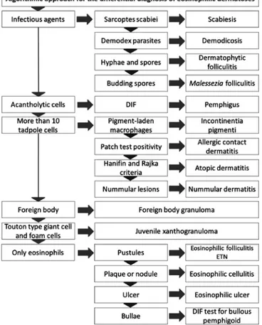
Figure 1. Algorithmic approach for eosinophilic dermatosis diagnoses according to the Tzanck smear test and clinical and laboratory findings
ETN: Erythema toxicum neonatorum, DIF: Direct immunofluorescence test
Results
Between January 2010 and January 2015, we evaluated 45.496 patients in our outpatient dermatology clinic, and performed Tzanck smear test in 3.214 patients (7%) who could not be confidently diagnosed solely by clinical evaluation.
Abundant eosinophils were observed in 95 patients. Of those 95 patients, seven were excluded from this study because a definitive diagnosis could not be made. From 3.214 patients, this study included 88 patients [2.7%; 53 women (60.2%) and 35 men (39.8%)] in whom Tzanck smear test was performed over a five-year period. Patients were between one month and 82 years old (mean, 32 years). Of the lesions, 22 lesions (25%) were vesicular, 21 lesions (23.9%) were erosive, 21 lesions (23.9%) were pustular, 14 lesions (15.9%) were bullous, five lesions (5.6%) were nodular, three lesions (3.4%) were ulcerative, and two lesions (2.3%) were plaque. The number of skin diseases,
lesion types, and characteristic cytologic findings are shown in Table 1.
To determine a final diagnoses in our study patients, after laboratory examinations were performed, 36 patients (40.9%) received histopathologic examination, 36 patients (40.9%) received patch test, 13 patients (17.8%) received direct and indirect immunofluorescence tests, and 10 patients (11.4%) had fungal cultures.
According to the final diagnoses in the 88 study patients, eosinophilic lesions in 44 patients (50%) were diagnosed as spongiotic dermatitis, lesions in 17 patients (19.3%) were diagnosed as infectious diseases, lesions in 13 patients (14.8%) were autoimmune bullous disorders, lesions in 11 patients (12.5%) were idiopathic eosinophilic diseases, and lesions in 3 patients (3.4%) were granulomatous diseases.
Findings in Patients with Spongiotic Dermatitis
Cytologic examination identified 44 patients (50%) of 88 with spongiotic dermatitis (21 with allergic contact Table 1. Number of skin diseases, lesion types, characteristic cytologic findings, and final diagnostic methods
Skin disease Number of patients Lesion types (n) Other cytologic findings Diagnostic method Spongiotic dermatitis 44
Allergic contact dermatitis 21 Vesicle (8)
erosion (7) bulla (6)
Tadpole cells Patch test with the European
standard
Atopic dermatitis 16 Erosion (9) vesicle (7) Tadpole cells and mast cells Hanifin and Rajka criteria
Nummular dermatitis 5 Vesicle (5) Tadpole cells Nummular plaque
Incontinentia pigment 2 Vesicle (2) Tadpole cells and pigment incontinence Histopathology
Infectious diseases 17
Malassezia folliculitis 6 Pustule (6) Budding spores Fungal culture
Demodicosis 5 Pustule (5) Demodex parasites Histopathology
Dermatophytic folliculitis 4 Pustule (4) Hyphae and spores Fungal culture
Scabies 2 Nodule (2) Sarcoptes scabiei Potassium hydroxide
examination
Autoimmune bullous disorders 13
Bullous pemphigoid 8 Erosion (2)
bulla (6) A few tadpole cells Histopathology, direct and indirect immunofluorescence tests
Pemphigus 5 Erosion (3)
bulla (2) Acantholytic cells
Idiopathic eosinophilic diseases 11
Erythema toxicum neonatorum 4 Pustule (4) Clinical follow up and
cytology
Eosinophilic ulcer 3 Ulcer (3) Histopathology
Eosinophilic folliculitis 2 Pustule (2) Histopathology
Eosinophilic cellulitis 2 Plaque (2) Histopathology
Granulomatous diseases 3
Foreign body granuloma 2 Nodule (2) Foreign body giant cells and foreign
body Histopathology
Juvenile xanthogranuloma 1 Nodule (1) Touton-type giant cells and foam cells Histopathology
dermatitis, 16 with atopic dermatitis, five with nummular dermatitis, and two with incontinentia pigment) (Figure 2a). Tzanck smears showed more than 10 tadpole-like cells (at x100 magnification) in all lesions of spongiotic dermatitis (Figure 2b). Cytology revealed plenty eosinophils (Figure 2c, 2d). Pigment-laden macrophages and melanocytes were detected in two lesions of incontinentia pigment (Figure 2e, 2f). Histopathologic examinations of these two patients confirmed incontinentia pigment. Patch testing with the European standard series showed positivity in 21 patients of allergic contact dermatitis. Hanifin and Rajka criteria were positive in 16 patients of atopic dermatitis, but these criteria and patch tests were negative in five patients of nummular dermatitis.
Findings in Patients with Infectious Diseases
In patients with infectious causes, abundant footprint-shaped spores adhering to keratinocytes were observed in six lesions of Malassezia folliculitis (Figure 3a, 3b).
Papulopustular lesions were located on the back of three patients and on the face of three patients. All patients with
Malassezia folliculitis also had pruritus. Malassezia globosa
was identified in fungal culture. Patients with Malassezia
folliculitis were treated with oral itraconazole capsule (200
mg/d for two wk). Papulopustular lesions and pruritus improved completely.
Eosinophilic dermatophytic folliculitis could be detected by cytologic examination in 4 patients. Fungal culture identified anthropophilic Trichophyton rubrum in four patients with dermatophytic folliculitis. Pustular lesions were located on the groin areas of three patients and on the leg of one patient (Figure 3c). In two patients, the lesions were localized in erythematous patches or plaques. Cytology showed neutrophils, eosinophils, hyphae, and spores in four lesions of dermatophytic folliculitis (Figure 3d). The patients with dermatophytic folliculitis were treated with terbinafine tablets (250 mg/d for four wk).
Demodicosis was identified in papulopustular lesions of five patients. The lesions were located on the face areas of all patients (Figure 3e). The specimen stained with May-Grunwald-Giemsa showed unstained Demodex parasites with four pairs of legs. Acid-fast test was positive for these parasites in a specimen stained with Ehrlich-Ziehl-Neelsen (Figure 3f). Lesions disappeared with two-month treatment with permethrin 5% cream.
Abundant eosinophils were detected in two lesions of nodular scabies (Figure 3g). KOH examination confirmed the scabies diagnoses (Figure 3h). Lesions disappeared with permethrin 5% cream.
Findings in Patients with Autoimmune Bullous Diseases
Abundant eosinophils and acantholytic cells were detected in five patients with pemphigus (3 with pemphigus vulgaris and 2 with pemphigus herpetiformis) (Figure 4a, 4b). Direct immunofluorescence on smears showed immunoglobulin G deposition around the cells in five lesions of pemphigus. Complete blood count of all patients with pemphigus revealed mild hypereosinophilia (ranging from 6%-9%). Diagnoses showed eight patients with bullous pemphigoid, one of them was infantile bullous pemphigoid due to vaccination (combined tetanus, diphtheria, Bordetella pertussis, and poliovirus), and others were adult patients (Figure 4c). Cytology revealed a few tadpole-like cells and abundant eosinophils without acantholytic cell (Figure 4d). Complete blood count of one patient with infantile bullous pemphigoid revealed leukocytosis (43.000 cells/mL) and intense hypereosinophilia (41%). In adult patients with bullous pemphigoid, complete blood count showed mild hypereosinophilia (ranging from 8%-11%). Histopathologic examination and direct and indirect immunofluorescence tests were compatible with bullous pemphigoid.
Idiopathic Eosinophilic Diseases
In 4 neonatal patients, facial and truncal pustular lesions were detected (Figure 5a). Cytology revealed only eosinophils, and erythema toxicum neonatorum was considered (Figure 5b). Pustular lesions resolved spontaneously within five to 10 days.
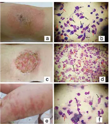
Figure 2. Clinical and cytologic findings of patients with spongiotic dermatoses, 2a. Allergic contact dermatitis due to topical antibiotics (nitrofurazone), 2b. Tzanck smear test result of vesicular lesion shows tadpole-like cells (black arrows) and eosinophils (red arrows), 2c. Nummular erythematous crusted plaque on the leg, 2d. Tzanck smear test of nummular dermatitis reveals tadpole-like cells (black arrows) and numerous eosinophils (red arrows), 2e. Vesicular lesions on the leg associated with incontinentia pigment, 2f. Cytologic examination of vesicular lesion due to incontinentia pigment shows tadpole-like cell (black arrow), eosinophil (red arrow), and melanocyte (green arrow), May-Grunwald-Giemsa stain used in b, d, and f (original magnification, x1000)
Eosinophilic ulcers were detected in three patients. These painful ulcerative lesions were located on the lip of two patients and on the perianal region of one patient (Figure 5c). Cytology showed only dense accumulation of eosinophils without infectious agent (Figure 5d). Diagnoses of eosinophilic ulcers were confirmed by histopathologic
examination. Complete blood count of two patients with eosinophilic ulcers revealed hypereosinophilia (at 11% and 13%). Lesions resolved with topical corticosteroid cream within two to four weeks.
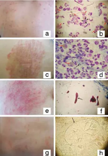
Figure 3. Clinical and cytologic findings of patients with infectious diseases, 3a. Erythematous papules and pustules on the back due to Malassezia folliculitis, 3b. Cytology shows budding/footprint-shaped Malassezia spores (black arrows) and eosinophils (red arrows) in a patient with Malassezia folliculitis, 3c. Multiple pustules, scales, and crusts on an erythematous background on the thigh of a patient with dermatophytic folliculitis, 3d. Tzanck smear test reveals hyphae (black arrows) and eosinophil (red arrows) in a patient with Trichophyton rubrum folliculitis, 3e. Multiple follicular pustules on an erythematous background on the face of a patient with Demodex folliculitis, 3f. Demodex parasite in the patient with Demodex folliculitis, 3g. Vesicles and pustules on erythematous background on the back of patient with scabies, 3h. Potassium hydroxide examination shows larvae of Sarcoptes scabiei in the egg, May-Grunwald-Giemsa stain used in b and d (original magnification, ×1000); h has original magnification of ×400, Ehrlich-Ziehl-Neelsen stain used in f (original magnification, ×100)
Figure 4. Clinical and cytologic findings of patients with pemphigus and bullous pemphigoid, 4a. Herpetiform vesicles on the lumbar region in patient with pemphigus herpetiformis, 4b. Tzanck smear test reveals acantholytic cells (black arrows) and eosinophils (red arrows), 4c. Vesicles and bullae on the erythematous background in a patent with infantile bullous pemphigoid, 4d. Cytology shows numerous eosinophils (arrows), May-Grunwald-Giemsa stain used in b and d (original magnification, ×1000)
Figure 5. Clinical findings of patients with idiopathic eosinophilic dermatoses, 5a. Multiple pustules on an erythematous background on the abdominal region of a patient with erythema toxicum neonatorum, 5b. Perianal eosinophilic ulcer, 5c. Vesicles, pustules, and yellow crusts on scalp of a patient with infantile eosinophilic folliculitis, 5d. Erythematous and indurated and excoriated plaque on the leg of a patient with eosinophilic cellulitis
Pustular lesions on the scalp, trunk, and extremities were detected in two pediatric patients with eosinophilic folliculitis. There was no history of drug use. Viral serology tests for hepatitis C virus, toxocariasis, and human immunodeficiency virus infection were negative. Cytology showed abundant eosinophils, a few tadpole-like cells without any infectious agent, and acantholytic cell. Lesions completely resolved after treatment with topical corticosteroids within three to four weeks.
Eosinophilic cellulitis could be detected by cytology in two patients, both confirmed histopathologically. Complete blood count of two patients revealed hypereosinophilia (16% and 12%). The lesions were located on the leg of one patient and on the thigh of the other patient. Lesions disappeared with treatment with topical corticosteroids and oral indomethacin.
Granulomatous Diseases
This study included 2 patients with foreign body granulomas, with 1 due to postoperative suture on the sternum and the other associated with stinging thorns (Figure 6a). Cytology showed granulomas, eosinophils, and foreign materials in foreign body giant cells and extracellularly (Figure 6b). Histopathology confirmed the diagnosis of foreign body granuloma.
In one patient with a solitary nodular lesion, cytology showed eosinophils, foamy cells, foreign body, and Touton-type giant cells (Figure 6c, 6d). Diagnosis of juvenile xanthogranuloma was confirmed histopathologically.
Discussion
Eosinophils can infiltrate various tissues and can lead to different clinical findings. Eosinophilic skin infiltration develops in many diseases and is commonly called eosinophilic dermatoses. Histopathologic examination is often used for differential diagnoses (1). However, histopathologic examination may pose a diagnostic challenge for dermatopathologists as an identical histologic picture can be produced by several different causes. In such cases, cytology can be useful for diagnosis (5,9-11). In this study, we presented the cytologic findings of 88 patients with eosinophilic dermatoses.
In half of patients with eosinophilic dermatoses, the diagnosis was spongiotic dermatitis. For most patients (95.5%) with spongiotic dermatitis, the diagnosis showed different forms of dermatitis. The presence of more than 10 tadpole-like cells (at x100 magnification) is highly specific (99.3%-100%) for acute spongiotic dermatitis (6,12). Patients with acute spongiotic dermatitis not only develop contact dermatitis, but spongiotic dermatitis can occur in many inflammatory diseases such as dyshidrotic dermatitis, atopic dermatitis, reactions, drug eruptions, insect bite reactions, incontinentia pigment, and dermatophyte infections. In addition to cells having tadpole-like cells and presence of eosinophils, pigment-laden macrophages and melanocytes were detected in only 2 patients with incontinentia pigment. Patch test for allergic contact dermatitis, KOH examination for dermatophyte infections, drug history for drug allergy, and Hanifin and Rajka criteria for atopic dermatitis are important techniques for the differentiation of dermatitis (1).
In this study, another important cause of cutaneous eosinophilic infiltration was fungal and parasitic infections (19.3%). Infectious eosinophilic folliculitis can occur with scabies, Demodex, Pseudomonas infections, and larva migrans, Malassezia species, and in dermatophytic folliculitis, especially Trichophyton rubrum as in our patients (9-11,13,14). A definitive diagnosis of cutaneous infectious diseases should be based on detecting the cause through cytology, histopathologic examination, culture techniques, or molecular diagnostic methods such as polymerase chain reaction. If infectious causes cannot be readily identified, histopathology can mimic eosinophilic folliculitis. Most causes of infectious folliculitis can be detected with careful cytologic examination. Demodex and scabies parasites usually fall during fixation on routine cytologic examination. For this reason, KOH examination should also be performed (5). In this study, May-Grunwald-Giemsa-stained specimens showed only eosinophils in patients with scabies; however, KOH examination revealed Sarcoptes scabiei parasites.
In autoimmune blistering diseases, pemphigus and bullous pemphigoid may cause eosinophilic infiltration. Cytology is very important to distinguish between the two autoimmune blistering diseases (15). In pemphigus, cytology reveals numerous single or loosely adherent clumps of acantholytic cells (16). If the test is positive for acantholytic cells, direct immunofluorescence test on smear can be performed (17). In our study, five of 13 autoimmune bullous lesions were
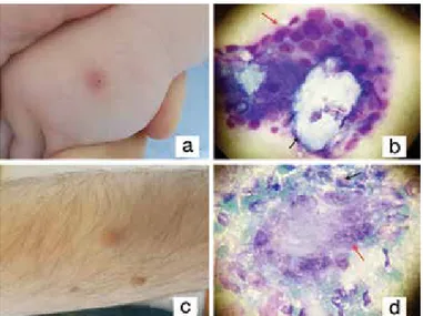
Figure 6. Clinical and cytologic findings of patients with granulomatous eosinophilic dermatoses, 6a. Erythematous nodule on the hand of a patient with foreign body granuloma, 6b. Tzanck smear test reveals foreign body (black arrow) and foreign body giant cell (red arrow), 6c. Yellowish nodular lesion on the arm of a patient with juvenile xanthogranuloma, 6d. Cytology of juvenile xanthogranuloma shows Touton-type giant cell (red arrow) and eosinophil (black arrow) May-Grunwald-Giemsa stain used in b and d (original magnification, x1000)
pemphigus. Direct immunofluorescence on smear showed immunoglobulin G deposition around the acantholytic cells.
This study included 11 patients with idiopathic eosinophilic dermatoses. Idiopathic eosinophilic dermatoses included erythema toxicum neonatorum, eosinophilic ulcer, eosinophilic folliculitis, and eosinophilic cellulitis. Although eosinophilic infiltration was seen in all patients with idiopathic eosinophilic dermatoses, their clinical manifestations were different. There are 3 forms of the eosinophilic folliculitis: the classic type, the infantile type, and the immunosuppression-associated type (1). In our study, 17 of 19 lesions of eosinophilic folliculitis were infectious; the remaining two patients had infantile eosinophilic folliculitis. Eosinophilic folliculitis may also develop secondary to hepatitis C virus infection, toxocariasis, drug, and vaccine use (18-23). Viral serology and drug use history were negative in our 2 cases with infantile eosinophilic folliculitis.
Granulomatous group included foreign body granuloma and juvenile xanthogranuloma. Diagnosis of these patients can be made by cytologic analyses. Foreign body granuloma shows granuloma formation, foreign body-type giant cells, eosinophils, and foreign body materials (24). On the other hands, cytology reveals histiocytic cells with lipid-like vacuoles, and Touton-type multinucleated giant cells in juvenile xanthogranuloma (25). In early stages of juvenile xanthogranuloma, abundant eosinophils can be seen as in our case (1).
We propose an algorithmic approach for eosinophilic dermatoses that is essentially based on the cytologic findings that we observed and the specific Tzanck smear findings reported previously (Figure 1). With the Tzanck smear test and the algorithmic approach that we propose, dermatologists can diagnose many eosinophilic skin diseases such as spongiotic dermatitis, infectious eosinophilic dermatoses, pemphigus, foreign body granuloma, and juvenile xanthogranuloma.
Study Limitation
Limitations of this study include the retrospective study design and inclusion of patients from only 1 treatment center. In our dermatology clinic, we do not perform Tzanck smear tests for urticarial lesions, maculopapular eruptions, and vasculitic lesions. For this reason, this study did not include pruritic urticarial papules and plaques of pregnancy, maculopapular drug eruptions, and cutaneous vasculitis.
Conclusion
The Tzanck smear test is a cost-effective, simple, and rapid test for outpatient dermatology practices. Along with its range of use in a number of skin diseases, dermatologists may also benefit from using this valuable test in the clinical diagnosis of certain eosinophilic skin lesions.
Ethics
Ethics Committee Approval: This study was approved by the Başkent University Institutional Review Board (Project No. KA16/86), Informed Consent: It was taken.
Peer-review: Internally peer-reviewed.
Financial Disclosure: The authors declared that this study received no financial support.
References
1. Long H, Zhang G, Wang L, et al. Eosinophilic Skin Diseases: A Comprehensive Review. Clin Rev Allergy Immunol 2016;50:189-213.
2. Nahm JH, Yoon G, Do SI, et al. Squash smear cytology of Langerhans cell histiocytosis. Int J Clin Exp Pathol 2015;8:7998-8007.
3. Das DK, Sheikh ZA, Alansary TA, et al. A case of Langerhans’ cell histiocytosis associated with Hodgkin’s lymphoma: Fine-needle aspiration cytologic and histopathological features. Diagn Cytopathol 2016;44:128-32.
4. Van Praag MC, Van Rooij RW, Folkers E, et al. Diagnosis and treatment of pustular disorders in the neonate. Pediatr Dermatol 1997;14:131-43. 5. Durdu M, Ilkit M. First step in the differential diagnosis of folliculitis: cytology.
Crit Rev Microbiol 2013;39:9-25.
6. Durdu M, Baba M, Seçkin D. The value of Tzanck smear test in diagnosis of erosive, vesicular, bullous, and pustular skin lesions. J Am Acad Dermatol 2008;59:958-64.
7. Durdu M, Baba M, Seçkin D. More experiences with the Tzanck smear test: cytologic findings in cutaneous granulomatous disorders. J Am Acad Dermatol 2009;61:441-50.
8. Durdu M, Baba M, Seçkin D. Dermatoscopy versus Tzanck smear test: a comparison of the value of two tests in the diagnosis of pigmented skin lesions. J Am Acad Dermatol 2011;65:972-82.
9. Cannavo SP, Guarneri F, Guarneri C. Strongyloidiasis histologically mimicking eosinophilic folliculitis. Eur J Dermatol 2004;14:264-5.
10. Opie KM, Heenan PJ, Delaney TA, et al. Two cases of eosinophilic pustular folliculitis associated with parasitic infestations. Australas J Dermatol 2003;44:217-9.
11. Dyall-Smith D, Mason G. Fungal eosinophilic pustular folliculitis. Australas J Dermatol 1995;36:37-8.
12. Pariser RJ. Diagnosis of spongiotic vesicular dermatitis by Tzanck smear: the “tadpole cell”. J Am Acad Dermatol 1983;8:519-22.
13. Nara T, Katoh N, Inoue K, et al. Eosinophilic folliculitis with a Demodex folliculorum infestation successfully treated with ivermectin in a man infected with human immunodeficiency virus. Clin Exp Dermatol 2009;34:e981-3.
14. Brenner S, Wolf R, Ophir J. Eosinophilic pustular folliculitis: a sterile folliculitis of unknown cause? J Am Acad Dermatol 1994;31:210-2.
15. Ruocco V, Ruocco E. Tzanck smear, an old test for the new millennium: when and how. Int J Dermatol 1999;38:830-4.
16. Ruocco E, Brunetti G, Del Vecchio M, et al. The practical use of cytology for diagnosis in dermatology. J Eur Acad Dermatol Venereol 2011;25:125-9. 17. Durdu M, Seçkin D, Baba M. The Tzanck smear test: rediscovery of a practical
diagnostic tool. Skinmed 2011;9:23-32.
18. Gesierich A, Herzog S, Grunewald SM, et al. Eosinophilic folliculitis in a Caucasian patient: association with toxocariasis? J Eur Acad Dermatol Venereol 2006;20:1317-21.
19. Gul U, Kilic A, Demiriz M. Eosinophilic pustular folliculitis: the first case associated with hepatitis C virus. J Dermatol 2007;34:397-9.
20. Kaufmann D, Pichler W, Beer JH. Severe episode of high fever with rash, lymphadenopathy, neutropenia, and eosinophilia after minocycline therapy for acne. Arch Intern Med 1994;154:1983-4.
21. Kimura K, Ezoe K, Yokozeki H, et al. A case of eosinophilic pustular folliculitis (Ofuji’s disease) induced by patch and challenge tests with indeloxazine hydrochloride. J Dermatol 1996;23:479-83.
22. Ooi CG, Walker P, Sidhu SK, et al. Allopurinol induced generalized eosinophilic pustular folliculitis. Australas J Dermatol 2006;47:270-3. 23. Roos TC, Albrecht H. Foscarnet-associated eosinophilic folliculitis in a
patient with AIDS. J Am Acad Dermatol 2001;44:546-7.
24. Chung YE, Kim EK, Kim MJ, et al. Suture granuloma mimicking recurrent thyroid carcinoma on ultrasonography. Yonsei Med J 2006;47:748-51. 25. Barroca H, Farinha NJ, Lobo A, et al. Deep-seated congenital juvenile
xanthogranuloma: report of a case with emphasis on cytologic features. Acta Cytol 2007;51:473-6.