CASE REPORT – OPEN ACCESS
International Journal of Surgery Case Reports 8 (2015) 124–126Contents lists available atScienceDirect
International Journal of Surgery Case Reports
j o u r n a l h o m e p a g e :w w w . c a s e r e p o r t s . c o mCombined use of alveolar distraction osteogenesis and segmental
osteotomy in anterior vertical ridge augmentation
Elif Öncü
a,∗,1, Kubilay Isik
b,1, E. Emine Alaaddino˘glu
c,2, Sina Uc¸kan
d,2 aNecmettin Erbakan University, Departmant of Periodontolgy, Karaci˘gan Mah, Ankara Cad. No. 74/A Karatay, Konya, TurkeybNecmettin Erbakan University, Departmant Of Oral And Maxillofacial Surgery, Karaci˘gan Mah., Ankara Cad. No. 74/A Karatay, Konya, Turkey cBaskent University, Departmant Of Periodontolgy, Bahc¸elievler mah. No. 16 C¸ankaya, Ankara, Turkey
dBaskent University, Departmant Of Oral And Maxillofacial Surgery, Bahc¸elievler mah. No. 16 C¸ankaya, Ankara, Turkey
a r t i c l e i n f o
Article history:
Received 2 December 2014
Received in revised form 22 January 2015 Accepted 23 January 2015
Available online 28 January 2015 Keywords:
Dental implants Distraction osteogenesis Segmental osteotomy Vertical ridge augmentation Connective tissue graft
a b s t r a c t
INTRODUCTION: Vertical defects of the anterioral veolar ridge are challenging cases in implant dentistry. Various techniques, such as onlay bone grafting, segmental osteotomy (SO) oral veolar distraction osteo-genesis (ADO), have been suggested to manage those situations. ADO has an advantage of being capable of enhancing both hard and soft tissue simultaneously.
PRESENTATION OF CASE: One of the possible complications of ADO is rotation ortilting the transport segment (TS). In this report, we present a 30-year old woman who had a severe anterior vertical deficiency. ADO was started to manage the case, but advancement of the TS lagged on the left side and the segment rotated. A SO was planned and the lagged side was corrected. Two years after the surgery, hard and soft tissue gains were found to be preserved.
DISCUSSION: Vertical alveolar bone deficiencies are challenging cases for dental implantology. Alveolar DO promotes soft tissue along with hard tissue, and the bone regeneration process and shows lower infection rates and greater stability over the long term. However, the technique has some disadvantages and can lead to complications, such as breaking of the distraction device, nerve injury or paresthesia, fracture of transport bone, hematoma, wound dehiscence, severe bleeding, and even jaw fractures. Deviation of the TS from the distraction path is another undesired situation. The rigidity of the device, the width of the mucosa, the volume of the transport and anchor segments, and the amount of augmentation can affect vector deviation.
CONCLUSION: We suggest that SO can be used in similar cases in which TS could not be distracted on a straight vector line.
© 2015 Published by Elsevier Ltd. on behalf of Surgical Associates Ltd. This is an open access article under the CC BY-NC-ND license (http://creativecommons.org/licenses/by-nc-nd/4.0/).
1. Introduction
In many cases, reconstruction of oral hard and soft tissue defi-ciencies aims to achieve both an adequate soft and hard tissue profile, in which dental implants could be placed later. Horizon-tal defects of the maxilla and mandible can generally be treated with traditional grafting techniques, with predictable results. Ver-tical defects, however, tend to have a higher risk for complications, such as soft tissue dehiscence, infection, inadequate bone volume, and loss of the graft. Numerous techniques have been advocated to
∗ Corresponding author. Tel.: +90 5053468207.
E-mail addresses:[email protected](E. Öncü),[email protected](K. Isik), cirit [email protected](E.E. Alaaddino˘glu),cirit [email protected](S. Uc¸kan).
1 Tel.: +332 220 00 26; fax: +332 220 00 45. 2 Tel.: +312 222 25 26.
solve the functional and esthetic problems, such as onlay grafting, bone regeneration and alveolar distraction as well[1]. Frequently, soft tissue contours are more important for the patient[1].
Distraction osteogenesis (DO) is a surgical technique used for new bone formation between bone segments that are gradually separated by incremental traction. When distraction forces are applied to the callus tissues between bone segments, the tension stimulates new bone formation parallel to the vector of distraction. A distraction device is used to gradually transport the mobilized bone segment. When the desired repositioning is achieved, the distraction device is left in place for an inactive period to act as a fixation tool[1].
DO for the craniofacial skeleton have been popularized by Chin and McCarthy[2,3]. Movement of the bone results in expansion of the soft tissue adjacent to the bone segment. Thus, the entire piece is enlarged in a single, simultaneous process, which is a major benefit. Other advantages are minimal risk for infection, relatively short http://dx.doi.org/10.1016/j.ijscr.2015.01.038
2210-2612/© 2015 Published by Elsevier Ltd. on behalf of Surgical Associates Ltd. This is an open access article under the CC BY-NC-ND license (http://creativecommons.org/licenses/by-nc-nd/4.0/).
Downloaded for Anonymous User (n/a) at Baskent University from ClinicalKey.com by Elsevier on June 24, 2019. For personal use only. No other uses without permission. Copyright ©2019. Elsevier Inc. All rights reserved.
CASE REPORT – OPEN ACCESS
E. Öncü et al. / International Journal of Surgery Case Reports 8 (2015) 124–126 125
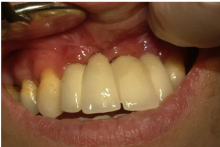
Fig. 1. Maxillary four incisors were found to be grade 3 mobile according to Miller’s
classification.
treatment time, little or no resorption of bone, and fairly predictable results[1].
In DO technique, a minimum of 6–7 mm of bone height above vital structures, such as neurovascular bundles or sinus cavity is needed[1]. In some cases, in which distraction is used, additional bone grafting or soft tissue augmentation, either before or after the procedure, may be required.
2. Presentation of case
The patient was a medically healthy, non-smoker, 30-year old woman who had a generalized aggressive periodontitis. Her periodontal treatment was completed and all pockets were elim-inated, but maxillary four incisors were found to be grade 3 mobile according to Miller’s classification (Fig. 1). We planned to extract these teeth and to rehabilitate the patient with dental implants. However, there was a vertical hard tissue deficit that needed to be improved. Thus, we intended to use alveolar DO for this augmentation.
Extraction of the anterior four teeth, which had very little hard tissue support but also were no longer periodontally diseased, was delayed to maintain the attached gingiva and they were endodon-tically treated. The placement of the distractor (MODUS, MDO 1.5; Medartis, Basel, Switzerland) was done under local anesthesia. A full thickness mucoperiosteal flap was reflected. The device was adapted to the operative site and then it was pre-fixed by two tita-nium screws on each side. After the osteotomy line was traced, a trapezoidal osteotomy was done using a surgical saw between maxillary canines. Special precaution was taken to preserve the palatal soft tissue for adequate blood supply. After the osteotomy, the distractor was fixed using 1.5-mm screws, and the mobility and path of the transport bone segment were confirmed through the activation of the distractor’s rod. Due to anatomical restrictions,
Fig. 2. Lag of the TS on the left side. Please note that the anterior teeth had been
abraded.
Fig. 3. Three months later from implant surgery, there were no radiographic
com-plications. The patient also received maxillary premolar and molar implants.
the distractor was placed on the right maxillary canine region, not in the midline. Right arm of the lower distractor plates (i.e., the one on the moving segment’s side) was cut off in order to make the device fit into its place.
After a latency period of one week after surgery, distraction pro-cess was initiated at a daily rate of 1 mm (two activations of 0.5 mm) according to the manufacturer’s recommendation. During the dis-traction period, the patient was invited weekly and the teeth on the transport segment (TS) were abraded with burs to avoid premature contacts with opposite teeth.
After one month, it was visible that the advancement of the TS had lagged on the left side (Fig. 2). Thus, the area was accessed again and the osteotomy on the left side was re-created. The left side of the segment was mobilized, approximately 4 mm advanced and fixed with one L-shaped miniplate. Since the right side did not reach to the desired point yet, the distraction process was continued for eight weeks in the right side. At the end of the distraction, the device was left for eight weeks ensure the consolidation of hard tissue, and then the distractor and the miniplates were removed.
One month later, four maxillary anterior teeth were extracted, as it had been planned at the beginning of the treatment. Since the teeth did not have a sound hard tissue support, no consider-able extraction defects were created. At the same time, two dental implants (AstraTechTM, Sweden) were placed into the sockets of right lateral and left central teeth, in where most available alveolar bone was present.
The implants were left to integrate for three months (Fig. 3). However, in the second stage surgery it was seen that the right implant exposed through the soft tissue. The area was surgically exposed and a bone defect of several millimeters on the buccal
Fig. 4. The implants were loaded after six months with a fixed prosthetic restoration.
Downloaded for Anonymous User (n/a) at Baskent University from ClinicalKey.com by Elsevier on June 24, 2019. For personal use only. No other uses without permission. Copyright ©2019. Elsevier Inc. All rights reserved.
CASE REPORT – OPEN ACCESS
126 E. Öncü et al. / International Journal of Surgery Case Reports 8 (2015) 124–126 side was found. It was repaired with a hard tissue graft(Osseo-biol TecnossTM, Italy) a resorbable collagen membrane (Osseobiol TecnossTM, Italy) and a free soft tissue graft harvested from the palatinal area. The implants were loaded after six months with a fixed prosthetic restoration (Fig. 4).
Two years later, there were no clinical or radiographic com-plications. The implants were stabile, hard and soft tissue gains were maintained and there were not any discrepancies between two sides of the anterior segment.
3. Discussion
Vertical alveolar bone deficiencies are challenging cases for den-tal implantology. They create esthetic and functional problems associated with increasing crown-to-implant ratio of the prosthe-sis. A range of surgical techniques have been suggested to address these problems, including guided bone regeneration, autogenous particulate or block bone grafts, and DO[4].
Since McCarthy et al.[5]first used DO in the craniofacial area, alveolar DO has been extensively employed in vertically insufficient alveolar ridges[2]. It allows the improvement of vertical defects measuring up to 15 mm[6]. Moreover, it also promotes soft tis-sue along with hard tistis-sue[6], and the bone regeneration process using alveolar DO shows lower infection rates and greater stability over the long term[7]. However, the technique has some disadvan-tages and can lead so some complications, such as breaking of the distraction device, nerve injury or paresthesia, fracture of transport bone, hematoma, wound dehiscence, severe bleeding, and even jaw fractures[8].
Deviation of the TS from the distraction path is another unde-sired situation. The rigidity of the device, the width of the mucosa, the volume of the transport and anchor segments, and the amount of augmentation can affect vector deviation[8]. In our case, most plausible reason can be failing the placement of the device on middle of the TS. Although we made the distraction path fairly parallel to the midline, applying the force away from the center could have rotated the TS. Another explanation might be that the soft tissue was thicker or more rigid on the left side and that hin-dered the segment’s advancement. It could also be claimed that the osteotomies on the left side were not complete and probably that hampered the advancement of the segment. However, the TS was fully mobilized during the distraction osteotomies and the TS was not immobile during the whole distraction process. The lag at the left side occurred only after some advancement had already been observed.
This combined use of DO and osteotomy could be critized that if the left side was able to been brought into a proper position using an osteotomy, so was the distraction process unnecessary? Could the case be managed from the start by employing only an osteotomy and advancement? The point here is that although the left side had lagged, it still made progress and expanded some keratinized gingival cover. This improvement and soft tissue gain made the surgical advancement of the left side possible, which might not be achievable otherwise.
In the beginning of the treatment, if we happened to consider the rotation of the TS quite likely, then we might plan to place two distractors (i.e., one device on the right side and another one on the left side). Another option could be using an intraosseous distractor. Since an intraosseous distractor is a more delicate device, it would allow to be placed in the center of the TS.
4. Conclusion
During the alveolar DO, if the TS shows an undesired rotation, a surgical correction as described above can be made, as long as soft tissue envelope permits this second operation.
Conflicts of interest
All co-authors have seen and agree with the contents of the manuscript and there is no financial interest to report. We certify that the submission is original work and is not under review at any other publication.
Funding
This case study has not a sponsor. We did not need a funding.
Ethical approval
This is a case report and ethics committee approval is not required for this paper.
Author contribution
Elif Öncü, Kubilay Is¸ik, Sina Uc¸kan, E. Emine Alaaddino˘glu.
Consent
Written informed consent was obtained from the patient for publication of this case report and accompanying images. A copy of the written consent is available for review by the Editor-in-Chief of this journal on request.
Guarantor
Elif Öncü.
References
[1] S.C. Bagheri, R.B. Bell, H.A. Khan, et al., Dental implant prosthetic rehabilitation: vertical distraction osteogenesis, in: S.C. Bagheri, R.B. Bell, H.A. Khan (Eds.), Current Therapy in Oral and Maxillofacial Surgery, Elsevier Saunders, 2012, pp. 163–166.
[2] M. Chin, B.A. Toth, Distraction osteogenesis in maxillofacial surgery using internal devices: review of five cases, J. Oral Maxillofacial Surg. Off. J. Am. Assoc. Oral Maxillofacial Surg. 54 (1996) 45–53 (Discussion 54). [3] J.G. McCarthy, E.J. Stelnicki, B.J. Mehrara, M.T. Longaker, Distraction
osteogenesis of the craniofacial skeleton, Plastic Reconstr. Surg. 107 (2001) 1812–1827.
[4] J.W. Kim, M.H. Cho, S.J. Kim, M.R. Kim, Alveolar distraction osteogenesis versus autogenous onlay bone graft for vertical augmentation of severely atrophied alveolar ridges after 12 years of long-term follow-up, Oral Surg. Oral Med. Oral Pathol. Oral Radiol. 116 (2013) 540–549.
J.G. McCarthy, J. Schreiber, N. Karp, C.H. Thorne, B.H. Grayson, Lengthening the human mandible by gradual distraction, Plastic Reconstr. Surg. 89 (1992) 1–8 (Discussion 9–10).
[6] M. Chiapasco, U. Consolo, A. Bianchi, P. Ronchi, Alveolar distraction osteogenesis for the correction of vertically deficient edentulous ridges: a multicenter prospective study on humans, Int. J. Oral Maxillofacial Implants 19 (2004) 399–407.
[7] M. Chiapasco, M. Zaniboni, L. Rimondini, Autogenous onlay bone grafts vs. alveolar distraction osteogenesis for the correction of vertically deficient edentulous ridges: a 2–4-year prospective study on humans, Clin. Oral Implants Res. 18 (2007) 432–440.
[8] F. Ugurlu, B.C. Sener, G. Dergin, H. Garip, Potential complications and precautions in vertical alveolar distraction osteogenesis: a retrospective study of 40 patients, J. Cranio–Maxillo–Facial Surg. Off. Publ. Eur. Assoc.
Cranio–Maxillo–Facial Surg. 41 (2013) 569–573.
Open Access
This article is published Open Access atsciencedirect.com. It is distributed under theIJSCR Supplemental terms and conditions, which permits unrestricted non commercial use, distribution, and reproduction in any medium, provided the original authors and source are credited.
Downloaded for Anonymous User (n/a) at Baskent University from ClinicalKey.com by Elsevier on June 24, 2019. For personal use only. No other uses without permission. Copyright ©2019. Elsevier Inc. All rights reserved.