Monitoring of the immune response to B. abortus S19 conjunctival
vaccine in cattle
Oktay GENÇ
1, Gülnur SERDAR
2, Özlem BÜYÜKTANIR YAŞ
1, Evrim GENÇ
11Department of Microbiology, Faculty of Veterinary Medicine, Ondokuz Mayıs University, Kurupelit, 55200, Samsun, Turkey; 2Samsun Veterinary Control Institute, Samsun, Turkey.
Summary: Brucella abortus S19strain is one of the most preferred strains in vaccines against brucellosis in cattle. However, monitoring of the B. abortus S19 vaccine is difficult due to non availability of sustainable immunoreactive antigen and accurate test method. In this study, the humoral and the cellular immune responsesto S19 vaccine in one year old heifers and calves were monitored on post vaccination days (pvd) of 46, 85 and 169. Thus, the levels of Immunoglobulin (Ig)G and IgA isotypes against lipopolysaccharide (LPS) for humoral immunity and interferon gamma (IFNg) against brucellergen for cellular immune response were investigated by home-made ELISAs. In this study, significant IgG positivity was observed on pvd 46 in calves (100%) and heifers (96.6%), but IgA positivity and IFNg levels were not over 50%. Moreover, percentage of positive animals for IFNg (13.3-43.3%) and IgA (0-44%) have shown no significance for monitoring the vaccine throughout the study. Therefore, IgG levels can be used to monitor the efficiency of
Brucella abortus S19 conjunctival vaccine in cattle. On the other hand, novel antigen combinations along with brucellergen and LPS
for monitoring the immunity would enhance the sensitivity of the test and could be recommended for future investigations. Keywords: B. abortus S19 vaccine, conjunctival route, immunity.
Sığırlarda Brucella abortus S19 konjunktival aşısına karşı immün yanıtın izlenmesi
Özet: Brucella abortus S19 suşu sığırlarda brusellozise karşı en çok tercih edilen aşı suşlarından biridir. Bununla birlikte, B.
abortus S19 aşısına bağlı immün yanıtın izlenmesi immünoreaktif bir antijeninin ve test yönteminin belirlenememesi nedeniyle zordur.
Bu çalışma ile düve ve buzağılarda bu aşıya karşı gelişen humoral ve hücresel immün yanıtın aşılama sonrası örneklemenin yapıldığı 46, 85 ve 169. günlerde izlenmesi amaçlandı. Bu nedenle, humoral immünite için, lipopolisakkarit (LPS) antijenine karşı immünoglobulin izotip IgG ve IgA'nın ve hücresel immün yanıt için ise brusellerjen antijenine karşı hücresel immünyanıt göstergelerinden interferon gamma (IFNg) home-made ELISA yöntemleri ile araştırıldı. Bu çalışmada, aşılama sonrası 46. günde buzağılarda (%100) ve düvelerde (%96.6) belirgin IgG pozitifliği gözlendi, fakat IgA pozitifliği ve IFNg düzeyleri %50’nin üzerine çıkmadı. Ayrıca, IFNg tespiti için %13.3-43.3 ve IgA tespiti için %0-44 aralığında belirlenen pozitif hayvan yüzde oranları aşı takibi için yetersiz olarak belirlendi. Bu nedenle, sığırlarda B. abortus S19konjunktival aşısının etkinlik takibinde IgG düzeyleri kullanılabilir. Diğer taraftan, immüniteyi izlemek için brusellerjen ve LPS ile birlikte yeni antijen kombinasyonlarının kullanılması durumunda testlerin duyarlılığı artacak olup, gelecekteki araştırmalar için önerilmektedir.
Anahtar sözcükler: B. abortus S19 aşısı, immünite, konjuktival aşı.
Introduction
Brucellosis is a bacterial zoonosis of worldwide importance that causes devastating losses to the livestock industry including livestock holders (12). Many countries managed to control bovine brucellosis by implementing the test and slaughter policy, practicing sanitary conditions and vaccination (6). B. abortus S19 used as vaccine is a stable smooth attenuated organism with high immunogenicity and antigenicity. Therefore, S19 vaccine has been used to prevent brucellosis for more than seven decades (1, 9, 19). Although generation and persistence of antibody response depends on age, dose, and route of vaccination (6), the conjunctival vaccination overcomes
disadvantages like abortion and persistent antibody titers (6, 18).Studies in mice have shown that S19 and RB51 induce a strong Th1 cell-mediated immune response producing IFNg and IL-2 in immunized animals (2, 9). A useful method to reveal the presence of a cell-mediated immune response against B. abortus is production of gamma interferon following lymphocyte stimulation with the specific antigen. Previous studies demonstrated that
Brucella spp. are able to elicit a cellular response through
the production of IFNg by the stimulated T lymphocytes both in mice (20) and cattle infected with B. abortus (23). Therefore, in vitro IFNg or other cytokine detection methods that depend on the use of specific antigens are
preferred to the complicating tests that require radioactivity and also to in vivo counterparts. Brucellin and other recombinant proteins (BP26, heat shock proteins and others) are selected to determine the best proteins for induction of in vivo and in vitro stimulation (7, 10, 23). Outer Membrane Proteins (OMPs) are the choice of immunogen for IFNg production. They were shown to be more antigenic than that of total live bacterial cell (5)and
B.abortus and B. melitensis strains those have common
antigens on cellular immune responses (3).
The purposes of this study were to investigate the humoral and cellular immune response triggered by conjunctival B. abortus S19 vaccine and to determine the period of immune responses in calves and heifers using home-made ELISAs, based on major antigens.
Materials and Methods
Sera: A panel of Brucella abortus positive and
negative blood sera were obtained from the collection of Genç et al. (13) and sandwich ELISA serum references were from Genç et al. (14).
Antigens: Brucellergen (Brucellergen OCB) antigen
was used for stimulation of blood cells in vitro and LPS antigen was used for the detection of anti-BrucellaIgG and IgA isotype antibodies.
Secondary Antibodies: Anti-bovine IgG (Novus,
NB776) and IgA (Biorad, AAI20AB) and streptavidin conjugates were used in iELISA, sandwich ELISA and competetive ELISAs (cELISAs). Anti-mouse IgG (Sigma A-2429) and anti-rabbit IgG (Sigma, A3687) conjugates were also used in competetive ELISA.
Substrates: pNPP (para-nitro-phenyl-phosphate)
(Amresco,0617) and streptavidin alkalen phosphatase (Code 3310-8, Mabtech ab) were used for the detection of both direct-indirect and cELISAs.
Animals and samples: Thirty Holstein calves, 3-5
months age (Group 1), and 30 Holstein heifers (1 year old, Group 2) were provided from a dairy cattle farm located in Tokat Province, Turkey. Calves and heifers were vaccinated with S19 vaccine by conjunctival route and then heifers were boosted. Blood samples were taken twice from each animal before vaccination and at pvd of 46, 85 and 169. Blood samples were taken into two sets of tubes with anticoagulant for plasma and without anticoagulant for serum samples.
All procedures that were done for obtaining animal sera were authorized by the scientific and animal experiments committee of 19 Mayis University.
Vaccination: The freze-dried S19 vaccine (Brupen
A, Istanbul, Turkey) was manufactured at Pendik Veterinary Control and Research Institute, Turkey. The vaccine was used to vaccinate 3-5 months old calves and 1 year old heifers. Heifers were then boosted when they
were 15-17 months old with the same dose of the vaccine.
The booster dose of S19 vaccine comprising of 5x10⁹ CFU
per 0.05 ml was administered on the conjunctiva as prescribed in the OIE manual (16).
Preparation of plasma: Plasma separation and blood
cell induction were performed as follows (11, 23). In the first step, blood samples in heparin containing tubes were transferred to cell culture plates (TPP, 92024) and induced with PBS, Concanavalin A (ConA, Sigma C5275) and brucellergen. Blood induced with the brucellergen and the
controls were incubated at 37C, 5% CO2 incubator
(Nuaire DH Autoflow) for 18 hours to induce lymphocytes to produce and release IFNg. After 18 hours of incubation, each blood sample was centrifugated at 1000 g for 10 minutes to separate plasma. Plasma samples were freeze-dried until used. In the second step, the levels of IFNg in each blood aliquot were determined using the sandwich ELISA (14).
IFNg Sandwich ELISA: Sandwich ELISA modified
by Genç et al. (14) was used for evaluation of the plasma samples.
Home-made ELISAs: B. abortus LPS antigen was
prepared from B. abortus S19 strain by hot phenol-water method as previously described by Caroff et al. (4). A total of 240 sera were screened by ELISAs according to the procedure for anti-BrucellaIgG detection by Genç et al. (13) and a modified procedure was developed in this study for anti-BrucellaIgA detection. The cut-off values of IgG and IgA ELISAs based on LPS were determined and the performance of the tests were evaluated according to sensitivity and specificity. Specificity of both tests was found 93.3%, while sensitivity of the tests were 96.7% and 93.3% for IgG and IgA, respectively.
Competitive ELISA: In order to eliminate
cross-reactivity to Y. enterocolitica O:9 and E. coli O157:H7, positive samples by IgG and IgA pertaining to the prevaccination period of group-1 was tested by cELISA using BrucellaLPS. Accordingly, the protocol was applied as outlined in OIE (16).
Data Analysis: For sandwich ELISA, index value
was calculated as mean value of brucellergen induced bIFNg divided by mean value of PBS stimulated bIFNg at the same dilution. In this study, results were interpreted according to SI score (1,15, 17) and were evaluated as positive when SI was found 2.5. For competitive ELISA, percent inhibition of the sera between the range of 0.3-0.7 OD was accepted as Brucella positive. Chi-square test was used for comparison of IFNg, IgG and IgA results and P value was calculated by using the SPSS 23.0 program package (SPSS Inc, Chicago, IL, USA). In all the statistical analyses made in the study, p values under 0.05 were considered to be statistically significant.
Results
Home-made IgG and IgA ELISA: Maximum IgG
proportions were obtained on 46 pvd in both calves (100%) and boosted-heifers (96.6%). The percentage of positive animals for IgG were decreased in following periods and it did not reach to 90%, which is an acceptable diagnostic level. These results with IgG shown that only 46 pvd can be valuable for detecting the immune responses in cattle vaccinated with S19 vaccine. Sufficient lgG levels were not detected later the time points.
Competitive ELISA: Seven sera belonging to the
prevaccination period in group 1 were found reactive for IgG and 5 sera were positive for IgA, for that reason, these samples were then checked for any cross reactivity with Y.
enterocolitica O:9 and E. coli O157:H7 by cELISA. In the
test, 5 sera positive by both IgG and IgA ELISA were found reactive to Y. enterocolitica O:9 and only 2 IgG
positive sera were reactive to E. coli O157:H7 and they were excluded from the study.
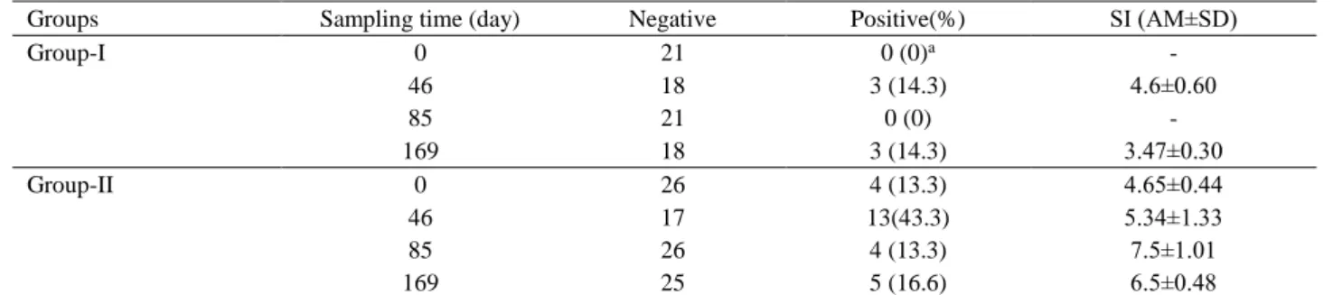
Cellular immunity: The level of IFNg was measured
and evaluated as positive according to Stimulation Index (SI) 2.5-9. Percentage of positivity for IFN-γ were shown as mean and mean of the standard deviations of the SI was demanstrated in Table 2. A total of nine blood samples, of seven reactive to LPS and 2 haemolysed, from group-1 were tested with Y. enterocolica O:9 and E. coli O:157:H7 and excluded from the study. As the level of both IgA and IFNg responses were lower than IgG and the level of immunity was prevailed under 45% (Table 1). Although, percentages of positivity to IFNg was between 13.3 and 43.3%, SI was between 3.47±0.30 and 7.5±1.01 (Table 2). This table shows that cell-mediated immunity is moderate but the proportion is low for estimating cellular immunity at S19 vaccinated cows.
Table 1. Level of anti-IgG and anti-IgA in sera of heifers boosted after primary vaccination with B. abortus S19 conjunctival vaccine based on various sampling times.
Tablo 1. Çeşitli örnekleme zamanlarına dayalı konjuktival B. abortus S19 aşısı ve ardından rapel doz uygulanan buzağı ve düvelerin kan serumlarında anti-IgG ve anti-IgA seviyeleri.
Sampling time (post vac day)
IgG, GI test results Sampling time (post vac day)
IgG, GII test results
neg pos % pos neg pos % pos
0 23 0 0a 0b 14 16 53.3
46 0 23 100 46 01 29 96.6
85 18 05 21.8 85 15 15 50
169 13 10 43.5 169 09 21 70
Sampling time (post vac day)
IgA, GI test results Sampling time (post vac day)
IgA, GII test results
neg pos % pos neg pos % pos
0 25 0 0c 0d 26 04 13.3
46 19 6 24 46 27 03 10
85 17 8 32 85 30 0 0
169 11 14 44 169 20 10 33.3
IgG samples; in Group-I (GI) from calves vaccinated with conjunctival route of B. abortus S19 vaccine at 3-5 months of age, in Grup-II (GGrup-II) from heifers boosted conjunctivally with the same vaccine after 1 year postvaccination.
a: As 7 sera were found reactive with competitive ELISA, they were not included in the study.
b,d: Sampling time in group II denotes the beginning of post vaccination time after boosting in heifers previously vaccinated at calfhood period.
c: As 5 samples were found reactive with competitive ELISA, these sera were not included in the study.
Table 2. Results of bovine IFNg-ELISA according to Stimulation Index. Tablo 2. Stimülasyon Indeksine Göre Sığır IFNg-ELISA Sonuçları.
Groups Sampling time (day) Negative Positive(%) SI (AM±SD)
Group-I 0 21 0 (0)a - 46 18 3 (14.3) 4.6±0.60 85 21 0 (0) - 169 18 3 (14.3) 3.47±0.30 Group-II 0 26 4 (13.3) 4.65±0.44 46 17 13(43.3) 5.34±1.33 85 26 4 (13.3) 7.5±1.01 169 25 5 (16.6) 6.5±0.48
bIFNg results; in Group-I, calves conjunctivally vaccinated at 3-5 months age, in Group-II, heifers boosted conjunctivally with B.
abortus S19 vaccine after 1 year postvaccination.
Discussion and Conclusion
The availability of diagnostic tests capable of detecting vaccinated animals is essential for the determination of immunogenicity of the vaccine. Due to this issue and also to increase the performance of the tests to detect the immune response for longer periods or at defined time limits, different native and recombinant antigens have been preferred (8, 22). Data on the cellular immunity triggered after infection with Brucella spp, a cornerstone in the protection, is more limited, particularly regarding the vaccine. Having some practical advantages and easy evaluation of the test results and higher sensitivity, IFNg test is preferred to in vivo Delayed Type Hypersensitivity (DTH) test. Brucellin and other specific proteins from rough strains such as B. abortus RB51 and
B. melitensis B115 are selected and used as stimulation
antigens in in-vitro tests (8,21, 23). In this study, commercially available brucellergen as the most immunogenic antigen was used as the induction antigen because B. abortus and B. melitensis have both remarkably similar antigenic moiety which is different from R strains that are devoid of Smooth LPS (S-LPS) (1) and OMPs which are constituents of brucellergen (5,22). In this sudy, IFNg response was evaluated in blood of calves and heifers evoked with brucellergen after vaccination with the conjunctival B. abortus S19 vaccine in terms of the cellular immunity. Humoral immunity to Brucella antigens associated with this vaccine have not been cited in extensively, but in this study, it was planned to show the level of antibody responses and if any, targeted to found out which immunoglobulin is acting in immunity. As there is no strict consensus on the detection time, types of test and the antigens, different suggestions are made for monitoring the immune response. In a study, IFNg response by Perez-Sancho et al. (17) was between 10-50% against B. melitensis Rev-1 vaccination during 60 to 180 days postvaccination in sheep. The immune response was followed for 42 days after challenging through one year. IFNg response was detected as 40-70% at the last day of the study, which is similar to the first sampling time of the boosted group. This result is higher than that of this study particularly in calfhood vaccinated group (0-14.3%) but approximate in heifer vaccinated group (13.3-40.3%) (Table1) and therefore, it can be concluded that some fluctiations can occur one month after the vaccianation. Nevertheless, the fact that this method can detect infection very early in time and in high proportion, it is suggestive that this test can significantly contribute to the existing eradication program for bovine brucellosis (23). However, in this study it wasn’t possible to estimate the results for the first 46 days. The results in our study were obtained at different time points of 46, 85 and 169 days postvaccination. The highest response in IFN-γ has been
recorded on 46 pvd in adult vaccinated group (43.3%), the responses on other periods in the group were lower than 20%. The fluctuations in test sensitivity limit the possibility of detecting animals vaccinated with S19 and do not preclude the use of the test as a monitoring method to identify vaccinated herds with S19 vaccine. Discrepancies on the results with the other authors may come from differences of the sampling times and differences in the antigen content of brucellergen. To explain this situation, study with the S19 vaccine should be monitored from the beginning of the postvaccination to thereafter 45 days. Cha et al. (5) showed that peak response was detected during first 5 weeks. Dorneles et al. (9), observed significant IFNg response on day 28, that were decreased after a year. IFNg has been presented to be detected at a very early time during the infection and determined in a high proportion in infected animals (7), however in this study IFNg was detected very low. Moreover, these results are not compatible with that of Weynants et al. (23) for the periods of 80 days based on the brucellergen induction.
Another approach monitoring the immune response is based on the serology. For this purpose, different serological tests and antigens such as SLPS, (Rough LPS
(RLPS), O polsaccharide (OPS), native hapten
polysaccharide, OMP, cytosolic proteins, BP26 have been proposed for the diagnosis of brucellosis in vaccinated and infected animals to increase the specificity of the test especially in ELISA with different monoclonal Abs (15). In this study, immunoglobulin responses to major
Brucella specific LPS antigen were evaluated by ELISAs
based on the serum samples throughout the study. Although IgA response didn’t give any conclusive datas for diagnostic perspective, the result was meaningful for IgG. The maximum IgG positivity was detected 100% in calves and 96.6% in adults. According to the IgG results, antibody responses can be detectable in vaccinated animals during that period. However, these results could not be interpreted in terms of IgA isotype. In a similar way, Chand et al. (6), monitored the antibody response to conjunctival B. abortus S19 vaccine for 3 months. In their study, sera obtained in 3 weeks intervals for 4 periods were evaluated by Rose Bengal Test (RBT) and maximum positivity was determined at 3 weeks postvaccination period as 79.2%. In 6 weeks, it was reduced to 29.2% and after 9 weeks it was 4.2% and 12 weeks later no antibody was detected. In another study, Perez-Sancho et al. (17) detected IgG response in B. melitensis Rev-1 vaccinated sheep at 60 to 180 days postvaccination period. In the study, RBT and Complement Fixation Test (CFT) results were at the range of 20-60%. In the same study one year after challenging, immune response was followed for 42 days. Humoral immunity was over 90% at this time. It
could be concluded that as both studies had the results with the same percentages (6, 17), there can be some fluctuations after the period of one month. Based on the time and the immune response, IgG results could be detectable for up to 46 pvd, but IgA level was lower than expected for screening of the immune status of vaccinated calves. Humoral immunity was detected in calves as 100% by CFT in another study (19), which supports the results of 6 weeks period.
These overall results show that it is not possible to monitor the vaccine status according to the post vaccination times with high detection rate. It would be better to monitor the immunity from pvd 7 to 180 and accumulating datas for detecting different antibody classes and cellular immunity markers based on different antigens either recombinant or native. Besides, our results should be evaluated to produce a standart protocol for vaccine monitoring.
Acknowledgment
This work was supported by the Scientific Research Projects’ Unit of Ondokuz Mayıs University, Samsun, Turkey (project no: VET.1904.14.004) and Tokat Vasfi Diren dairy cattle farm.
References
1. Aragon V, Diaz R, Moreno E, et al. (1996):
Characterization of B. abortus and B. melitensis Native Haptens as Outer Membrane O-Type Polysaccharides Independent from the Smooth Lipopolysaccharide. J
Bacteriology, 178(4), 1070–1079.
2. Barquero-Calvo E, Chaves-Olarte E, Weiss DS, et al. (2007): B. abortus uses a stealthy strategy to avoid
activation of the innate immune system during the onset of infection. PLoS One. 2:e631.
3. Brooks-Worrell BM, Splitter GA (1992):Sodium dodecyl
sulfate and salt extracted antigens from various Brucella species induce proliferation of bovine lymphocytes. Infect
Immun, 60(5), 2136-2138.
4. Caroff M, Bundle DR, Perry MB, et al. (1984):Antigenic
S-type lipopolysaccharide of Brucella abortus 1119-3.
Infect Immun; 46, 384-388.
5. Cha S-B, Rayamajhi N, Kang M-L, et al. (2010):
Comparative study of gamma interferon production in mice immunized with outer membrane proteins and whole bacteria of Brucella abortus. Jpn J Infect Dis, 63, 49-51.
6. Chand P, Chhabra R, Nagra J (2015): Vaccination of
adult animals with a reduced dose of Brucella abortus S19 vaccine to control brucellosis on dairy farms in endemic areas of India. Trop Anim Health Prod, 47, 29-35.
7. Chukwu CC (1986): Comparison of the brucellin skin test
with the lymphocyte transformation test in bovine brucellosis. J Hyg. Camb, 96, 403-413.
8. Denoel PA, Vo TK-O, Weynants VE, et al. (1997):
Identification of the major T cell antigens present in the
Brucella melitensis B115 protein preperation, Brucellergene OCM. J Med Microbiol, 46, 801-806.
9. Dorneles EMS, Sriranganathan N, Lage AP (2015):
Recent advances in Brucella abortus vaccines. Vet Res;
46(76), DOI 10.1186/s13567-015-0199-7.
10. Duncombe L, Commander NJ, Erdenliğ S, et al. (2013):
Investigating the Use of Protein Saver Cards for Storage and Subsequent Detection of Bovine Anti-Brucella abortus Smooth Lipopolysaccharide Antibodies and Gamma Interferon. Clin Vaccine Immunol, 20(11), 1669 –1674.
11. Duran-Ferrer M, Leon L, Nielsen K, et al. (2004):
Antibody response and antigen-specific gamma-interferon profiles of vaccinated and unvaccinated pregnant sheepexperimentally infected withBrucella melitensis.Vet
Microbiol, 100, 219-231.
12. Franc KA, Krecek RC, Hasler BN, et al. (2018):
Brucellosis remains a neglected disease in the developing world: a call for interdisciplinary action. BMC Public
Health, 18 (125), 1-9.
13. Genç O, Büyüktanır Ö, Yurdusev N (2011): Development
of an individual rapid test based on enzymatic immunofiltration assay for detection of anti–Brucella abortus antibody in bovine sera. J Vet Diagn Invest, 23, 49–
56.
14. Genç O, Büyüktanır Ö, Serdar G, et al. (2015):
Development and validation of sandwich quantitative ELISA prototype based on the bovine IFNg for the detection of cellular immunity. Turk J Vet Anim Sci, 39(6), 724-729.
15. Nielsen K, Smith P, Widdison J, et al. (2004): Serological
relationship between cattle exposed to Brucella abortus Yersinia enterocoliticaO:9 and Escherichia coliO157:H7.
Vet Microbiol, 100, 25-30.
16. Office International des Epizooties (OIE) (2009): Bovine
brucellosis, Section 2.4.3. In OIE Manual of standards for diagnostic tests and vaccines, OIE, Paris, 2009.
17. Perez-Sancho M, Duran-Ferrer M, Garcio-Seco T, et al. (2014): Interferon-gamma responses in sheep exposed to
virulent and attenuated Brucella melitensis strains. Vet
Immun Immunopathol, 160, 123-128.
18. Plommet M, Fensterbank R(1976):Vaccination against
bovine brucellosis with a low dose of strain 19 administered by the conjunctival route. Serological response and immunity in the pregnant cow. Annals Vet Res, 7, 9-23.
19. Stevens MG, Hennager SG, Olsen SC, et al. (1994):
Serologic Responses in Diagnostic Tests for Brucellosis in Cattle Vaccinated with Brucella abortus 19 or RB51. J Clin
Microbiol, 32(4), 1065-1066.
20. Stevens MG, Pugh GW, Tabatabai LB (1992): Effects of
gamma interferon and indomethacin in preventing Brucella
abortus infections in mice. Infect. Immun, 60, 4407-4409.
21. Tittarelli M, De Massis F,Bonfini B, et al. (2009): An
ELISA for the evaluation of gamma interferon production in cattle vaccinated withBrucella abortusstrain RB51.Vet Ital,
45(2), 347-54.
22. Wareth G, Melzer F, Weise C, et al. (2015):
Proteomics-based identification of immunodominant proteins of Brucella using sera from infected hosts points towards
enhanced pathogen survival during the infection. Biochem
and Biophys Res Comm, 456, 202-206.
23. Weynants V, Godfroid J, Limbourg B, et al. (1995):
Specific bovine brucellosis diagnosis based on in vitro antigen specific gamma interferon production. J Clin
Microbiol, 33, 706-712.
Geliş tarihi : 12.12.2017 / Kabul tarihi : 26.03.2018
Adress for correspondence:
Prof. Dr. Oktay GENÇ University of Ondokuz Mayıs, Faculty of Veterinary Medicine Department of Microbiology, 55200 Kurupelit-Samsun-Turkey e-mail:[email protected]