• Sonuç bulunamadı

Choroidal thickness in macular holes

N/A
N/A
Protected

Academic year: 2021

Share "Choroidal thickness in macular holes"

Copied!
7
0
0

Yükleniyor.... (view fulltext now)

Tam metin

(1)

O R I G I N A L P A P E R

Choroidal thickness in macular holes

Sabahattin Sul .Go¨khan Gurelik .S¸afak Korkmaz.Cemal Ozsaygili

Received: 23 April 2018 / Accepted: 29 April 2019 / Published online: 7 May 2019 Ó Springer Nature B.V. 2019

Abstract

Purpose To determine the association between choroidal thickness (CT) and anatomic success in closed and open macular holes (MHs) following surgery.

Methods One hundred and thirty-six eyes of 136 patients who underwent surgery due to primary MH were included in this study. Choroidal thickness was measured from various points (subfoveal, temporal, nasal, superior and inferior 1500 lm from the center of the fovea) in both eyes with MH and fellow eyes. We determined associations among the duration of symptoms, MH dimensions and CTs from various points with anatomic success and correlations between CT and MH dimensions and duration of symptoms.

Results Choroidal thickness was significantly lower in eyes with MH than fellow eyes in both open and closed MHs. Base diameter [p = 0.025, odds ratio (OR) = 0.428], minimum hole diameter (p = 0.030, OR = 0.211) and duration of symptoms [p = 0.034, OR = 0.443] were significantly associated with ana-tomic success. However, CTs from various points were not associated with anatomic success despite a significant preoperative subfoveal CT difference between open and closed MHs (198 ± 21 lm in open MHs and 230 ± 30 lm in closed MHs; p \ 0.001). Preoperative subfoveal CT was moderately correlated with base diameter (r = - 0.505, p \ 0.001), mini-mum hole diameter (r = - 0.518, p \ 0.001) and duration of symptoms (r = - 0.510, p \ 0.001). Conclusions Failed MHs were associated with larger hole dimensions (base diameter and minimum hole diameter) and longer duration of symptoms. Preoper-ative subfoveal CT was thinner in open MHs, but there was no association with anatomic success. Choroidal thinning may be linked to larger and chronic MHs. Keywords Macular hole Choroidal thickness  Anatomic success Macular hole dimensions

S. Sul (&)

Department of Ophthalmology, Mug˘la Sıtkı Koc¸man University, 48000 Mug˘la, Turkey

e-mail: drsulgoz@gmail.com G. Gurelik

Department of Ophthalmology, Gazi University Medical School, Ankara, Turkey

S¸. Korkmaz

Department of Ophthalmology, Du¨zce State Hospital, Du¨zce, Turkey

C. Ozsaygili

Department of Ophthalmology, Kayseri Training and Research Hospital, Kayseri, Turkey

(2)

Introduction

Macular hole (MH) formation was proposed by Gass as anteroposterior and tangential vitreal traction on the foveal area, which has been demonstrated in optical coherence tomography (OCT) [1, 2]. Pars plana vitrectomy (PPV) and internal limiting membrane (ILM) peeling which removes the anteroposterior and tangential traction, become the standard surgical treatment in these cases [3, 4]. However, MH can develop in the absence of vitreomacular tractional forces such as in complete posterior vitreous detach-ment or vitrectomized eyes [5, 6]. Cystoid macular edema, progressive outer retinal degeneration, epireti-nal membrane formation and choroidal thinning have been suggested to be other preceding conditions. [6,7].

Aras et al. [7] found a reduction in foveal choroidal blood flow, and, Zhang et al. [8] found decreased foveal choroidal thickness (CT) with enhanced depth image (EDI) OCT in cases of MH, and they concluded that CT may play a role in the pathogenesis of MH. The choroid is an important metabolic supplier for the fovea. Therefore, reduction in the choroidal blood flow may alter the healing process and contribute to failed MH closure. On the other hand, choroidal thinning may be thought a marker of MH character-istics such as chronicity and hole dimensions which have been demonstrated to be the predictors of anatomic outcomes of surgery [9–14], because decreased foveal choroidal blood flow may be a consequence of the loss of overlying retinal tissue, which has been strongly suggested to control choroidal blood flow [15].

The aim of this study was to evaluate and compare MH characteristics (MH dimensions and duration of symptoms) and preoperative CTs between closed and open MHs and to determine whether there was a relationship among CT, anatomic success and MH characteristics.

Methods

We retrospectively reviewed the records of patients who underwent surgical intervention for the treatment of primary idiopathic MH between 2014 and 2017 at the Gazi University Faculty of Medicine (Ankara, Turkey). The study was approved by the Ethics

Committee and adhered to the tenets of the Declara-tion of Helsinki.

All of the patients applied to hospital due to loss of their vision. Patients who had MH due to other etiologies (e.g., myopia, trauma, cystoid macular edema), patients with ocular pathologies such as diabetic retinopathy or retinal vascular diseases, patients with previous ocular surgery history, hyper-opia or myhyper-opia greater than 1 diopter, axial length lower than 22 mm or greater than 24 mm or systemic diseases that may have effected the measurements, were excluded from the study.

Surgical procedure

After the entrance of transvitreal 23 gauge vitreoreti-nal instruments, core vitrectomy was completed. The posterior hyaloid was stained with triamcinolone and separated and cleaned using ocutome probe. The ILM was stained using brilliant blue dye and peeled with ILM forceps up to the arcuates. After fluid air exchange, a 23 gauge, silicone-tipped cannula was placed over the MH without touching the retinal pigment epithelium to drain the remaining fluid. The air was changed to gas (20% sulphur hexafluoride) endotamponade. The patients were asked to take facedown positioning for 1 week.

The preoperative hole base diameter, the minimum hole diameter and duration of the symptoms were evaluated and determined as MH characteristics. Preoperative and postoperative (1 month after sur-gery) OCT measurements were performed using Spectralis (Heidelberg Engineering, Heidelberg, Ger-many) OCT. The EDI mode of Spectralis was performed for the CT measurements. The CT estima-tions were performed by two independent expert OCT readers who were not involved in the data analysis. The estimations of the readers were compared and averaged for the analysis. The CT was defined as the distance between the retinal pigment epithelium line (the outermost hyperreflective line) and the choroi-doscleral boundaries. The subfoveal, inferior, supe-rior, nasal and temporal thickness 1500 lm away from the fovea were measured. Patients who were examined between 9 AM and 12 PM were included in the study to avoid the effects of diurnal variation. To adjust for potential compounding factors, age, sex, refraction and axial length (AL) matched patients were included in this study.

(3)

Statistical analysis was performed using SPSS version 16 (SPSS, Chicago, IL,USA). An independent t test was used to compare the continuous data including the duration of symptoms, the mean hole base diameter, the mean minimum hole diameter and preoperative CTs between closed and open MHs. A paired t-test was used to compare CTs between eyes with MH and fellow eyes. Continuous data, which were statistically significant in independent t-test, were included in a multilogistic regression model to better delineate the association of these data with anatomic success. Furthermore, Pearson’s correlation test was to investigate correlations between the duration of symptoms, MH dimensions (mean hole base and minimum hole diameter) and CTs in all patients. A p value less than 0.05 was considered to indicate statistical significance.

Results

There were 254 eyes of 254 patients who underwent surgical intervention for the treatment of idiopathic MH between 2014 and 2017. In total, 230 eyes (90.5%) underwent a successful MH surgery. One hundred and thirty-six eyes of 136 patients who satisfied the inclusion criteria were included in this study. Mean age was 65.4 ± 3.4 years (range 60–69) in open MHs and 65.3 ± 3 years (range 60–69) in closed MHs (p = 0.907). All of the patients underwent a standard surgical procedure (PPV, large ILM peeling, gas (20% sulphur hexafluoride) endotampon-ade and facedown positioning). There were 36 men (26.5%) and 100 women (73.5%). The MH remained open in 16 eyes (11.7%) and closed in 120 eyes (88.3%). Thirty-three of 36 men (91.7%) and 87 of 100 women (87%) had anatomic success following surgery (p = 0.559). Duration of symptoms, mean hole base

diameter and minimum hole diameter were signifi-cantly higher in open MHs than closed MHs (Table1). The mean CTs of the fellow eyes were significantly higher than eyes with MHs in both open and closed MHs (Table2). In open MHs, the preoperative subfoveal CT and CT at the temporal 1500 lm from the center of the macula were significantly lower than closed MHs (Table3). The mean AL was 23.03 ± 0.6 mm (range 22–24 mm) in open MHs and 23.2 ± 0.58 mm (range 22–24 mm) in closed MHs; the difference was not statistically significant (p = 0.331).

Based on the multilogistic regression analysis, the duration of symptoms, mean hole base diameter and mean minimum hole diameter were significantly associated with anatomic success (Table4). There was a moderate correlation between subfoveal CT and duration of symptoms and macular hole dimensions (mean hole base and minimum hole diameter) (Table 5).

Discussion

The closing mechanisms of MHs are not yet fully understood. Therefore, some preoperative parameters were used to predict the closure of the MHs. Chronicity, minimal hole diameter and hole base diameter are the most commonly used parameters to predict anatomic success [9, 10]. Our findings were consistent with the literature that open MHs had significantly larger dimensions and longer duration of symptoms than closed MHs.

Vitreomacular tractional forces are not thought to be the sole factor in the pathogenesis of MH. Alteration in choroidal layer has been suggested to be a possible contributing factor to the pathogenesis of MH. Aras et al. [7] found a decreased choroidal blood

Table 1 Duration of symptoms, mean base and minimum hole diameter in open and closed MHs

Parameter Open Closed p value

Average ± SD Range Average ± SD Range

Duration of symptoms (months) 9.4 ± 3.2 5–15 5.4 ± 2 2–12 \ 0.001

Base diameter (l) 1237 ± 185 725–1441 864 ± 225 426–1542 \ 0.001

Minimum diameter(l) 539 ± 135 265–800 327 ± 107 140–655 \ 0.001

(4)

flow in eyes with MH, and other authors such as Xu et al. [16], Reibaldi et al. [17] and Zeng et al. [18] found a decreased CT in eyes with MH. Furthermore, Michalewska et al. [19] demonstrated irregularities in

outer choroidoscleral boundaries in MH patients. We found significant choroidal thinning in eyes with MH compared with their fellow eyes in both groups, which was consistent with the findings of the reports noted above.

The choroid is the main blood supply of fovea; retinal capillary blood flow is significantly lower than in other retinal locations, especially the fovea [20]. Therefore, the choroid plays an important role in the metabolic activity of central fovea. Decreased chor-oidal blood flow and altered metabolic activity might alter the healing process and contribute to failed MH closure. In this study, however, when we analyzed MH characteristics and subfoveal CT together in the logistic regression analysis, we did not find any association between CT and anatomic success.

Table 2 Mean CTs of the eyes with MHs and fellow eyes

Mean choroidal thickness (lm)

Open MH Closed MH

Eyes with MH Fellow eye p value Eyes with MH Fellow eye p value

Average ± SD (range) Average ± SD (range) Average ± SD (range) Average ± SD (range) Subfoveal 198 ± 21 lm (165–233) 242 ± 22 lm (220–295) \ 0.001 230 ± 30 (145–300) 251 ± 17 (220–300) \ 0.001 Temporal 1500 206 ± 21 (169–240) 240 ± 13 (220–260) \ 0.001 227 ± 36 (137–323) 251 ± 19 (230–300) \ 0.001 Nasal 1500 179 ± 40 (128–254) 210 ± 29 (170–253) \ 0.001 195 ± 36 (100–290) 215 ± 21 (172–272) \ 0.001 Superior 1500 224 ± 35 (175–281) 253 ± 30 (210–300) \ 0.001 234 ± 37 (120–320) 260 ± 26 (198–305) \ 0.001 Inferior 1500 211 ± 46 (151–281) 241 ± 36 (181–290) \ 0.001 220 ± 39 242 ± 24 (195–280) \ 0.001

CT choroidal thickness, MH macular hole

Table 3 Mean CTs and axial lengths in closed and open MHs

Mean choroidal thickness (l) Open Closed p value

Average ± SD Range Average ± SD Range

Subfoveal 198 ± 21 165–233 230 ± 30 145–300 < 0.001 Temporal 1500 l 206 ± 21 169–240 227 ± 36 137–323 0.026 Nasal 1500 l 179 ± 40 128–254 195 ± 36 100–290 0.146 Superior 1500 l 224 ± 35 175–281 234 ± 37 120–320 0.351 Inferior 1500 l 211 ± 46 151–281 220 ± 39 131–320 0.438 Axial length 23.1 ± 5.7 22–24 23.3 ± 5.6 22–24 0.657

Subfoveal and temporal CTs were significantly lower in open MHs than closed MHs are given in bold CT choroidal thickness, MH macular hole

Table 4 Multivariate logistic regression analysis of factors

associated with anatomic success

Odds ratio p value

Subfoveal CT 1.892 0.186

Temporal 1500 l CT 0.522 0.079

Minimum hole diameter 0.211 0.030

Hole base diameter 0.428 0.025

Duration of symptoms 0.443 0.034

MHs with larger dimensions and longer duration significantly associated with anatomic success are given in bold

(5)

Conversely, we found that larger and chronic holes contributed to failed hole closure, which has been noted by previous studies [9, 10]. In other words, preoperative subfoveal choroidal thinning, which was encountered in open MHs, may have been the result of chronic and larger MHs. Indeed, there was a moderate

correlation between subfoveal CT and MH dimensions and duration of symptoms. The patients who had larger MHs and longer durations of symptoms tended to have thinner preoperative subfoveal choroids (Fig.1a, b) than patients who had small MHs and shorter duration of symptoms (Fig.2a, b). A

Table 5 Correlation of MH dimensions (minimum hole diameter and hole base diameter) and duration of symptoms with CT from

various points

Choroidal thickness Minimum hole diameter Hole base diameter Duration of symptoms

r p value r p value R p value

Subfoveal - 0.518 \ 0.001 - 0.505 \ 0.001 - 0.510 \ 0.001

Temporal 1500 l - 0.343 0.005 - 0.294 0.016 - 0.389 0.005

Nasal 1500 l - 0.309 0.011 - 0.276 0.024 - 0.366 0.008

Superior 1500 l - 0.265 0.030 - 0.279 0.022 - 0.275 0.051

Inferior 1500 l - 0.364 0.002 - 0.309 0.011 - 0.303 0.031

Subfoveal CT significanly correlated with MH dimensions and duration of symptoms are given in bold MH macular hole, CT choroidal thickness

Fig. 1 Preoperative and postoperative EDI-OCT images of a

64-year-old patient who had complaints for 14 months. MH had flat margins. a Hole base diameter: 1400 lm, minimum hole

diameter: 800 lm, axial length: 24 mm and subfoveal choroidal thickness: 204 lm. b Macular hole was not closed after surgery. Subfoveal choroidal thickness: 210 l

(6)

correlation of subfoveal choroidal thinning with chronic and larger MHs might be thought as data supporting the findings of Flu¨gel et al. These authors suggested that overlying retinal tissue regulated choroidal blood flow [8]. In addition, Teng et al. [21] recently reported OCT angiographic findings in MHs. These authors found a negative correlation between choriocapillaris circulation and hole diame-ter. Furthermore, Allen et al. [22] demonstrated decreased choroidal perfusion following laser-induced MH formation.

In conclusion, preoperative subfoveal CTs appear to be thinner in open MHs. However, subfoveal choroidal thinning was not associated with anatomic success. Subfoveal choroidal thinning likely is the result of larger and chronic MHs.

Compliance with ethical standards

Conflict of interest The authors declare that they have no

conflict of interest.

References

1. Gass JD (1995) Reappraisal of biomicroscopic classification of stages of development of a macular hole. Am J Oph-thalmol 119(6):752–759

2. Altaweel M, Ip M (2003) Macular hole: improved under-standing of pathogenesis, staging, and management based on optical coherence tomography. Semin Ophthalmol 18:58–66

3. Wendel RT, Patel AC, Kelly NE, Salzano TC, Wells JW, Novack GD (1993) Vitreous surgery for macular holes. Ophthalmology 100(11):1671–1676

4. Haritoglou C, Gass CA, Schaumberger M, Gandorfer A, Ulbig MW, Kampik A (2002) Long-term follow-up after macular hole surgery with internal limiting membrane peeling. Am J Ophthalmo 134(5):661–666

5. Gordon LW, Glaser BM, Ie D, Thompson JT, Sjaarda RN (1995) Full-thickness macular hole formation in eyes with a pre-existing complete posterior vitreous detachment. Oph-thalmology 102:1702–1705

6. Lee SH, Park KH, Kim JH, Heo JW, Yu HG, Yu YS et al (2010) Secondary macular hole formation after vitrectomy. Retina 30:1072–1077

7. Aras C, Ocakoglu O, Akova N (2004) Foveolar choroidal blood flow in idiopathic macular hole. Int Ophthalmol 25:225–231

Fig. 2 Preoperative and postoperative EDI-OCT images of a

65-year-old patient who had complaints for 2 months. a Base diameter: 1542 lm, minimum diameter: 221 lm, axial length:

24 mm and subfoveal choroidal thickness: 243 lm. b Macular hole was closed after surgery. Subfoveal choroidal thickness: 254 l

(7)

8. Zhang P, Zhou M, Wu Y, Lu B, Li T, Zhao J et al (2017) Choroidalthickness inunilateal idiopathic macular hole: a

cross-sectional study and meta-analysis. Retina

37(1):60–69

9. Willis AW, Garcia-Cosio JF (1996) Macular hole surgery: comparison of longstanding versus recent macular holes. Ophthalmology 103:1811–1814

10. Ip MS, Baker BJ, Duker JS, Reichel E, Baumal CR, Gangnon R et al (2002) Anatomical outcomes of surgery for idiopathic macular hole as determined by optical coherence tomography. Arch Ophthalmol 120(1):29–35

11. Wakely L, Rahman R, Stephenson JA (2012) Comparison of several methods of macular hole measurement using optical coherence tomography, and their value in predicting anatomical and visual outcomes. Br J Ophthalmol 96(7):1003–1007

12. Ullrich S, Haritoglou C, Gass C, Schaumberger M, Mw Ulbig, Kampik A et al (2002) Macular hole size as a prog-nostic factor in macular hole surgery. Br J Ophthalmol 86:390–393

13. Kusuhara S, Teraoka Escano MF, Fujii S, Nakanishi Y, Tamura Y, Nagai A et al (2004) Prediction of postoperative visual outcome based on hole configuration by optical coherence tomography in eyes with idiopathic macular holes. Am J Ophthalmol 138:709–716

14. Ruiz-Moreno JM, Staicu C, Pinero DP, Montero J, Lugo F, Amat P (2008) Optical coherence tomography predictive factors for macular hole surgery outcome. Br J Ophthalmol 92:640–644

15. Flu¨gel C, Tamm ER, Mayer B, Lutjen-Drecoll E (1994) Species differences in choroidal vasodilative innervation: evidence for specific intrinsic nitrergic and VIP-positive

neurons in the human eye. Investig Ophthalmol Vis Sci 35:592–599

16. Xu LT, Srivastava SK, Ehlers JP, Kaiser PK (2015) Chor-oidal thickness in macular holes: a case-control study. Ophthalmic Surg Lasers Imaging Retina 46(1):33–37 17. Reibaldi M, Boscia F, Avitabile T, Uva MG, Russo V,

Zagari M et al (2011) Enhanced depth imaging optical coherence tomography of the choroid in idiopathic macular hole: a cross-sectional prospective study. Am J Ophthalmol 151(1):112–117

18. Zeng J, Li J, Liu R, Chen X, Pan J, Tang S et al (2012) Choroidal thickness in both eyes of patients with unilateral idiopathic macular hole. Ophthalmology 119:2328–2333 19. Michalewska Z, Michalewski J, Nawrocka Z,

Dulczewska-Cichecka K, Nawrocki J (2015) The outer choroidoscleral boundary in full-thickness macular holes before and after surgery—a swept-source OCT study. Graefes Arch Clin Exp Ophthalmol 253:2087–2093

20. Rawji MH, Flanagan JG (2001) Intraocular and interocular symmetry in normal retinal capillary perfusion. J Glaucoma 10:4–12

21. Teng Y, Yu M, Wang Y, Liu X, You Q, Liu W (2017) OCT angiography quantifying choriocapillary circulation in idiopathic macular hole before and after surgery. Graefes Arch Clin Exp Ophthalmol 255(5):893–902

22. Allen RD, Brown J, Zwick H (2004) Laser-induced macular holes demonstrate impaired choroidal perfusion. Retina 24:92–97

Publisher’s Note Springer Nature remains neutral with

regard to jurisdictional claims in published maps and institutional affiliations.

Şekil

Table 1 Duration of symptoms, mean base and minimum hole diameter in open and closed MHs
Table 3 Mean CTs and axial lengths in closed and open MHs
Table 5 Correlation of MH dimensions (minimum hole diameter and hole base diameter) and duration of symptoms with CT from
Fig. 2 Preoperative and postoperative EDI-OCT images of a

Referanslar

Benzer Belgeler

The aim of this thesis is to study the effect of the hole size diameter on different aspects of multi stage incremental forming or hole flanging like thickness distribution

In terms of MPV, although there was no significant difference between the ARF patients in the acute stage and those in remission; the MPV/platelet ratio was significantly lower

Results: Mean myometrial thickness overlying scar pregnancy was significantly lower in the group with isthmocele formation, and the mean gestational age of scar pregnancy was

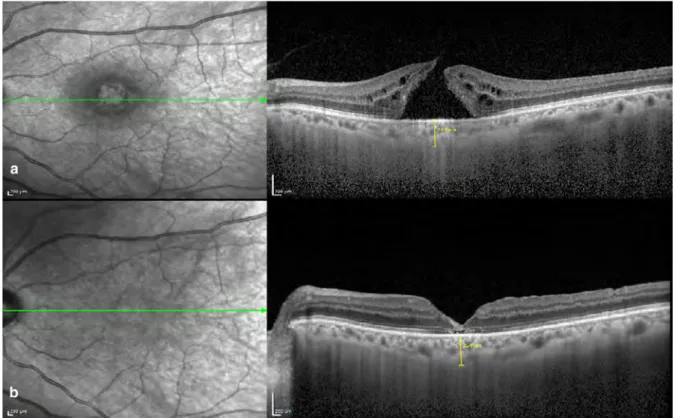
Our case was also noteworthy for having the highest axial length (34.97 mm) among the spontaneous closures reported to date. 9 reported a 55-year-old woman with an axial length

The other patients received intravitreal gas injection as well as 2-3 rows of endolaser around 9 lattice degenerations, 3 retinal holes, 1 lattice degeneration + retinal hole, and

Similar to our study, they showed that the choroidal thickness of Type 2 DM patients was thicker than the control group, but there was no statistically significant difference

The patients were divided into two groups according to the timing of vitrectomy and cataract surgery: the combined surgery group consists of patients who underwent

graph. Iu figw·e 2, the data set is skewed to the right. Here, the mode is still at the highest · oıı the graph, but the median lies to the right of this point and the mean falls to