Distraction lengthening of the proximal phalanx
in distal thumb amputations
Correspondence: Eren Cansü, MD. Marmara Üniversitesi Tıp Fakültesi,
Ortopedi ve Travmatoloji Anabilim Dalı, İstanbul, Turkey. Tel: +90 532 – 274 25 56 e-mail: [email protected]
Submitted: August 19, 2014 Accepted: December 12, 2014 ©2015 Turkish Association of Orthopaedics and Traumatology
Available online at www.aott.org.tr doi: 10.3944/AOTT.2015.14.0292 QR (Quick Response) Code
doi: 10.3944/AOTT.2015.14.0292
Eren CANSÜ1, Mehmet Bekir ÜNAL2, Fatih PARMAKSIZOĞLU3, Serkan GÜRCAN4 1Marmara University Faculty of Medicine, Department of Orthopaedics and Traumatology, İstanbul, Turkey
2Medipol University Faculty of Medicine, Department of Orthopaedics and Traumatology, İstanbul, Turkey 3Yeni Yüzyıl University Faculty of Medicine, Department of Orthopaedics and Traumatology, İstanbul, Turkey
4Medistate Hospital Kavacık, Department of Orthopaedics and Traumatology, İstanbul, Turkey
Objective: Thumb amputation is a major cause of hand dysfunction, and the treatment for distal thumb amputations remains controversial. Although finger reconstruction methods using distraction lengthening are known to restore finger length and function, we found no reports in the literature regarding phalangeal lengthening in thumb amputations. We aimed to evaluate proximal phalangeal lengthening in thumb amputations at or near the interphalangeal (IP) joint.
Methods: We retrospectively evaluated patients who had undergone distraction lengthening of the proximal phalanx of the thumb. All patients underwent osteotomy, either during the initial procedure or as a second-stage procedure. Distraction began 10 days after osteotomy with the use of an external fixator that remained in place until ossification of the gap occurred without bone grafting. Patients were evaluated using the QuickDASH score.
Results: Fourteen patients with a mean age of 27 years and a mean follow-up period of 7 years were enrolled. The mean phalangeal lengthening achieved was 20 mm. Ossification occurred at all distrac-tion sites, and the fixators were maintained for a mean of 85 days. The mean healing index was 42.5 days/cm. All 14 patients achieved the desired amount of phalangeal lengthening without major com-plications such as nonunion, premature union, or gross infection.
Conclusion: For reconstruction in cases of distal thumb amputations, distraction lengthening of the proximal phalanx can be used to improve absolute length, web space, and grip distance. The technique is safe and effective, improves functionality/cosmesis, and offers a low complication risk.
Keywords: Distraction lengthening; phalangeal lengthening; thumb amputation; thumb lengthening; thumb reconstruction.
Thumb amputations create significant hand dysfunc-tion, and morbidity is higher with when the amputation level is more proximal. From simple local procedures to complex microsurgical operations, many reconstructive
procedures and techniques have been reported for every amputation level.
Treatment of thumb amputations at or near the in-terphalangeal (IP) joint remains controversial. Primary
closure, advancement or island flaps, and deepening of the first web are some of the reconstructive procedures that are now being used.
Although the first metacarpal is a more common lengthening site for thumb reconstruction, lengthen-ing of the proximal phalanx of the thumb has not been widely mentioned in the literature. It was first present-ed by Matev as a single case report.[1] However, to our knowledge, there has been no specific research on pha-langeal lengthening in thumb amputations. For this level of thumb amputation, we prefer to lengthen the proximal phalanx if the amputated part is not suitable for replan-tation, or if the replantation attempt fails. Accordingly, to assess procedure outcomes, we analyzed the data for consecutive patients with thumb amputations who un-derwent distraction lengthening of the proximal phalanx.
Patients and methods
Between December 2001 and June 2012, we treated 14 patients (13 men, 1 woman; mean age; 27 years, range: 18–43 years) who had sustained thumb amputations at or near the IP joint level with distraction lengthening of the proximal phalanx (Table 1). All were manual labor-ers who had sustained their injuries during industrial accidents. The amputation level varied from the base of the distal phalanx to the middle of the proximal phalanx (Figure 1). Osteotomies were performed during the ini-tial procedure in 8 patients (Figure 2) and as a second-stage procedure in 6 patients.
Before surgery, all patients were provided with a
complete explanation of the risks, benefits, limitations, and potential complications of amputation, osteotomy, distraction lengthening, and alternative procedures, as well as the expected outcomes of the procedures. All provided written acknowledgment of the explanation and of their consent to undergo the procedures.
Osteotomies were performed through a longitudinal mini-incision lateral to the extensor tendon, through a periosteal window, without cutting the periosteum. Be-fore the osteotomy, mini-Schanz pins were inserted per-cutaneously under fluoroscopy. We cut the bone with an osteotome after drilling multiple times with a Kirschner wire or an oscillating saw. After verifying that the os-teotomy was complete, we constructed the distraction device. We used a unilateral miniature distraction device (TST®-Turkey) with 2 mini-Schanz pins for each frag-ment. A shorter bone stump could be osteotomized by inserting 1 of the proximal mini-Schanz pins into the base of the proximal phalanx and the other into the first metacarpal head.
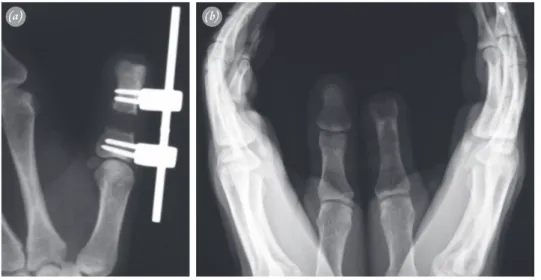
Ten days after the osteotomy, we began the distrac-tion at an interval of 1 mm/day (2×0.5 mm), stopping when the desired length was achieved (Figure 3a). The middle of the contralateral distal phalanx was the goal for the maximum lengthening level (Figure 3b); in addi-tion, we decided on a maximum elongation gap of 100% of the remaining bone. When the elongated callus ma-tured, the distraction device was extracted without the need for anesthesia. The patients’ functional statuses were evaluated using the QuickDASH score.[2,3]
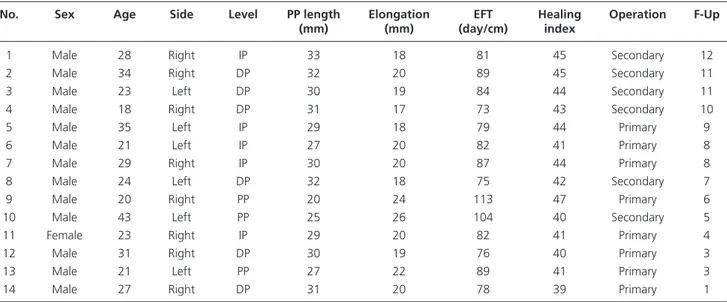
Table 1. Patient data.
No. Sex Age Side Level PP length Elongation EFT Healing Operation F-Up
(mm) (mm) (day/cm) index
1 Male 28 Right IP 33 18 81 45 Secondary 12 2 Male 34 Right DP 32 20 89 45 Secondary 11 3 Male 23 Left DP 30 19 84 44 Secondary 11 4 Male 18 Right DP 31 17 73 43 Secondary 10 5 Male 35 Left IP 29 18 79 44 Primary 9 6 Male 21 Left IP 27 20 82 41 Primary 8 7 Male 29 Right IP 30 20 87 44 Primary 8 8 Male 24 Left DP 32 18 75 42 Secondary 7 9 Male 20 Right PP 20 24 113 47 Primary 6 10 Male 43 Left PP 25 26 104 40 Secondary 5 11 Female 23 Right IP 29 20 82 41 Primary 4 12 Male 31 Right DP 30 19 76 40 Primary 3 13 Male 21 Left PP 27 22 89 41 Primary 3 14 Male 27 Right DP 31 20 78 39 Primary 1
PP length, length of the proximal phalanx; EFT: External fixator time; Op.: Operation; F-Up: Follow-up (years); IP: Interphalangeal joint; DP: Distal phalanx; PP: Proxi-mal phalanx.
Results
The mean follow-up period was 7 years (range: 1–12 years). In 6 patients, the stumps were reconstructed with local flaps (using a neurovascular island flap from the ulnar side of the middle finger in 2 patients) or closed during the primary procedure. When there was not enough healthy soft tissue to close the wound during the primary procedure, we used local flaps to avoid excessive bone shortening. After the soft tissues healed in those 6 patients, we performed the osteotomy and lengthen-ing as a second-stage procedure. For the remainlengthen-ing 8 pa-tients, the lengthening procedure was performed as part of a single-stage procedure in which the soft tissues were treated at the same time (Figure 2). The mean amount of lengthening of the phalanges was 20 mm (range: 17–26 mm).
In all patients, we maintained the external fixator in place until ossification occurred, and we did not perform any bone-grafting procedures. All distraction sites ossi-fied. The mean time from osteotomy to the extraction of the distraction device was 85 days (range: 72–113 days).
There were no instances of nonunion, malunion, pre-mature union, fracture of the elongated callus, or gross infection necessitating the early removal of the pins. There was 1 instance of pin-tract infection, which was treated with local care and oral antibiotics. We observed thinning of the proximal phalanx in 2 patients and pro-truding proximal phalanx condyles in 1 patient. There was no soft-tissue breakage at the tip of the phalanx
during or after the lengthening procedure in any patient. All patients had good results from the lengthening pro-cedure in terms of the pain level and discomfort, and all patients were compliant. There were no requests for distraction discontinuation before the desired amount of lengthening was achieved. A study limitation may be that we did not evaluate pinch strength or metacarpo-phalangeal joint movements in these patients. The aver-age QuickDASH score was 1.62 (range: 0–6.8).
Discussion
The thumb accounts for nearly 40% of hand func-tions, and amputation below the IP joint level results in marked limitations in function. Opposition is the dis-tinctive movement of the thumb. Proper thumb length along with thumb stability, strength, and mobility are major requirements for thumb opposition.[4] The carpo-metacarpal joint plays a major role in thumb movements, and adequate function can be preserved despite a lack of mobility in the metacarpophalangeal and IP joints. However, sensation is another issue that must be ad-dressed to allow thumb functionality.
Although there may be different procedure options for thumb reconstructions, replantation is always the first choice because it results in the best functionality with an almost-normal appearance, as reported by many authors.[4–6] However, when replantation is not appro-priate or if replantation attempts fail, many alternative procedures can be considered depending on the
ampu-Fig. 1. Loss of the distal phalanx.
Fig. 2. During primary closure of the wound, an external fixator was
applied after the osteotomy of the proximal phalanx. [Color figure can be viewed in the online issue, which is available at www.aott.org.tr]
tation level. These procedures include revision amputa-tions, the use of simple local flaps, and other complex and demanding microvascular operations such as free toe transfers;[7] each of these procedures has its own ad-vantages and disadad-vantages.
After a distal thumb amputation, the decision about the reconstruction method and even the need for re-construction remain controversial.[4,7,8] Some surgeons choose primary wound closure, whereas others prefer to use various types of local flaps, especially when there is extensive tactile surface loss. Some may suggest that no lengthening is needed when the distal phalanx of the thumb is lost. However, there has been a report of a 6-mm lengthening of the distal phalangeal stump of an amputated thumb,[9] and others prefer to use even more complex techniques such as toe or partial-toe transfers for this amputation level.[10] We believe that patients should be given information about the various options available for restoring thumb length.
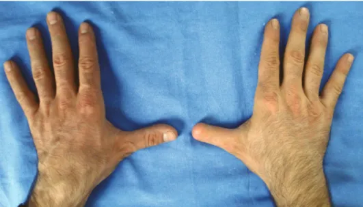
Because length is of prime importance for opposabil-ity of the thumb, restoring length ameliorates difficulties with both function and cosmesis by absolute deepening and widening of the first web space (Figure 4). Length-ening the bones of the hand via distraction is a widely-used method for both traumatic and congenital defects. After its first presentation by Matev in 1967,[11] this method has been safely used by many hand surgeons for a variety of indications such as lengthening the stumps of distal phalanges or even replanted fingers.[9,12–16] We find that this technique has many advantages:
i. It is easy to perform and has a short learning curve.
ii. It is inexpensive because it does not require micro-surgery, expertise, or even special training; it also does not require special equipment such as micro-scopes or other micro-instruments.
iii. Unlike some other procedures, the duration of sur-gery for this procedure is short, and it can be done with local or regional anesthesia.
iv. In cases with multiple injuries, the technique can be applied to multiple fingers at the same time.
v. Although patients must wear a fixator for up to 3 months, they are still able to freely use their hands in daily life during the treatment.
vi. There is no donor-site morbidity because only local sources (bone and soft tissues of the individual fin-ger) are used; this is a main advantage of this tech-nique. These local sources have qualities similar to the lost tissues. The skin also provides original sensa-tion and thus does not require cortical adaptasensa-tion, as with use of an island flap, or microsurgical neuror-rhaphy, as with free tissue transfers.
vii. There is also no risk of losing the lengthened finger, as there is with toe transplantation.
The first metacarpal is historically the first site for dis-traction lengthening of the thumb.[11] A common prob-lem with first metacarpal lengthening is the elevation of the first web space base, which diminishes the effect of the procedure.[17] This occurs because the intrinsic tendons of the thumb insert into the base of the proximal phalanx, a point which is distal to the osteotomy site.[18] Therefore, additional surgery (deepening of the first web space and/
Fig. 3. (a) Lengthening of the distal phalanx was initiated 10 days after the osteotomy; it was continued
until the tip of the proximal phalanx reached the middle of the opposite distal phalanx. (b) The
acquired osseous length after full consolidation of the gap, as seen in this radiograph at the final follow-up evaluation.
or proximal transfer of the adductor pollicis brevis) may be required to alleviate this problem. Salom et al reported that they had performed deepening procedures for the first web in all cases and additional proximal transfers for the adductor pollicis brevis in 2 cases in their series of 6 patients who underwent first metacarpal lengthening.[19] Finsen and Russwurm have treated 9 patients with first metacarpal lengthening after traumatic thumb amputa-tions.[20] In 6 of these patients, the proximal transfer of the adductor pollicis brevis and deepening of the first web were performed as an additional treatment.
With proximal phalangeal lengthening, however, there is no elevation of the base of the first web space, because the osteotomy site is distal to the insertions of the intrinsic tendons of the thumb.
Another drawback of first metacarpal lengthening in comparison with proximal phalangeal lengthening is that flexion contracture of the proximal phalangeal stump in the former.[17, 20] To avoid this complication, the proximal phalangeal stump can be fixed to the first metacarpal or it can be integrated into the external fix-ator, as done by Finsen and Russwurm.[20] We believe that lengthening the proximal phalanx may be more convenient if its length allows the mounting of an ex-ternal fixator.
Another method involves osteoplastic reconstruc-tion, in which a piece of bone from the iliac crest and surrounding skin are transferred to the amputation site. Resorption of the transferred bone is a well-known complication with this method, and the skin of the new thumb has no sensation.[4]
The main disadvantages of distraction lengthening of the proximal phalanx are the overall procedure dura-tion (nearly 3 months until solid bone consolidadura-tion is achieved) and the requirement that the patient wear a fixator during the entire process. If an osteotomy can be performed during the initial procedure, the total treat-ment time will be shortened, as it was for 8 of our 14 patients. Bone grafting of the gap is another option to decrease the treatment time. Although this allows the treatment to be completed several weeks earlier, it also carries the risks of another procedure: donor-site mor-bidity or graft-site nonunion. Similar to other surgeons, we prefer to leave the external fixator in place until the elongated callus matures.[12,13,15,16] The absence of the thumb’s nail may be an important cosmetic factor for some patients. These patients will also have some limita-tions in picking up thin objects.[21]
Another issue associated with this method is the extent of lengthening required. According to Matev, al-though 100% lengthening is possible for the hand skel-eton, the elongated thumb should be shorter than its normal counterpart to provide better function, because it lacks a joint.[17] He has advised lengthening the in-jured thumb until it is equal in length to the middle of the uninjured thumb’s distal phalanx. We have followed his suggestions for our lengthening procedures, setting a goal of allowing the most distal level of the injured thumb to reach the middle of the contralateral distal phalanx. For achieving the maximum elongation gap, we do not exceed 100% of the remaining bone so that we can avoid destroying its regeneration potential.
Fig. 4. Improved absolute deepening of the first web space, which was achieved by the distraction. [Color
Finally, the possibilities and limitations of this meth-od and of alternative procedures must be explained thor-oughly to patients. Patients must be informed that there will be no joint or nail in the lengthened finger; these can be achieved only by replantation or with free toe trans-plantation. In a mentally healthy patient who is aware of these risks and benefits, this lengthening procedure may be done during the initial procedure. For other pa-tients, making a decision may not be easy immediately after such an accident; therefore, it may be appropriate to give the patient some time, and to allow postponement of the procedure until the second procedure. Tolerating a fixator might be problematic for some patients at first. However, it has been our experience that most learn to function well with the fixator after education about its care and after they discover that they can function easily with it in daily life.
Conflics of Interest: No conflicts declared.
References
1. Matev IB. Thumb reconstruction after amputation at the interphalangeal joint by gradual lengthening of the proxi-mal phalanx. A case report. Hand 1979;11:302–5. CrossRef
2. Institute for Work & Health. The Quick DASH. Toronto, Ontario, Canada: The DASH Outcome Measure; 2013. Available from: http://dash.iwh.on.ca/.
3. Düger T, Yakut E, Öksüz Ç, Yörükan S, Bilgütay BS, Ayhan Ç ve ark. Kol, Omuz ve El Sorunları (Disabilities of the Arm, Shoulder and Hand - DASH) Anketi Türkçe uyarlamasının güvenirliği ve geçerliği. Fizyoter Rehabil 2006;17:99–107.
4. Azari KK, Andrew Lee WP. Thumb reconstruction. In: Wolfe SW, Hotchkiss RN, Pederson WC, Kozin SH, editors. Green’s Operative Hand Surgery. 6th ed. Vol. 2., Philadelphia, PA: Elsevier; 2011. p. 1839–82. CrossRef
5. Koman LA, Poehling GG, Price JL Jr. Thumb reconstruc-tion-an algorithm. Orthopedics 1986;9:873–8.
6. Muzaffar AR, Chao JJ, Friedrich JB. Posttraumatic thumb reconstruction. Plast Reconstr Surg 2005;116:103–22. CrossRef
7. Heitmann C, Levin LS. Alternatives to thumb replanta-tion. Plast Reconstr Surg 2002;110:1492–505. CrossRef
8. Ratliff AH. Amputations of the distal part of the thumb. Hand 1972;4:190–3. CrossRef
9. Sawaizumi T, Ito H. Lengthening of the amputation stumps of the distal phalanges using the modified Ilizarov method. J Hand Surg Am 2003;28:316–22. CrossRef
10. Del Piñal F, Moraleda E, de Piero GH, Ruas JS, Galin-do C. Onycho-osteo-cutaneous defects of the thumb re-constructed by partial hallux transfer. J Hand Surg Am 2014;39:29–36. CrossRef
11. Matev IB. The bone-lengthening method in hand re-construction: twenty years’ experience. J Hand Surg Am 1989;14(2 Pt 2):376–8. CrossRef
12. Brooks D, Buncke GM, Buncke HJ. Distraction osteogen-esis after toe-to-thumb transplantation. Plast Reconstr Surg 2002;109:2434–8. CrossRef
13. Lee JW, Chiu HY, Hsu HY. Distraction lengthening of a replanted digit. Plast Reconstr Surg 1995;96:1438–41. CrossRef
14. Lesley NE, Pirela-Cruz MA. Distraction lengthening of the thumb following replantation. J Reconstr Microsurg 2005;21:161–5. CrossRef
15. Seitz WH Jr, Froimson AI. Digital lengthening using the callotasis technique. Orthopedics 1995;18:129–38. 16. Ünal MB, Cansü E, Parmaksizoğlu F. Lengthening of a
thumb distal phalanx replanted to its metacarpus because of loss of the proximal phalanx: case report. J Hand Surg Am 2011;36:661–4. CrossRef
17. Matev I. Thumb metacarpal lengthening. Tech Hand Up Extrem Surg 2003;7:157–63. CrossRef
18. Parmaksizoğlu F, Beyzadeoğlu T. Lengthening of the pha-langes by callus distraction in traumatic amputations of the fingers. [Article in Turkish] Acta Orthop Traumatol Turc 2004;38:60–6.
19. Salom M, Aroca JE, Chover V, Alonso R, Vilar R. Dis-traction-lengthening of digital rays using a small external fixator. J Hand Surg Br 1998;23:781–4. CrossRef
20. Finsen V, Russwurm H. Metacarpal lengthening after traumatic amputation of the thumb. J Bone Joint Surg Br 1996;78:133–6.
21. Sommer NZ, Brown RE. The Perionychium. In: Wolfe SW, Hotchkiss RN, Pederson WC, Kozin SH, editors. Green’s Operative Hand Surgery. 6th ed. Vol. 1., Philadel-phia, PA: Elsevier; 2011. p. 333–54. CrossRef