AGE-RELATED SYNAPSE LOSS IN HIPPOCAMPAL CA3 IS NOT
REVERSED BY CALORIC RESTRICTION
M. M. ADAMS,a,b,c* H. S. DONOHUE,aM. C. LINVILLE,a E. A. IVERSEN,aI. G. NEWTONa,dAND
J. K. BRUNSO-BECHTOLDa,b a
Departments of Neurobiology and Anatomy, Wake Forest University School of Medicine, Medical Center Boulevard, Winston-Salem, NC 27157-1010, USA
b
Roena Kulynych Center for Memory and Cognition Research, Wake Forest University School of Medicine, Medical Center Boulevard, Win-ston-Salem, NC 27157-1010, USA
c
Department of Psychology, Bilkent University, 06800 Bilkent, Ankara, Turkey
d
Department of Radiology, University of California, San Diego, CA 92103, USA
Abstract—Caloric restriction (CR) is a reduction of total ca-loric intake without a decrease in micronutrients or a dispro-portionate reduction of any one dietary component. While CR attenuates age-related cognitive deficits in tasks of hip-pocampal-dependent memory, the cellular mechanisms by which CR improves this cognitive decline are poorly under-stood. Previously, we have reported age-related decreases in key synaptic proteins in the CA3 region of the hippocampus that are stabilized by lifelong CR. In the present study, we examined possible age-related changes in the functional mi-crocircuitry of the synapses in the stratum lacunosum-mo-leculare (SL-M) of the CA3 region of the hippocampus, and whether lifelong CR might prevent these age-related alter-ations. We used serial electron microscopy to reconstruct and classify SL-M synapses and their postsynaptic spines. We analyzed synapse number and size as well as spine sur-face area and volume in young (10 months) and old (29 months) ad libitum fed rats and in old rats that were calori-cally restricted from 4 months of age. We limited our analysis to SL-M because previous work demonstrated age-related decreases in synaptophysin confined to this specific layer and region of the hippocampus. The results revealed an age-related decrease in macular axo-spinous synapses that was not reversed by CR that occurred in the absence of changes in the size of synapses or spines. Thus, the benefits of CR for CA3 function and synaptic plasticity may involve other bio-logical effects including the stabilization of synaptic proteins levels in the face of age-related synapse loss. © 2010 IBRO. Published by Elsevier Ltd. All rights reserved.
Key words: dietary restriction, electron microscopy, serial reconstruction, synapses, hippocampus, rat.
Progressive cognitive impairment is a hallmark of the aging process in rodents. In particular, cognitive functions medi-ated by the hippocampus decline across lifespan. Interest-ingly, old (29 –32 months) rodents that have been calori-cally restricted starting from 4 months of age demonstrate better performance on the Morris Water Maze test of spa-tial learning and memory than their ad libitum-fed counter-parts (Adams et al., 2008; Markowska and Savonenko, 2002). Although a broad range of anatomical, physiologi-cal, and biochemical studies have evaluated the effects of caloric restriction (CR) on the brain, a complete under-standing of the interaction between the aging-related and CR-mediated changes that underlie the attenuation of cog-nitive decline in old CR animals has remained elusive.
Glutamate receptors, synaptic proteins that underlie excitatory transmission and are linked to hippocampal-dependent learning and memory, are stabilized by CR. Specifically, in contrast to ad libitum fed rats, tissue levels of the N-methyl-D-aspartate (NMDA) and alpha-amino-3-hydroxy-5-methyl-4-isoxazole proprionic acid (AMPA) sub-units of the glutamate receptor in rats fed 60% of ad libitum calories (40% CR) are stabilized in dentate gyrus, CA1, and CA3 of the hippocampus across lifespan (Adams et al., 2008; Newton et al., 2007; Shi et al., 2007). CR also prevents age-related deficits in long-term potentiation (LTP), an NMDA-dependent process believed to underlie learning and memory (Eckles-Smith et al., 2000). In addi-tion to stabilizing hippocampal funcaddi-tion and glutamate recep-tor proteins, CR has been shown to positively regulate genes involved in energy metabolism, oxidative stress responses, and cell death, as well as growth factor levels such as BDNF (Martin et al., 2007, 2008; Newton et al., 2005). Although quantitative electron microscopic studies did not reveal an effect of CR on synapse number in the middle molecular layer of dentate gyrus (Newton et al., 2007) or in stratum radiatum of CA1 (Shi et al., 2007), no age-related synaptic decline was detected in those layers. Interestingly, in CA3 overall tissue levels of the essential synaptic protein synaptophysin de-clined with age and were significantly higher in CR than in ad libitum fed old rats (Adams et al., 2008).
Age-related synaptic changes in CA3, reflected as a decline in synaptophysin, have been associated with def-icits in spatial learning and memory (Adams et al., 2008; Smith et al., 2000). Importantly, those changes were cir-cuit-specific. When compared to young and aged-unim-paired rats, aged-imaged-unim-paired rats demonstrated a signifi-cantly lower level of synaptophysin immunoreactivity that was limited to stratum lacunosum-moleculare (SL-M), but not present if the immunoreactivity was averaged across different individual layers (Smith et al., 2000). Declines in *Correspondence to: M. M. Adams, Psychology Department, Bilkent
University, 06800 Bilkent, Ankara, Turkey. Tel:⫹90-312-290-1090; fax:⫹90-312-290-2561.
E-mail address:[email protected](M. M. Adams).
Abbreviations: CR, caloric restriction; LTD, long-term depression; LTP, long-term potentiation; PSD, postsynaptic density; SL-M, stratum la-cunosum-moleculare.
0306-4522/10 $ - see front matter © 2010 IBRO. Published by Elsevier Ltd. All rights reserved. doi:10.1016/j.neuroscience.2010.09.022
synaptophysin have been associated with numerical de-creases in synapses (Cabalka et al., 1992; Goto and Hirano, 1990; Heinonen et al., 1995; Masliah et al., 1993, 1994, 1995), but could also be due to other factors such as a reduction in synapse size. Although synapse number can be determined from serial pairs of thin sections using unbi-ased stereology (Newton et al., 2007; Shi et al., 2007), the synaptic microcircuitry cannot. Importantly, the size and mor-phology of synapses and the structural features of their den-dritic targets can only be determined from three-dimensional (3-D) reconstructions of serial thin sections. In the present study, we used 3-D serial reconstruction to explore the rela-tionship between CR and age-related changes in the synap-tic microcircuitry associated with cognitive function. Specifi-cally, we have evaluated synapse number, size, and com-plexity as well as postsynaptic spine morphology in the SL-M of CA3 in young and old rats fed ad libitum and in old rats that were calorically restricted from 4 months of age.
EXPERIMENTAL PROCEDURES
The present study was designed to assess the functional architecture of the microcircuitry in the SL-M in the CA3 region of the hippocam-pus. SL-M was chosen for study because a selective decrease in synaptophysin correlated with an age-related cognitive decline has been reported in this layer of CA3 (Smith et al., 2000). In addition, we previously reported an age-related loss of CA3 synaptic proteins, including synaptophysin, which is prevented by CR (Adams et al., 2008). Accordingly, we used 3-D serial reconstruction to analyze the synaptic microcircuitry of SL-M including a quantitative assessment of synapse number as well as the size of synapses and postsynaptic spines. The advantage of this technique is that it permits evaluation of multiple parameters in a defined tissue volume. For example, we were able to determine if a decrease in synapse number was ac-companied by a compensatory increase in the size of synapses or spines or if synapse morphology changed with age or diet.
Experimental subjects
Experimental animals for this study consisted of F1 male Fischer 344⫻Brown Norway (F344⫻BN) rats obtained from the National Institute of Aging Caloric Restriction Colony (Harlan Laboratories, Indianapolis, IN, USA). The F344⫻BN hybrid is a widely-used model for studies of aging and CR (Adams et al., 2008; Mayhew et al., 1998; Newton et al., 2005, 2007; Poe et al., 2001; Ramsey et al., 2004; Shi
et al., 2002, 2007; Wetter et al., 1999) and demonstrates CR-medi-ated stabilization of learning and memory ability between middle and old age (Adams et al., 2008; Markowska and Savonenko, 2002). The present study included three groups of F344⫻BN rats (n⫽5/group): ad libitum fed young (Y-AL, 10 months old) and old (O-AL, 29 months old) rats and calorically restricted old rats (O-CR, 29 months old). Rats were housed individually to monitor daily food intake. The animal facility at the Wake Forest University School of Medicine is fully accredited by the American Association for Accreditation of Laboratory Animal Care and complies with all Public Health Service, National Institutes of Health and institutional policies and standards for laboratory animal care. All experiments were conducted in accor-dance with Guidelines for the Care and Use of Experimental Animals, using protocols approved by the Institutional Animal Care and Use Committee at the Wake Forest University School of Medicine.
In the NIA Colony, all rats were fed ad libitum (NIH-31 diet) from weaning until 14 weeks of age. At 14 weeks, caloric intake for CR rats was reduced by 10% per week over 4 weeks, reaching a full 40% reduction by 17 weeks of age. The vitamin-fortified NIH-31 diet fed to CR rats provided 60% of the calories and 100% of the vitamins consumed by AL rats. The same dietary restriction regimen for the CR group was continued at our facility. All rats were fed daily, shortly before dark cycle onset, and had free access to water. Rats were weighed weekly, before feeding.
Electron microscopy and three-dimensional reconstruction
Details of tissue processing for electron microscopy have been pub-lished previously (Donohue et al., 2006; Stewart et al., 2005). Briefly, rats were anesthetized with ketamine/xylazine and perfused tran-scardially with fixative (2% paraformaldehyde/2% glutaraldehyde). Trapezoidal blocks of CA3 were cut from 200m coronal vibratome sections through the dorsomedial hippocampus (Fig. 1A). The blocks were processed for electron microscopy, embedded in Araldite resin (Araldite 502 Embedding Kit, Ted Pella, Incorporated, Redding, CA, USA), and trimmed to include the full extent of the apical dendrites and the pyramidal cell layer of CA3 (Fig. 1B). Ultrathin sections (70 nm) were cut using a Leica UCT ultramicrotome (Leica, Bannock-burn, IL, USA). A ribbon of serial ultrathin sections from each animal was collected on formvar-coated copper slot grids and stained with uranyl acetate and lead citrate. Serial fields were photographed at 8,000⫻ magnification using a Zeiss 10-C transmission electron mi-croscope (Zeiss, Peabody, MA, USA).
Corresponding regions on serial electron photomicrographs of the CA3 SL-M were identified using fiducial markers. Electron micro-graph negatives were digitally scanned and aligned using the Reconstruct program (software available from: http://synapses.
Fig. 1. Hippocampal regions and the location of stratum lacunosum-moleculare (SL-M) within CA3. (A) Low power photomicrograph of a coronal section
through the dorsal hippocampus illustrating the dentate gyrus (DG), CA3, and CA1 regions in the Fischer 344⫻Brown Norway rat. (B) High power photomicrograph of a coronal section depicting the layers of hippocampal area CA3. Quantification of synapses and neurons was confined to SL-M. SL-M, stratum lacunosum-moleculare; SR, stratum radiatum; SL, stratum lucidum. * indicates pyramidal cell layer. Scale bars: (A) 300m and (B) 150 m.
M. M. Adams et al. / Neuroscience 171 (2010) 373–382 374
clm.utexas.edu/). For each serial section, an 8m⫻8 m sampling frame was placed in corresponding locations on the aligned images. Within these defined frame areas, all synapses were identified in each section in the series. Using this method, the entirety of individ-ual synapses could be visindivid-ualized and were reconstructed across serial sections.Fig 2illustrates an example of a macular synapse, visualized in serial sections and reconstructed along with the den-dritic spine (asterisk) on which it is located. In this manner, all syn-apses contained within a volume of 150 –200m3in SL-M were
reconstructed and quantified. Reconstruction of the synapses and dendritic spines permitted synapse classification by morphology and target as well as determination of the postsynaptic density (PSD) area and spine surface area and volume. Synapses were classified as (1) either axo-spinous or axo-dendritic based on whether they terminated on a dendritic spine or dendritic shaft (Fig. 3), and (2) either macular or complex based on whether the reconstructed PSD demonstrated a simple, disk-shaped form or contained either partial or complete discontinuities (Fig. 4). Complex synapses included perforated, segmented, and partitioned PSDs (Donohue et al., 2006; Ganeshina et al., 2004a,b; Mezey et al., 2004; Stewart et al., 2005; Toni et al., 2001). Due to the very small numbers of these individual
types of complex synapses, all three types were combined for the statistical analysis in order to increase the statistical power.
Quantification of all of the synapses in our 3-D reconstructed samples yielded the number of synapses for a known tissue volume. In order to control for the unlikely possibility of differential tissue shrinkage between groups, we normalized the number of synapses in each classification to the number of pyramidal cells. Pyramidal cell number was determined stereologically at the light microscopic level with StereoInvestigator software (MicroBrightField, Inc., Colchester, VT, USA) from dissector pairs of toluidine blue stained 1m sections cut from the same tissue block and distrib-uted in a systematically random manner throughout SL-M as described previously (Newton et al., 2007; Shi et al., 2007).
Statistical analysis
Statistical analyses were performed with SPSS software (version 16.0 for Windows, SPSS Inc., Chicago, IL, USA). A one-way analysis of variance (ANOVA) with a Bonferroni post hoc test was used to assess the effect of group on the dependent variables among groups. Statistical significance was defined as P⬍0.05 and Fig. 2. Serial reconstruction of a spine in CA3 SL-M. The series of 12 electron photomicrographs shown in (A–L) represent a portion of a typical serial
series used to reconstruct the synaptic microcircuitry in SL-M. The asterisks in (B–J) indicate a spine that was reconstructed in this series; four different views of that reconstructed spine and its postsynaptic density (red) are shown at the bottom of the figure. Scale bar: 0.5m.
a Bonferroni adjustment was used for multiple comparisons to control for a type I error. Correlational analysis (Pearson’s r) was performed to explore relationships between discrete dependent variables within different individual subjects.
RESULTS
Body weight
Food consumption for all animals was determined daily, and body weight was checked weekly to monitor health. Consistent with our previous reports (Adams et al., 2008; Newton et al., 2007; Shi et al., 2007), mean body weight varied significantly with both age and diet (F(2,14)⫽195.36,
P⬍0.0001). For AL rats, the average weight at 29 months
(558 g⫾20) was higher than that at 10 months (425 g⫾26; P⬍0.0001). In addition, the average weights of both the young (425 g⫾26) and old AL (558 g⫾20) groups were significantly higher than that of the old CR rats (311 g⫾8; both P-values⬍0.0001). Although CR rats were leaner than AL rats, they appeared healthy and had full coats of fur.
Tissue ultrastructure in AL and CR rats
Qualitative examination of the ultrastructure of tissue sam-ples across groups revealed comparable quality in all groups with no evidence of gross abnormalities (Fig. 3). During all phases of the present study, the experimenter was blind to the age and dietary group of the animals from Fig. 3. Axo-spinous and axo-dendritic synapses in CA3 SL-M of young AL, old AL, and old CR rats. Electron micrographs illustrating tissue
ultrastructure and representative examples of axo-spinous and axo-dendritic synapses in the SL-M of young AL, old AL and old CR rats. Both types of synapses were observed in all groups and no gross abnormalities were observed. Y-AL, young ad libitum fed; O-AL, old ad libitum fed; O-CR, old caloric restricted. Red arrows indicate synapses. Scale bar: 0.5m.
M. M. Adams et al. / Neuroscience 171 (2010) 373–382 376
which tissue samples were taken and in no case were differences in the tissue quality among groups detectable. All axo-spinous and axo-dendritic synapses (Fig. 3) were quantified within a defined volume of SL-M (approx-imately 200m3
). Synapses were identified by a darkly stained membrane corresponding to the PSD and the presence of at least two synaptic vesicles in the presyn-aptic bouton (Peters and Palay, 1991). Synapses were classified according to whether they were located on den-dritic spines (axo-spinous;Fig. 3) or dendritic shafts (axo-dendritic;Fig. 3). Each axo-spinous and axo-dendritic syn-apse was reconstructed and the PSD area determined. Based on morphology, each synapse was further classified either as macular (Fig. 4A, C, E) or complex (including perforated, segmented, and all other non-macular con-tacts;Fig. 4B, D, F). In addition, the postsynaptic spine of each axo-spinous synapse was reconstructed and the
spine surface area and volume determined. Qualitatively, we observed a similar range of synapse and spine mor-phologies in all animal groups (representative examples for each group are shown inFig. 4).
CA3 synapse numbers
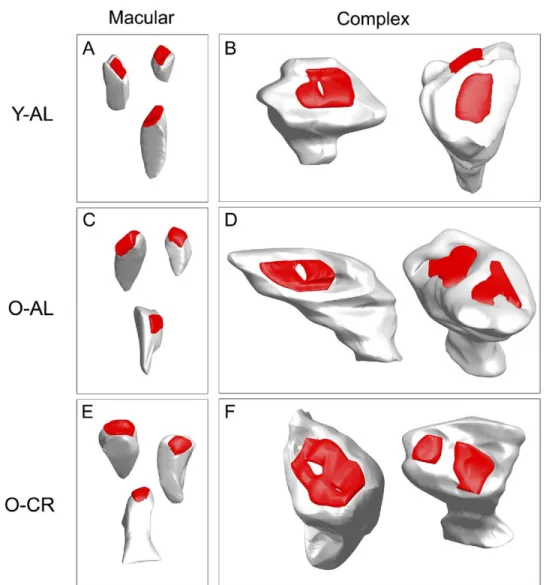
Only by using 3-D serial reconstruction is it possible to reliably distinguish macular from complex synapses that have been associated with age-related learning impair-ments (Nicholson et al., 2004) and functional plasticity such as LTP and long-term depression (LTD) (Geinisman et al., 1991, 1993, 1996; Mezey et al., 2004). Once the total number of synapses of each type in the reconstructed tissue sample was determined, synapse numbers were normalized to the stereologically determined number of neurons in that tissue sample as described in the Methods. Fig. 4. Serial reconstructions of postsynaptic densities (PSD) and spines in CA3 SL-M of young AL, old AL, and old CR rats. Based on the PSD
morphology, synapses were classified as macular or complex. Macular synapses demonstrated a simple, disk-shaped form; complex synapses demonstrated either partial or complete discontinuities (see Methods). Both types of synapses [macular (A, C, E) and complex (B, D, F)] and a similar range of spine morphologies were found in the all groups. Y-AL, young ad libitum fed; O-AL, old ad libitum fed; O-CR, old caloric restricted. Scale bar: 0.5m.
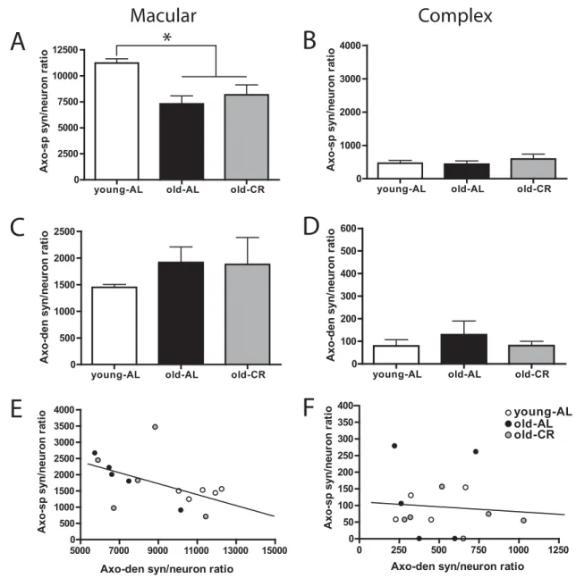
Fig 5illustrates the resulting synapse per neuron ratio for macular and complex synapses terminating on spines and dendritic shafts for each of the three groups in our study. Our data revealed a significant main effect of group for macular synapses terminating on spines (F(2,14)⫽7.584,
P⬍0.01;Fig. 5A). Post hoc analysis indicated a significant effect of age such that both old AL and CR rats had significantly fewer macular axo-spinous synapses than young AL fed animals (both P-values ⬍0.05; Fig. 5A). Moreover, there was no difference between old AL and CR fed animals (P⫽1.00; Fig. 5A). In contrast to the
age-related decline in macular axo-spinous synapses, no dif-ference among groups was detected for complex axo-spinous synapses (F(2,14)⫽0.544, P⬎0.5; Fig. 5B),
sug-gesting that complex synapses contacting spines are more stable across lifespan than are macular synapses, and also that the decline in macular synapses is not accompa-nied by an increase in complex synapses.
Interestingly, the pattern of change in macular axo-dendritic synapses appeared to mirror that seen across lifespan for macular synapses contacting spines. Although no significant group effect was detected (F(2,14)⫽0.595,
young-AL old-AL old-CR
0 2500 5000 7500 10000 12500 A x o -s p syn/ neur on ra ti o
young-AL old-AL old-CR
0 1000 2000 3000 4000
young-AL old-AL old-CR
0 500 1000 1500 2000 2500 Axo-den s y n /n e u ro n r a ti o
young-AL old-AL old-CR
0 100 200 300 400 500 600 5000 7000 9000 11000 13000 15000 0 500 1000 1500 2000 2500 3000 3500 4000 0 250 500 750 1000 1250 0 50 100 150 200 250 300 350 400 young-AL old-AL old-CR
A
C
E
B
D
F
*
Axo-sp s y n /n e u ro n r a ti o Axo-den s y n /n e u ro n r a ti o Axo-sp s y n /n e u ro n r a ti o Axo-sp s y n /n e u ro n r a ti oAxo-den syn/neuron ratio Axo-den syn/neuron ratio
Macular
Complex
Fig. 5. Quantification of macular and complex synapses terminating on spines and dendrites in CA3 SL-M of young AL, old AL, and old CR rats. The
total number synapses of each type in the reconstructed volume of S-LM was normalized to the stereologically determined number of neurons in that tissue block. An ANOVA revealed a main effect on macular axo-spinous synapses (A), but not complex axo-spinous synapses (B). Post hoc analysis indicated that the number of macular axo-spinous synapses decreased with age (P⬍0.05; * indicates significant decrease from young-AL group), but was not reversed by CR. No significant differences were observed for macular (C) or complex (D) axo-dendritic synapses. Data are presented as mean⫾ standard error of the mean (n⫽5). Correlational analysis revealed a significant negative correlation between macular axo-spinous and axo-dendritic synapses (r⫽⫺0.51, P⫽0.051; E), indicating that axo-dendritic synapses increased as axo-spinous synapses decreased. No relationship was observed for the complex axo-spinous and axo-dendritic synapses (F). Young-AL, young ad libitum fed rats (open bars); Old-AL, old ad libitum fed rats (black bars); Old-CR, old caloric restricted rats (gray bars). Axo-sp, Axo-spinous; Axo-den, Axo-dendritic.
M. M. Adams et al. / Neuroscience 171 (2010) 373–382 378
P⬎0.5;Fig. 5C), a trend for macular synapses on dendritic shafts to be more numerous than on spines was observed in both old AL and CR rats compared to the young group. No effect of age or diet was detected or trend observed for complex synapses contacting dendritic shafts (F(2,14)⫽
0.493, P⬎0.6; Fig. 5D). In order to assess whether a relationship existed between the significant age-related decrease in macular axo-spinous synapses and the appar-ent age-related increase in macular axo-dendritic syn-apses, we performed a correlational analysis within indi-vidual animals.Fig 5E illustrates the results of that analysis and demonstrates a significant inverse relationship be-tween axo-spinous and axo-dendritic macular synapses (r⫽⫺0.51; P⫽0.051) indicating that more macular syn-apses contact dendritic shafts as the prevalence of mac-ular synapses contacting spines decreases with age. No such relationship exists for complex spinous and axo-dendritic synapses (r⫽⫺0.08; P⫽0.76;Fig. 5F).
CA3 synapse size
As changes in synapse number could be accompanied by compensatory changes in PSD area, and changes in PSD area have been associated with altered excitatory trans-mission and plasticity (Desmond and Levy, 1986; Ganeshina et al., 2004a,b; Nicholson et al., 2006; Nichol-son and Geinisman, 2009; Stewart et al., 2005; Takumi et al., 1999), we also measured the PSD surface area of all reconstructed synapses. The results demonstrated no ef-fect of age or diet on PSD size (all P-values⬎0.25;Table 1). Thus, as synapses decrease in number, there is no concomitant change in the surface area of the PSD sug-gesting that synapse size is independent of age and di-etary condition.
The majority of excitatory transmission in the hip-pocampus occurs at spinous synapses and previous re-ports have indicated that the morphology of hippocampal spines is plastic, fluctuating with changes in hormone lev-els, learning and memory, and environmental complexity (De et al., 2008; Jedlicka et al., 2008; Knott and Holtmaat,
2008; Leuner and Shors, 2004; Sala et al., 2008). There-fore, we determined surface area and volume of the each postsynaptic spine in our 3-D serial reconstruction of SL-M. The results revealed no significant differences be-tween groups in either measure (all P-values⬎0.85;Table 1), suggesting that spine morphometry, like PSD size, does not demonstrate a compensatory change in response to the age-related loss of macular axo-spinous synapses.
DISCUSSION
The present study investigated whether age and/or CR alter the functional microcircuitry of the SL-M in CA3 of the hippocampus. Using 3-D serial reconstruction, we quanti-fied macular and complex synapses that terminated on dendritic spines and shafts as well as determined the size of the PSDs of those synapses and their postsynaptic spines. Our results demonstrated a significant age-related decline in macular axo-spinous synapses in SL-M that was correlated with an increase in macular axo-dendritic syn-apses. Complex synapses did not decline across lifespan, and no effect of CR was detected for any of the synapse classes in the present study. Finally, neither age nor CR affected the size of PSDs or postsynaptic spines.
Although quantitative electron microscopy has been used to evaluate age-related changes in the hippocampus, this is the first study to specifically evaluate the SL-M of CA3 using this technique. A previous stereological study reported a 24% overall decrease in CA3 synapses early in life, between 3 and 12 months (De Groot and Bierman, 1987). In the middle and inner molecular layers of the dentate gyrus, axo-spinous synapses have been reported to decrease between 5 and 28 months of age (Geinisman et al., 1992). However, stereological quantification of syn-apses in the middle molecular layer indicated no synapse decline from 10 through 29 months of age (Newton et al., 2007) suggesting a developmental reduction in the number of synapses in the dentate gyrus followed by a synaptic stability from adulthood through old age. In CA1, no quan-titative change in synapses was detected in stratum radia-tum either between 6 and 29 months (Geinisman et al., 2004) or from 10 through 29 months (Shi et al., 2007). Similarly, stereological quantification of synapses at the electron microscopic level in stratum lucidum of CA3 re-vealed no change across lifespan (Poe et al., 2001). In contrast, the present observation of an age-related syn-apse loss in SL-M of CA3 suggests that this layer may be uniquely sensitive to aging. This observation corroborates the results of a light microscopic study suggesting circuit-specific synaptic changes in the hippocampus across the life span (Adams et al., 2008; Smith et al., 2000). In the latter study, using immunodetection of synaptophysin within the layers of each hippocampal regions as a synap-tic marker, SL-M was the only layer in hippocampus that demonstrated significantly lower levels of synaptophysin in old, learning impaired rats compared either to young or old, unimpaired rats.
The age-related decline in SL-M synapses parallels the well-documented cognitive decline that occurs across the
Table 1. Synapse and spine areas and volumes in young AL, old AL,
and old CR rats
Young AL Old AL Old CR Synapse size
Axo-spinous synapse surface area macular (m2)
0.049 0.050 0.051 Axo-spinous synapse surface
area complex (m2 )
0.193 0.161 0.181 Axo-dendritic synapse surface
area macular (m2 )
0.091 0.092 0.091 Axo-dendritic synapse surface
area complex (m2)
0.108 0.148 0.171 Spine size
Spine surface area (m2) 0.598 0.568 0.580 Spine volume (m3) 0.050 0.049 0.051 No significant group differences were observed in any measure of the synapse or spine size. Data are reported as the group mean.
life span. It has been reported that progressive cognitive impairment was attenuated by caloric restriction as old CR rats performed significantly better on the MWM test of spatial learning and memory than age-matched AL rats (Adams et al., 2008; Markowska and Savonenko, 2002). The present results, however, indicated that both old CR and AL rats had fewer synapses than the young group. These findings suggest that cellular mechanisms, rather than alterations in synapse number, may underlie the higher cognitive functioning found in old CR rats. Consis-tent with this notion, we have previously reported an age-related decline in glutamate receptor subunit levels in hip-pocampus that is prevented by CR (Adams et al., 2008; Newton et al., 2007; Shi et al., 2007). Moreover, both NMDA and AMPA subunits in CA3 are significantly more abundant in old CR than old AL rats (Adams et al., 2008), and future immunogold studies will assess whether these changes are found at the synapse. Interestingly, CR has been reported to prevent the age-related decline in LTP as well as in the hippocampal expression of the NR1 subunit of NMDA receptors (Eckles-Smith et al., 2000). Thus, CR may enhance synaptic plasticity by altering synaptic protein levels in the hippocampus to compensate for a synaptic loss.
The size of hippocampal synapses has been repeat-edly associated with measures of hippocampal function. For example, aged animals with impaired cognition have been reported to demonstrate reductions in the surface area of synapses (Nicholson et al., 2004), and the PSD area of specific types of synapses in young rats increased both as a function of learning (Geinisman et al., 2004) and with LTP induction in the hippocampus (Geinisman et al., 1991, 1993, 1996; Weeks et al., 1999, 2001). Previous re-ports also have indicated that the abundance of AMPA-type glutamate receptors is related to the size and complexity of the PSD (Ganeshina et al., 2004a,b; Nicholson et al., 2006; Nicholson and Geinisman, 2009). As old CR rats have both higher levels of AMPA receptor subunits in CA3 (Adams et al., 2008) and better cognitive performance than old AL rats (Adams et al., 2008; Markowska and Savonenko, 2002), we hypothesized that PSDs might be larger size in old CR than old AL rats in the present study. Nevertheless, our results demonstrated no greater PSD size or number of complex synapses in old CR rats.
Spine surface area and volume also did not differ between groups and thus provided no morphological evi-dence of spine plasticity in response to age or diet. Al-though no compensatory change in dendritic spine mor-phology was detected in SL-M, age-related or a CR-in-duced changes impacting cognitive function could occur in a different layer or region of the hippocampus. Such re-gion-specific dendritic changes in the hippocampus have been shown in response to manipulations such as ovarian steroids and stress induction. For example, estrogen in-creased spine number in the CA1 region of the hippocam-pus, but had no effect on the dentate gyrus or CA3 region (Woolley et al., 1990a). Moreover, exposure to excessive glucocorticoids altered dendritic morphology only in CA3 causing atrophy of the apical, but not basal, dendrites in the stratum oriens (Woolley et al., 1990b). These data
support the idea that even in a single hippocampal region, manipulations of the cellular microenvironment can pro-duce differential dendritic responses across layers.
Our data suggest an age-related shift in the relative abundance of axo-spinous and axo-dendritic synapses. In a previous study examining the effects of LTP and LTD on the functional microcircuitry of the dentate gyrus, it was observed that LTP of the medial perforant path led to an increase in axo-spinous synapses, and that LTD was as-sociated with an increase in axo-dendritic synapses (Mezey et al., 2004). That report supports the idea that the inverse correlation in macular axo-spinous and axo-dendritic synapse numbers observed here may be associated with functional alterations within the hippocampus. Consistent with this possibility are the observed age-related decreases in hippocampal LTP (Eckles-Smith et al., 2000; Molina et al., unpublished observations) and the propensity of aged ani-mals for increased LTD (Foster and Kumar, 2007; Kumar and Foster, 2005; Norris et al., 1996). An increase in the amount of LTD would likely keep the synapses from being able to be strengthened and alter the excitatory/inhibitory balance, and thus, could contribute to some of the memory losses ob-served with aging.
The results of this study of the microcircuitry of S-LM of CA3 provide the first ultrastructural evidence that macular synapses in this layer demonstrate a unique vulnerability to aging. Nevertheless, PSD size and spine surface area and volume are stable between young and old age. Moreover, despite the age-related loss of macular axo-spinous syn-apses and the previously reported CR-induced benefits for behavior and glutamate receptors in old animals, no effect of CR on the measures of S-LM microcircuitry evaluated in the present study were detected. Accordingly, the behavioral benefits of CR may be mediated by the effects of CR on synaptic protein levels in CA3 in conjunction with cellular and functional alterations elsewhere in the hippocampus. Acknowledgments—This work was supported by NIH grants: NIA PO1 AG11370, RO1 AG019886, and KO1 AG027828.
REFERENCES
Adams MM, Shi L, Linville MC, Forbes ME, Long AB, Bennett C, Newton IG, Carter CS, Sonntag WE, Riddle DR, Brunso-Bechtold JK (2008) Caloric restriction and age affect synaptic proteins in hippocampal CA3 and spatial learning ability. Exp Neurol 211: 141–149.
Cabalka LM, Hyman BT, Goodlett CR, Ritchie TC, Van Hoesen GW (1992) Alteration in the pattern of nerve terminal protein immuno-reactivity in the perforant pathway in Alzheimer’s disease and in rats after entorhinal lesions. Neurobiol Aging 13:283–291. De Groot DMG, Bierman EPB (1987) Numerical changes in rat
hip-pocampal synapses. An effect of “aging”? Acta Stereol 6:53–58. De RM, Klauser P, Garcia PM, Poglia L, Muller D (2008) Spine
dynamics and synapse remodeling during LTP and memory pro-cesses. Prog Brain Res 169:199 –207.
Desmond NL, Levy WB (1986) Changes in the postsynaptic density with long-term potentiation in the dentate gyrus. J Comp Neurol 253:476 – 482.
Donohue HS, Gabbott PL, Davies HA, Rodriguez JJ, Cordero MI, Sandi C, Medvedev NI, Popov VI, Colyer FM, Peddie CJ, Stewart MG (2006) Chronic restraint stress induces changes in synapse M. M. Adams et al. / Neuroscience 171 (2010) 373–382
morphology in stratum lacunosum-moleculare CA1 rat hippocampus: a stereological and three-dimensional ultrastructural study. Neuro-science 140:597– 606.
Eckles-Smith K, Clayton D, Bickford P, Browning MD (2000) Caloric restriction prevents age-related deficits in LTP and in NMDA re-ceptor expression. Brain Res Mol Brain Res 78:154 –162. Foster TC, Kumar A (2007) Susceptibility to induction of long-term
depression is associated with impaired memory in aged Fischer 344 rats. Neurobiol Learn Mem 87:522–535.
Ganeshina O, Berry RW, Petralia RS, Nicholson DA, Geinisman Y (2004a) Differences in the expression of AMPA and NMDA recep-tors between axospinous perforated and nonperforated synapses are related to the configuration and size of postsynaptic densities. J Comp Neurol 468:86 –95.
Ganeshina O, Berry RW, Petralia RS, Nicholson DA, Geinisman Y (2004b) Synapses with a segmented, completely partitioned postsynaptic density express more AMPA receptors than other axospinous synaptic junctions. Neuroscience 125:615– 623. Geinisman Y, Detoledo-Morrell L, Morrell F (1991) Induction of
long-term potentiation is associated with an increase in the number of axospinous synapses with segmented postsynaptic densities. Brain Res 566:77– 88.
Geinisman Y, Detoledo-Morrell L, Morrell F, Persina IS, Beatty MA (1996) Synapse restructuring associated with the maintenance phase of hippocampal long-term potentiation. J Comp Neurol 368:413– 423.
Geinisman Y, Ganeshina O, Yoshida R, Berry RW, Disterhoft JF, Gallagher M (2004) Aging, spatial learning, and total synapse number in the rat CA1 stratum radiatum. Neurobiol Aging 25: 407– 416.
Geinisman Y, Toledo-Morrell L, Morrell F, Heller RE, Rossi M, Parshall RF (1993) Structural synaptic correlate of long-term potentiation: formation of axospinous synapses with multiple, completely parti-tioned transmission zones. Hippocampus 3:435– 445.
Geinisman Y, Toledo-Morrell L, Morrell F, Persina IS, Rossi M (1992) Age-related loss of axospinous synapses formed by two afferent systems in the rat dentate gyrus as revealed by the unbiased stereological dissector technique. Hippocampus 2:437– 444. Goto S, Hirano A (1990) Synaptophysin expression in the striatum in
Huntington’s disease. Acta Neuropathol 80:88 –91.
Heinonen O, Soininen H, Sorvari H, Kosunen O, Paljarvi L, Koivisto E, Riekkinen PJ Sr (1995) Loss of synaptophysin-like immunoreac-tivity in the hippocampal formation is an early phenomenon in Alzheimer’s disease. Neuroscience 64:375–384.
Jedlicka P, Vlachos A, Schwarzacher SW, Deller T (2008) A role for the spine apparatus in LTP and spatial learning. Behav Brain Res 192:12–19.
Knott G, Holtmaat A (2008) Dendritic spine plasticity— current under-standing from in vivo] studies. Brain Res Rev 58:282–289. Kumar A, Foster TC (2005) Intracellular calcium stores contribute to
increased susceptibility to LTD induction during aging. Brain Res 1031:125–128.
Leuner B, Shors TJ (2004) New spines, new memories. Mol Neurobiol 29:117–130.
Markowska AL, Savonenko A (2002) Retardation of cognitive aging by life-long diet restriction: implications for genetic variance. Neurobiol Aging 23:75– 86.
Martin B, Pearson M, Brenneman R, Golden E, Keselman A, Iyun T, Carlson OD, Egan JM, Becker KG, Wood W III, Prabhu V, de Cabo R, Maudsley S, Mattson MP (2008) Conserved and differential effects of dietary energy intake on the hippocampal transcriptomes of females and males. PLoS One 3:e2398.
Martin B, Pearson M, Kebejian L, Golden E, Keselman A, Bender M, Carlson O, Egan J, Ladenheim B, Cadet JL, Becker KG, Wood W, Duffy K, Vinayakumar P, Maudsley S, Mattson MP (2007) Sex-dependent metabolic, neuroendocrine, and cognitive responses to dietary energy restriction and excess. Endocrinology 148:4318 – 4333.
Masliah E, Mallory M, Ge N, Alford M, Veinbergs I, Roses AD (1995) Neurodegeneration in the central nervous system of apoE-deficient mice. Exp Neurol 136:107–122.
Masliah E, Mallory M, Hansen L, DeTeresa R, Alford M, Terry R (1994) Synaptic and neuritic alterations during the progression of Alzhei-mer’s disease. Neurosci Lett 174:67–72.
Masliah E, Mallory M, Hansen L, DeTeresa R, Terry RD (1993) Quan-titative synaptic alterations in the human neocortex during normal aging. Neurology 43:192–197.
Mayhew M, Renganathan M, Delbono O (1998) Effectiveness of ca-loric restriction in preventing age-related changes in rat skeletal muscle. Biochem Biophys Res Commun 251:95–99.
Mezey S, Doyere V, De Souza I, Harrison E, Cambon K, Kendal CE, Davies H, Laroche S, Stewart MG (2004) Long-term synaptic morphometry changes after induction of long-term potentiation and long-term depression in the dentate gyrus of awake rats are not simply mirror phenomena. Eur J Neurosci 19:2310 –2318. Newton IG, Forbes ME, Legault C, Johnson JE, Brunso-Bechtold JK,
Riddle DR (2005) Caloric restriction does not reverse aging-related changes in hippocampal BDNF. Neurobiol Aging 26:683– 688. Newton IG, Forbes ME, Linville MC, Pang H, Tucker EW, Riddle DR,
Brunso-Bechtold JK (2007) Effects of aging and caloric restriction on dentate gyrus synapses and glutamate receptor subunits. Neu-robiol Aging 29:1308 –1318.
Nicholson DA, Geinisman Y (2009) Axospinous synaptic subtype-specific differences in structure, size, ionotropic receptor expres-sion, and connectivity in apical dendritic regions of rat hippocampal CA1 pyramidal neurons. J Comp Neurol 512:399 – 418.
Nicholson DA, Trana R, Katz Y, Kath WL, Spruston N, Geinisman Y (2006) Distance-dependent differences in synapse number and AMPA receptor expression in hippocampal CA1 pyramidal neu-rons. Neuron 50:431– 442.
Nicholson DA, Yoshida R, Berry RW, Gallagher M, Geinisman Y (2004) Reduction in size of perforated postsynaptic densities in hippocampal axospinous synapses and age-related spatial learn-ing impairments. J Neurosci 24:7648 –7653.
Norris CM, Korol DL, Foster TC (1996) Increased susceptibility to induction of long-term depression and long-term potentiation re-versal during aging. J Neurosci 16:5382–5392.
Peters A, Palay SL (1991) The fine structure of the nervous system, neurons, and their supporting cells. Oxford, England: Oxford Uni-versity Press.
Poe BH, Linville C, Riddle DR, Sonntag WE, Brunso-Bechtold JK (2001) Effects of age and insulin-like growth factor-1 on neuron and synapse numbers in area CA3 of hippocampus. Neuroscience 107:231–238.
Ramsey MM, Weiner JL, Moore TP, Carter CS, Sonntag WE (2004) Growth hormone treatment attenuates age-related changes in hip-pocampal short-term plasticity and spatial learning. Neuroscience 129:119 –127.
Sala C, Cambianica I, Rossi F (2008) Molecular mechanisms of den-dritic spine development and maintenance. Acta Neurobiol Exp Wars 68:289 –304.
Shi L, Adams MM, Linville MC, Newton IG, Forbes ME, Long AB, Riddle DR, Brunso-Bechtold JK (2007) Caloric restriction elimi-nates the aging-related decline in NMDA and AMPA receptor subunits in the rat hippocampus and induces homeostasis. Exp Neurol 206:70 –79.
Shi L, Poe BH, Constance LM, Sonntag WE, Brunso-Bechtold JK (2002) Caloric restricted male rats demonstrate fewer synapses in layer 2 of sensorimotor cortex. Brain Res 931:32– 40.
Smith TD, Adams MM, Gallagher M, Morrison JH, Rapp PR (2000) Circuit-specific alterations in hippocampal synaptophysin immuno-reactivity predict spatial learning impairment in aged rats. J Neu-rosci 20:6587– 6593.
Stewart MG, Medvedev NI, Popov VI, Schoepfer R, Davies HA, Mur-phy K, Dallerac GM, Kraev IV, Rodriguez JJ (2005) Chemically induced long-term potentiation increases the number of perforated
and complex postsynaptic densities but does not alter dendritic spine volume in CA1 of adult mouse hippocampal slices. Eur J Neurosci 21:3368 –3378.
Takumi Y, Ramirez-Leon V, Laake P, Rinvik E, Ottersen OP (1999) Different modes of expression of AMPA and NMDA receptors in hippocampal synapses. Nat Neurosci 2:618 – 624.
Toni N, Buchs PA, Nikonenko I, Povilaitite P, Parisi L, Muller D (2001) Remodeling of synaptic membranes after induction of long-term potentiation. J Neurosci 21:6245– 6251.
Weeks AC, Ivanco TL, Leboutillier JC, Racine RJ, Petit TL (1999) Sequential changes in the synaptic structural profile following long-term potentiation in the rat dentate gyrus. I. The inlong-termediate maintenance phase. Synapse 31:97–107.
Weeks AC, Ivanco TL, Leboutillier JC, Racine RJ, Petit TL (2001) Sequential changes in the synaptic structural profile following long-term potentiation in the rat dentate gyrus. III. Long-long-term mainte-nance phase. Synapse 40:74 – 84.
Wetter TJ, Gazdag AC, Dean DJ, Cartee GD (1999) Effect of calorie restriction on in vivo] glucose metabolism by individual tissues in rats. Am J Physiol 276:E728E738.
Woolley CS, Gould E, Frankfurt M, McEwen BS (1990a) Naturally occurring fluctuation in dendritic spine density on adult hippocam-pal pyramidal neurons. J Neurosci 10:4035– 4039.
Woolley CS, Gould E, McEwen BS (1990b) Exposure to excess glu-cocorticoids alters dendritic morphology of adult hippocampal py-ramidal neurons. Brain Res 531:225–231.
(Accepted 14 September 2010) (Available online 18 September 2010)
M. M. Adams et al. / Neuroscience 171 (2010) 373–382 382