CASE REPORT
A rare reason of foot drop caused by primary diffuse
large b-cell lymphoma of the sciatic nerve: case report
Serdar Kahraman&Hakan Sabuncuoglu&
Omer Gunhan&Mehmet Ali Gurses&Sait Sirin
Received: 8 November 2008 / Accepted: 19 March 2009 / Published online: 5 May 2009 # Springer-Verlag 2009
Abstract
Introduction Primary central nervous system lymphomas account for 2% of all malignant lymphomas. Although the involvement of peripheral nerves has been previously described as a dissemination of systemic lymphomas or a direct extension to the nerve trunk from contiguous lymphomas, primary involvement of the sciatic nerve is extremely rare.
Case To the best of our knowledge, the primary localiza-tion of lymphoma within sciatic nerve has been reported only nine times. We report, a very rare example of a primary diffuse large B-cell lymphoma of the sciatic nerve. Discussion The patient presented with atypical sciatica. Such symptoms can be misdiagnosed as lumbar disc pathology and magnetic resonance imaging and electro-physiological studies avoid this misinterpretation.
Keywords B cell . Lymphoma . Sciatic . Peripheral . Nerve
Introduction
Primary extranodal lymphomas have been described in virtually every site of the body. Primary central nervous system (CNS) lymphomas constitute 1% of all intracranial tumors and 2% of all malignant lymphomas [3, 4, 9]. Although the involvement of peripheral nerves has been previously described as a dissemination of a systemic lymphoma or as a direct extension into the nerve from a contiguous lymphoma, a primary lymphoma of the sciatic nerve is extremely rare [16].
Case report
A 63 year-old woman had complained severe pains in the back of her left thigh, radiating down to the ankle in a way very similar to sciatica. She was conservatively for a year. However, the initial neurological examination had shown a grade 1/5 muscle power of the left ankle dorsoflexion, a positive single leg raising test at 200. Numbness was found in left L5 dermatomal area. After a diagnosis of L4–5 herniation was made on lumbosacral magnetic resonance imaging (MRI), she underwent a L4 hemipartial laminec-tomy, L4–5 discectomy and left L5 foraminotomy. On the second day, she was found to have a loss of left leg flexion, foot dorsiflexion/supination, 2–5 toes extension, great toe extension, foot plantar flexion/supination and 2–5 terminal flexion and ankle jerk. Lumbosacral MRI showed only postoperative changes. Electrophysiological study showed a total denervation of the left sciatic nerve. Lumbosacral plexus T2 weighted MRI (Fig. 1), showed thickening and
Acta Neurochir (2010) 152:125–128 DOI 10.1007/s00701-009-0339-9
S. Kahraman
:
S. SirinDepartment of Neurosurgery, Gülhane Military Medical Academy, Etlik,
Ankara, Turkey H. Sabuncuoglu
Department of Neurosurgery, Ufuk University, Ankara, Turkey
O. Gunhan
Department of Pathology, Gülhane Military Medical Academy, Etlik,
Ankara, Turkey M. A. Gurses
Integra Advanced Diagnosis and Research Center, Ankara, Turkey
H. Sabuncuoglu (*)
Koza Sokak No:72/36 Ikizler Sitesi 06700 Gaziosmanpasa, Ankara, Turkey
hyperintensity of the left sciatic nerve. After IV gadolinium injection, diffuse contrast enhancement throughout the left sciatic nerve was seen on coronal T1 weighted MRI (Fig.2).
The patient’s history included sarcoidosis but there was no history of cancer, neurogenetic disorder nor use of immuno-suppressive medication. Despite that no abnormalities were found in tests of angiotensin converting enzyme, antinuclear antibody, antilymphocyte antibody or antiphosholipid anti-body. No abnormalities were found in WBC, erythrocyte sedimentation rate, C-reactive protein, HBsAg, Anti HIV, anti HCV, chest x-ray and PPD. Physical examination showed no evidence of cardiopulmonary, hepatorenal or splenic abnor-mality nor systemic lympadenopathy.
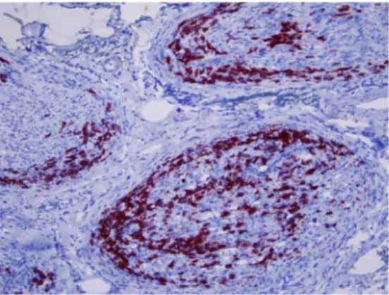
The patient was then referred to our department and exploration carried out. A 30 cm long vertical incision was made in the left gluteal area and fusiform expansion of sciatic nerve was seen in the piriform canal . There was not a clear border between the sciatic nerve and pathological soft tissue. A subtotal excision was performed and the incision was closed in a routine manner. Histopathological examination showed a diffuse large B cell lymphoma (DLBCL) with infiltration confined to the peripheral nerve fibers (Fig. 3). Tumor was composed of large atypical lymphoid cells having prominent nucleoli. The tumor cells expressed LCA, CD20 and CD 79A immunopositivity (Fig.4). There was no immunoreactivity cytokeratin, HMB 45, CD3, CD30, BCL-6 on myeloperoxidase.
Cranial, chest, abdominal CT, scintigraphy and lumbar puncture were performed but another primary focus of non-Hodgkin lymphoma (NHL) was not found. There was little improvement in the left leg and she was referred to the hematology department for additional therapy. After 10 months of chemotherapy protocol (cytoxan, hydroxy-daunorubicin, oncovin, and prednisone (CHOP) with rituximab), she had no recurrence but the muscle power remained 1/5 in the left leg.
Fig. 1 Axial MR examination demonstrates increased signal and thickening of the sciatic nerve in the fat-suppressed T2A images
Fig. 2 After IV contrast agent injection, coronal MR examinationde-monstrates that sciatic nerve is thickened and intense along its course, there is diffuse contrast accumulation in the fat-suppressed T1A images
Fig. 3 Large B cell lymphoma cells showing neurotrophic pattern of growth.HEX50
Fig. 4 The tumor was composed of cells express CD20 immunopo-sitivity. Avidin-biotin immunoperoxidase stainingX400
Discussion
Malignant NHLs are defined as the malignant proliferation of B or T lymphocytes [16]. DLBCL, the most common form, is responsible for one-third of NHLs [19].
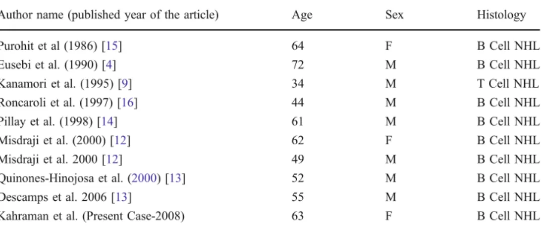
These fatal neoplasms are most common in adults with or without a history of immunosupression. Usually, they are present in lymph nodes rather than in extranodal sites. When lymphomatous infiltration of spinal nerve roots, dorsal root ganglia and peripheral nerves occurs, it is usually a consequence of hematogenous spread from systemic NHL or the result of invasion from an adjacent structure [16]. The involvement of the nervous system as extranodal site is seen in approximately 5% of the patients with NHL. They are usually large B-cell types and have a variable prognosis [16]. The term neurolymphomatosis has been used for circumstances in which multiple peripheral nerves are infiltrated by lymphoma [12]. To the best of our knowledge, there are few reports of neurolymphomatosis presenting with the tumor confined largely to the peripheral nervous system [12], and most of patients have shown concomitant central nervous system involvement [6,9,16]. There are 9 reports of a primary nerve lymphoma in the sciatic nerve (Table 1) and 5 more reports of a primary lymphoma in another peripheral nerve, including the brachial plexus, ulnar, radial and median nerves, and sympathetic chain [3].
Lymphomas that occur in the central nervous system are relatively uncommon but well documented. Although there has been an increase in primary CNS lymphomas, both in patients with acquired immunodeficiency syndrome and immunocompetent individuals [2, 5, 7, 8, 13], primary NHLs of the peripheral nervous system are highly unusual neoplastic conditions that occur exclusively in the sciatic nerve [3,4,14–16].
The relationship of primary lymphoma of the peripheral nerves to other extranodal lymphomas and the reason for the localization of lymphoma to the peripheral nerve are unclear. One theory is the protection from immunologic
surveillance by the blood-nerve barrier. Although endoneu-ral involvement of the lymphoma has not been demonstrat-ed in all patients, some cases have confirmdemonstrat-ed this [10,16]. Quinones-Hirojosa et al. postulated that B cells reside or originate in the sciatic nerve [12]. More likely, specific cell adhesion molecule expansion is the basis for tissue targeting, but these are yet to be identified. The absence of disease elsewhere suggests that primary lymphoma of a peripheral nerve may have unique characteristics [3].
Clinically, sciatic nerve involvement is characterized by progressive paresthesia, numbness and weakness of the lower limb that are associated with pain. The symptoms related to peripheral nervous system diseases or lumbar disc herniation are mostly similar. Hence, this often causes the patient to seek medical attention [12]. The grade of lymphoma does not affect the outcome. In polyradiculo-pathies due to infiltrations of the lumbosacral plexus, features can include back pain, weakness, sensory loss, decreased deep tendon reflexes, cauda equina syndrome, paresthesia and such findings as urinary frequency and constipation related to autonomic dysfunction [19].
Enlargement of the nerve due to a lymphoma is indistinguishable from a schwannoma on computed tomog-raphy (CT) or MRI scan. In the absence of evidence of a primary focus of NHL in other organs, in our patient, the pathogenesis may be extranodal development of lymphoma within the sciatic nerve. The original lymphoma might have arisen from B cells that normally course through or reside in the sciatic nerve. It is also possible that additional spread into CNS along peripheral nerve tracts and systemic organs may have occurred.
Although the optimal treatment for primary peripheral nerve lymphomas is still under debate, extended surgical treatment and additional local radiotherapy or systemic chemotherapy should be considered. In view of the high mortality of B-cell lymphoma, aggressive and prophylactic treatment of CNS may be necessary, even in the absence of initial involvement [12]. The initial treatment of all patients with DLBCL should be with a combination chemotherapy
Author name (published year of the article) Age Sex Histology
Purohit et al (1986) [15] 64 F B Cell NHL
Eusebi et al. (1990) [4] 72 M B Cell NHL
Kanamori et al. (1995) [9] 34 M T Cell NHL
Roncaroli et al. (1997) [16] 44 M B Cell NHL
Pillay et al. (1998) [14] 61 M B Cell NHL
Misdraji et al. (2000) [12] 62 F B Cell NHL
Misdraji et al. 2000 [12] 49 M B Cell NHL
Quinones-Hinojosa et al. (2000) [13] 52 M B Cell NHL
Descamps et al. 2006 [13] 55 M B Cell NHL
Kahraman et al. (Present Case-2008) 63 F B Cell NHL
Table 1 Clinical and histologi-cal specialities of reported ten cases of sciatic nerve lymphoma
regimen. The most popular regimen in the United States is cytoxan, hydroxydaunorubicin, oncovin, and prednisone (CHOP), sometimes in combination with rituximab, although a variety of other antracycline-containing combination chemotherapy regimens appear to be equally efficacious. Patients with stage I or nonbulky stage II can be effectively treated with three or four cycles of combination chemother-apy, and cure rates of 60 to 70% in stage II disease and 80 to 90% in stage I disease can be expected. For patients with bulky stage II, stage III or stage IV, six to eight cycles of a combination chemotherapy regimen such as CHOP often in combination with rituximab, are usually administered. Be-cause a large number of patients with diffuse large B cell lymphoma are either initially refractory to therapy or relapse after apparently effective chemotherapy, nearly half of patients will be candidates for salvage treatment at some point. Alternative combination chemotherapy regimens can induce complete remission in as many as 50%of these patients, but long term disease free survival is seen in less than 10% [1].
Lymphomas occasionally recur in the peripheral nervous system after treatment, with either single or multiple involvements. Recurrence in multiple peripheral nerves may be accompanied by CNS involvement. This suggests that the blood-nerve barrier may protect tumor cells from therapeutic agents [11,12,17,18]. In addition, sciatic nerve lymphomas, compared with CNS lymphomas, seem to behave more aggressively.
In conclusion, sciatic nerve involvement may be consid-ered as a rare selective occurrence site of a peripheral nerve lymphoma. In the presence of atypical sciatica, a sciatic nerve tumour should be in the differential diagnosis in order to avoid misdiagnosis as lumbar disc pathology. Hence, electrophysiological studies and extended MRI must be performed in order to establish the diagnosis.
References
1. Armitage JO, Lorgo DL (2005) Malignancies of lymhoid cells. In: Kasper DL, Braunwald E, Anthony SF, Hauser SL, Longo DL, Jameson JL (eds) Harrison’S principles of internal medicine, 16th edn. Mc Graw Hill, New York, pp 651–652
2. DeMario MD, Liebowitz DN (1998) Lymphomas in the immuno-compromised patient. Semin Oncol 25(4):492–502
3. Descamps MJ, Barret L, Yung L, Birch R, Murray NMF, Linch DC, Lunn MPT, Reilly MM (2006) Primary sciatic nerve lymphoma: a case report and review of the literature. J Neurol Neurourg Psychiatry 77:1087–1089
4. Eusebi V, Bondi A, Cancellieri A, Canedi L, Frizzera G (1990) Primary malignant lymphoma of sciatic nerve: Report of a case. The American Journal of Surgical Pathology 14(9):881–885 5. Feigal EG (1999) AIDS-associated malignancies: research
perspec-tives. Biochim Biophys Acta 1423(1):C1–9
6. Fine HA, Mayer RJ (1993) Primary central nervous system lymphoma. Ann Intern Med 119(11):1093–1104
7. Grant JW, Isaacson PG (1992) Primary central nervous system lymphoma. Brain Pathol 2(2):97–109
8. Jellinger KA, Paulus W (1992) Primary central nervous system lymphomas–an update. J Cancer Res Clin Oncol 119(1):7–27 9. Kanamori M, Matsui H, Yudoh K (1995) Solitary T-cell
lymphoma of the sciatic nerve: case report. Neurosurgery 36 (6):1205–1205
10. Kim J, Kim YS, Lee EJ, Kang CS, Shim SI (1998) Primary CD56-positive NK/T-cell lymphoma of median nerve: a case report. J Korean Med Sci 13(3):331–333
11. Krendel DA, Stahl RL, Chan WC (1991) Lymphomatous polyneuropathy; biopsy of clinically involved nerve and success-ful treatment. Arch Neurol 48(3):330–332
12. Misdraji J, Ino Y, Louis DN, Rosenberg AE, Chiocca EA, Harris NL (2000) Primary lymphoma of peripheral nerve: Report of four cases. Am J Surg Pathol 24(9):1257–1265
13. Quinones-Hinojosa A, Friedlander RM, Boyer PJ, Batchelor TT, Chiocca EA (2000) Solitary sciatic nerve lymphoma as an initial manifestation of diffuse neurolymphomatosis. Case report and review of the literature. J Neurosurg 92(1):165–169
14. Pillay PK, Hardy RW, Wilbourn AJ, Tubbs RR, Lederman RJ (1998) Solitary primary of the sciatic nerve: case report. Neurosurgery 23:370–371
15. Purohit DP, Dick DJ, Perry RH, Lyons PR, Schofield IS, Foster JB (1986) Solitary extranodal lymphoma of sciatic nerve. J Neurol Sci 74(1):23–34
16. Roncaroli F, Poppi M, Riccioni L, Frank F (1997) Primary non-hodgkin’s Lymphoma of the sciatic nerve followed by localization in the central nervous system: case report and review of the literature. Neurosurgery 40(3):618–21 discussion 621-2
17. Shoenfeld Y, Aderka D, Sandbank U, Gadot N, Santo M, Pinkhas J (1983) Fatal peripheral neurolymphomatosis after remission of histiocytic lymphoma. Neurology 33(2):243–245
18. van den Bent MJ, de Bruin HG, Beun GD, Vecht CJ (1995) Neurolymphomatosis of the median nerve. Neurology 45 (7):1403–1405
19. Yavasoglu A, Kadikoylu G, Ozkul A, Bolaman Z (2006) Foot-drop due to involvement of lumbosacral plexus in diffuse large B-cell lymphoma. Turk J Hematol 23(1):63–67