Yazışma Adresi/Address for Correspondence: Dr. Derya Kanza Gül, Medipol University School of Medicine Health, Istanbul, Turkey. E-mail:[email protected]
Geliş tarihi/Received: 01.12.2020 Kabul tarihi/Accepted: 24.12.2020 Çevrimiçi yayın/Published online: 30.12.2020
ARAŞTIRMA / RESEARCH
Can blood eosinophil to basophil ratio, eosinophil to lymphocyte ratio
and basophil to lymphocyte ratio predict the severity of intrahepatic
cholestasis in pregnancy?
Kan eozinofil-bazofil oranı, eozinofil-lenfosit oranı ve bazofil-lenfosit oranı değerleri
gebelikteki intrahepatik kolestazın şiddetini tahmin edebilir mi?
Derya Kanza Gül1 , Ayça Şolt Kırca2
1Medipol University School of Medicine Health, Istanbul, Turkey.
2Kirklareli University School of Health, Midwifery Depertmant, Kırklareli, Turkey Cukurova Medical Journal 2020;45(4):1742-1749
Abstract Öz
Purpose: The purpose of the study was to investigate the
relation between inflammatory markers used for allergic diseases and intrahepatic pregnancy cholestasis (ICP).
Materials and Methods: In this retrospective
case-control study, a total of 114 intrahepatic pregnancy cholestasis patients; 87 with mild cholestasis (group 1) and 27 with severe cholestasis (group 2) and 292 healthy pregnant women in the control group (group 3) were included in the study. The basophil, eosinophil, eosinophil-basophil ratio (EBR), eosinophil-lymphocyte ratio (ELR) and basophil-lymphocyte ratio (BLR), neutrophil-lymphocyte ratio (NLR), and platelet-lymphocyte ratio (PLR) values used in this study.
Results: Significant differences were detected between
groups in terms of ELR, NLR and PLR values. ELR, NLR, PLR values were significantly higher in the mild cholestasis group than in the control group. To predict the pregnancy cholestasis, receiver operating characteristic curve (ROC) analysis was made, which yielded 76.3% sensitivity and 56.1% specificity when ELR’s interrupt value was 0.2243, NLR interrupt value 11.27% when sensitivity was 70% and specificity 54,000 2%, and PLR cut-off value was 194.46 when sensitivity was 60% and specificity was 58%.
Conclusion: Serum inflammation markers and allergy
markers may be diagnostic in intrahepatic pregnancy cholestasis; however, they are not decisive in determining the severity of the disease.
Amaç: Alerjik hastalıklar için kullanılan inflamatuvar
belirteçler ile intrahepatik gebelik kolestazı (ICP) arasındaki ilişkiyi araştırmayı amaçladık.
Gereç ve Yöntem: Bu retrospektif vaka-kontrol
çalışmasına 87si hafif kolestazlı (grup 1) ve 27si şiddetli kolestazlı (grup 2) olmak üzere toplam 114 intrahepatik gebelik kolestazlı hasta ve kontrol grubunda 292 sağlıklı gebe (grup 3) çalışmaya dahil edildi. Bu çalışmada kullanılan bazofil, eozinofil, eozinofil-bazofil oranı (EBO), eozinofil-lenfosit oranı (ELO) ve bazofil-lenfosit oranı (BLO), nötrofi-lenfosit oranı (NLO) ve platelet-lenfosit oranı (PLO) değerleri herhangi bir tıbbi veya cerrahi işlem başlatılmadan önce alınan kanlardan değerlendirilmiştir.
Bulgular: ELO, NLO ve PLO değerleri açısından gruplar
arasında anlamlı bir fark olduğu saptandı. ELO, NLO, PLO değerleri hafif kolestaz grubunda kontrol grubuna göre anlamlı olarak daha yüksekti. Gebelik kolestazını tahmin etmek için, alıcı işletim karakteristik eğrisi (ROC) analizi kullanıldı. ELO'nın kesme değeri 0,2243 olduğu zaman %76,3 duyarlılık ve %56,1 özgüllük, NLO kesme değeri 11.27 olduğu zaman duyarlılığı 70% ve özgüllük 54,2%, PLO kesme değeri 194,46 olduğu zaman duyarlılığı 60% ve özgüllük 58%olarak saptandı.
Sonuç: Serum inflamasyon belirteçleri ve alerji belirteçleri
intrahepatik gebelik kolestazda tanı koydurucu olabilir ancak hastalığın şiddetini saptamada belirleyici değildir.
Keywords: Intrahepatic cholestasis pregnancy, allergy
INTRODUCTION
Intrahepatic cholestasis of pregnancy (ICP) is the most common liver disease during pregnancy and characterized with widespread itching, high serum bile acid levels (≥10 μmol /L) and/or high liver enzyme levels1. It is usually detected in the second & third trimesterof pregnancy2.
High bile acid levels lead to pregnancy complications such as preterm birth, fetal distress, fetal and maternal sudden death. The risk of complications is higher in patients with severe ICP. Intrauterine fetal mortality with cardiograph, fetal movements and cannot be predicted with Doppler Ultrasonography3. For this reason, predicting the diagnosis and severity of ICP is very important for the outcomes of pregnancy.
Environmental factors, diet habits, hormonal changes, genetic variations and uterine pressure were reported as the responsible factors for the pathogenesis of ICP4. An important cause responsible for the formation of pregnancy cholestasis is inflammation; however, the mechanism that initiates this inflammation is not known5. Inflammatory cells due to clogging cholestasis become activated, and cause damage to hepatocytes and bile ducts. The course of the disease varies depending on the local and intra-cell response of hepatocytes6.
Diagnostic bile acid test in ICP is costly and difficult to obtain and time-consuming, which makes it necessary to investigate cost-effective tests that can be easily applied to all segments of the society, including in primary healthcare organizations, in a short time period, and which can be easily interpreted by every healthcare worker.
When the literature was reviewed, the efficiency of inflammation markers obtained from CBC was examined in recurrent miscarriages7, preeclampsia8, intrauterine developmental retardation9, gestational diabetes10 and intrahepatic cholestasis of pregnancy11 Although there are studies that found that inflammatory markers are statistically significant in the diagnosis of ICP, there was no study examining hematologic allergy markers such as eosinophil to lymphocyte ratio (ELR), eosinophil to basophil ratio (EBR), basophil to lymphocyte ratio (BLR). For this reason, we aimed to determine the role of easily administered, calculated and cost-effective allergy markers in detecting ICP and clinically early prediction of its severity.
MATERIALS AND METHODS
The ethics committee approval was obtained from Medipol University for Non-Interventional Ethics Committee before the study (Reference number: 10840098-772.02-E.60609, Date:10/11/2020). The permission was obtained from the Private Nisa Hospital Then written permission was obtained from the participitants. All procedures were performed in the 1964 Helsinki declaration ethical standards.
Sample
This retrospective case control study reviewed the information obtained with the hospital files and computer registration system of patients admitted for pregnancy follow-up between January 1, 2015 and October 1, 2020 in Yenibosna Private Nisa Hospital Maternity Clinic. The study was based on chart reviews. A total of 13.937 patients were examined. A total of 114 patients with cholestasis, including 87 with mild and 27 with severe cholestasis, were included in the 3rd trimester healthy pregnant study with 292 patients that had no complications. Pregnant women between the ages of 18 and 35 with serum bile acid levels 10 μmol / L whose itching could not be explained with any other conditions were included in the case group. Those who were ≥ 37 weeks of live birth and between the ages of 18 and 35 were included in the control group. The participitans whose data were missing, twin pregnancies, liver disease and HELLP syndrome (hemolysis, elevated liver enzymes, low platelet), dermatological diseases that causes itching.
Procedure
The patients with ICP were divided into two groups as mild (<40 μmol / L, n = 87) and severe (≥ 40 μmol / L, n = 27). Medical records of the patients were examined, socio-demographic and obstetric characteristics were obtained. Serum bile acids are evaluated using an enzymatic analysis in our hospital. The tubes containing K3EDTA were used for CBC investigation. The quantity of CBC parameters were measured by automated hematology analyzer (XT2000i, Sysmex, Osaka, Japan).
The hemoglobin (Hb), white blood cell count (WBC), neutrophil count, number of lymphocytes, number of eosinophils, number of basophils, platelet (PLT) number, Red cell distribution width (RDW), platelet distribution width (PDW), mean platelet volume
1744
(MPV),plateletchrit (PCT), neutrophils lymphocyte ratio (NLR), Platelet lymphocyte ratio (PLR), eosinophil lymphocyte ratio (ELR), eosinophil-basophil ratio (EBR), eosinophil-basophil-lymphocyte ratio (BLR) values were obtained from the blood samples that were taken before any medical or surgical procedure was initiated. The primary result of the study was to determine the relations of these allergy markers with ICP severity.
Statistical analysis
The SPSS 22.0 software program was used for the analysis of the study data. Shapiro-Wilk test was used to examine whether the distribution of variables in this study was normal. The One-Way ANOVA test was used to compare the normal distribution data of the groups, Mann-Whitney U and Kruskal Wallis test
were used to compare the data that did not show normal distribution in groups, and post-hoc Tamhane Test was used in further analyses. A P value < 0.05 was taken as being statistically significant.
RESULTS
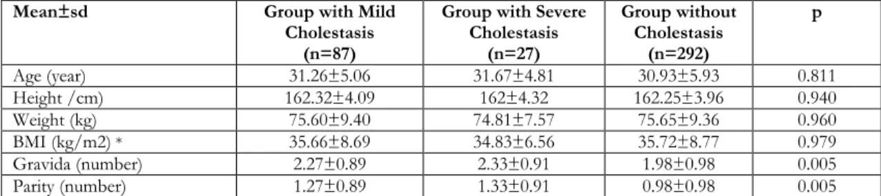
It was found that the groups had similar characteristics in terms of age, height, weight, body mass index (BMI) values (p>.05, Table 1). Gravida and parity values differ at statistically significant levels between groups (p< .05, Table 1). In the post-hoc analysis, a statistically significant difference was detected in gravida and parity in groups without cholestasis (p=0.013), and there wasnt difference in gravida and parity between groups with mild ICP and groups with severe ICP (p=1,000, p=0.137).
Table 1. Intergroup comparisons in terms of socio-demographic and obstetric characteristics
Mean±sd Group with Mild
Cholestasis (n=87)
Group with Severe Cholestasis (n=27) Group without Cholestasis (n=292) p Age (year) 31.26±5.06 31.67±4.81 30.93±5.93 0.811 Height /cm) 162.32±4.09 162±4.32 162.25±3.96 0.940 Weight (kg) 75.60±9.40 74.81±7.57 75.65±9.36 0.960 BMI (kg/m2) * 35.66±8.69 34.83±6.56 35.72±8.77 0.979 Gravida (number) 2.27±0.89 2.33±0.91 1.98±0.98 0.005 Parity (number) 1.27±0.89 1.33±0.91 0.98±0.98 0.005
Kruskal Wallis Test. BMI. * Body Mass Index
Table 2. Comparison of intergroup laboratory values
Mean±sd Group with Mild
Cholestasis (n=87)
Group with Severe Cholestasis (n=27) Group without Cholestasis (n=292) P WBC *(/mmᶾ×10ᶾ) 9.93±3.07 10.16±3.40 9.95±2.67 0.928 Hb(g/dL) 11.81±1.27 11.52±1.31 11.61±1.20 0.380 PLT(/mmᶾ×10ᶾ) 225.24±64.94 214.77±51.96 214.69±56.57 0.327 MPV(fL) 10.60±1.52 10.86±1.53 10.98±1.00 0.030 PCT(%) 0.233±0.064 0.236±0.054 0.23±0.055 0.957 RDW(%) 14.26±2.49 14.54±2.49 13.96±1.76 0.203 PDW(%) 15.18±3.37 15.07±4.01 13.68±2.59 0.001 Neutrophil*(×10ᶾ/uL) 7.13±2.71 7.50±3.32 6.95±2.15 0.751 Lymphocyte*(×10ᶾ/uL) 1.94±1.07 1.64±0.45 2.06±0.61 0.000 Eosinophil*(×10ᶾ/uL) 0.114±0.11 0.089±0.092 0.073±0.072 0.003 Basophil*(×10ᶾ/uL) 0.046±0.056 0.038±0.042 0.029±0.014 0.408 AST (U/L) 50.63±39.69 56.94±44.78 18.44±6.10 0.000 ALT (U/L) 78.43±72.93 91.21±70.19 20.31±6.01 0.000 TBA 17.47±10.19 72.74±30.97 - 0.000***
* Kruskal Wallis Test. **One-way anova test ***Mann-Whitney U) Hemoglobin (Hb), white blood cell count (WBC), Red cell distribution width (RDW), platelet (PLT) count, platelet distribution width (PDW), mean platelet volume (MPV), plateletkrit (PCT), AST: Aspartate aminotransferase, ALT: Alanine aminotransferase
When the study groups with pregnant women that had mild and severe ICP and those without ICP were examined in terms of laboratory markers, there werent differences in the groups in terms of WBC, HGB, PLT, and PCT values (p>0.05, Table 2), and there was a difference in the groups in terms of MPV value (p=0.030, Table 2).
When groups were examined in terms of RDW, Neutrophil and Basophil values, no differences were detected in groups with mild and severe ICP without ICP, PDW, Lymphocyte, Eosinophil, ALT and AST values, and there were differences between the groups (p<0.05, Table 2). According to the results of further analyses, there was no difference between groups with severe and mild ICP in terms of PDW value and groups with severe ICP and groups with no ICP, no differences were detected between the groups without ICP and the group with mild ICP (p=0.001). Although no differences were found between mild and severe ICP groups in terms of lymphocyte values, there was a statistically significant difference in the group without ICP and the group with mild and severe ICP (p=0.012, p=0.001). Although no differences were found in the groups with severe and mild ICP in terms of eosinophil values, there was a difference in the groups without ICP and the group with mild ICP (p=0.002). There were no differences in ALT values between groups with mild and severe ICP, and difference was detected between the group without ICP and the group with mild and severe ICP (p=0.000). There was no difference in terms of AST values between mild and severe ICP groups, and there was a difference between the group without ICP and the group with mild and severe ICP (p=0.000).In terms of blood values ratios, a significant difference was detected
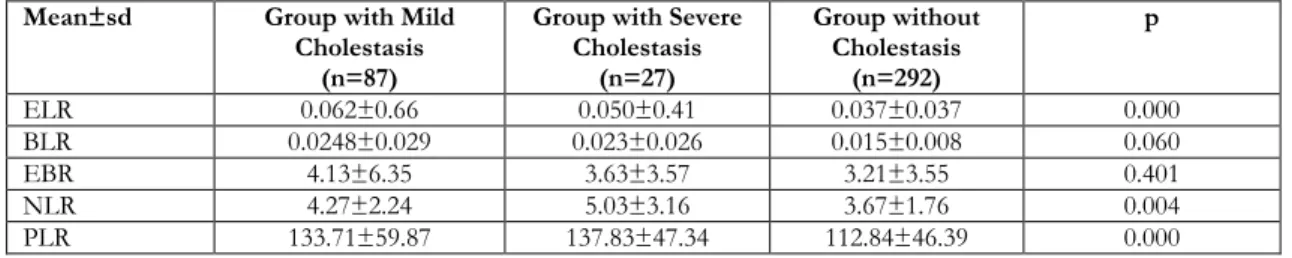
between the groups in terms of ELR, NLR and PLR values (p<0.05), and there were no differences in the groups in terms of BLR and EBR values (p=0.060, p=0.401, Table 3). Further analyses were made to determine which groups the difference originated from, and it was found that although there was a difference in terms of ELR between the no ICP group and the group with mild ICP (p=0.000), and there wasn’t difference in the groups with severe and mild ICP (p=0.993).
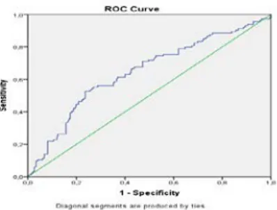
Figure 1. ROC analysis ( the effect of ELR value in predicting ICP)
In terms of NLR value, there wasnt difference between the groups of ICP (p=1.000), there was a difference between groups with mild ICP and no ICP and severe ICP (p=0.019, p=0.041). When PLR values were evaluated by groups, it was found that there was a difference between the group without ICP and the groups of ICP (p=0.007, p=0.000), and there were no differences between the groups of ICP (p=1.000). When groups with severe ICP and that with mild ICP were evaluated in terms of bile acid, there was a difference between the groups (p=0.000, Table 3).
Table 3. Comparison of intergroup laboratory ratio values Mean±sd Group with Mild
Cholestasis (n=87)
Group with Severe Cholestasis (n=27) Group without Cholestasis (n=292) p ELR 0.062±0.66 0.050±0.41 0.037±0.037 0.000 BLR 0.0248±0.029 0.023±0.026 0.015±0.008 0.060 EBR 4.13±6.35 3.63±3.57 3.21±3.55 0.401 NLR 4.27±2.24 5.03±3.16 3.67±1.76 0.004 PLR 133.71±59.87 137.83±47.34 112.84±46.39 0.000
Kruskal Wallis Test NLR (Neutrophil/Lymphocyte), PLR (PLT / Lymphocyte), ELR (Eosinophil / Lymphocyte), EBR (Eosinophil / Basophil) BLR (Eosinophil / Basophil), TBA (total bile acid)
When ROC analysis was made, the case of cholestasis status and absence of cholestasis was evaluated in patients, the best ELR interrupt value was 0.2243
with 76.3% sensitivity and 56.1% specificity (Area under curve AUC: 0.650±0.03, (95% Confidence Interval (CI) 95% CI: 0.592 -0.717) (Figure-1).
1746
When ROC analysis was made and when the case of cholestasis status and absence of cholestasis was taken into consideration, it was found that the best NLR interrupt value was 11.27. The results of the ROC analysis of NLR had 70% sensitivity and 54.2% specificity (Area under curve AUC: 0.600±0.03, (95% Confidence Interval (CI) 95% CI: 0.541-0.670) (Figure-2).
Figure 2. ROC analysis ( the effect of NLR value in predicting ICP)
When ROC analysis was made, when the case of cholestasis status and absence of cholestasis was taken into consideration in patients, the best PLR interrupt value was 194.46. The results of the ROC analysis of PLR had 60% sensitivity and 58% specificity (AUC: 0.650±0.031, (95% (CI) 95% CI: 0.583-0.705) (Figure-3).
Figure 3. ROC analysis (effect of PLR value in predicting ICP).
When only patients who developed cholestasis status and those with mild or severe cholestasis were considered in ROC analysis, ELR, NLR and PLR values were found to be unable to make the discrimination of patients for the severity of cholestasis. The AUC value, which is the area below the ROC Curve, was calculated to be 45% for each ratio and standard deviation was calculated as 6%, 54% and standard deviation as 6% and 53%, and standard deviation was 6%.
DISCUSSION
In this retrospective case control study investigating the relations between inflammatory markers used for allergic diseases and intrahepatic pregnancy cholestasis (ICP), serum inflammation markers and allergy markers were diagnosed in ICP but were not decisive to determine the severity of the disease. Previous studies reported that the most common laboratory abnormalities in ICP are high serum bile acid, AST and ALT concentrations. 12 Bile acid levels are used to group the severity of ICP. Bile acid level is 10-40 mmol / L light respectively, and > 40 mmol / L level is grouped into severe ICP. 13 In their studies, the mean bile acid level was 18± 5.9 mmol/L in the mild ICP group and 57.3± 28.6 mmol/L in the severe ICP group. 12 In this study, the mean bile acid level was determined as 17.47±10.19 mmol/L in the mild ICP group and 72.74±30.97 mmol/L in the severe ICP group.
Although the liver enzyme levels like ALT and AST increased significantly in women with ICP, no consensus exists on the diagnostic threshold level of liver enzymes13. In previous studies, statistically significant load values were found in groups with mild and severe ICP in terms of AST and ALT values11,12. The results of the study are parallel to other study results. AST 50.63±39.69 in the group with mild ICP. (U/L) ALT 78.43±72.93(U/L); In the severe ICP group, AST was 56.94±44.78. (U/L) ALT was ± 91.21.70.19(U/L).
When the literature was reviewed, studies were detected showing that maternal circulation is responsible for the immunity mechanisms and ICP pathogenesis of the oxidative inflammation that is itself caused by thrombosis11,14. Platelets also have tasks as strong immune modulators besides the clotting function, which are the most important components of CBC. MPV, PDW and PCT are markers of platelet activation. When the literature was reviewed, high levels of MPV and PDV were reported in inflammatory diseases like recurrent pregnancy loss7, gestational diabetes10, thyroid diseases15. In the study conducted by Abide et al., 84 pregnant cholestasis patients were included and it was found that MPV values were higher in the cholestasis group than in healthy controls7. MPV and PDW levels were found to be significantly higher in another study that evaluated 117 pregnant women with cholestasis14. In this study, MPV and PDW levels were found to be higher in ICP patients than in
controls. The results of the study were similar to other study results.
It was reported in the literature that the number of lymphocyte increases due to increased immune response17. NLR is an important marker in the diagnosis of inflammatory diseases, and vary in various systemic inflammatory diseases. NLR was also found to increase in the diagnosis of many inflammatory diseases and complicated pregnancies like hyperemesis gravidarum18, gestational diabetes10, preeclampsia8, intrahepatic pregnancy cholestasis11. In the study of Kırbaş et al. conducted with 65 cholestasis pregnant women, it was found that the levels of lymphocyte were higher in healthy controls, and neutrophil and NLR values were higher in patients with cholestasis. NLR value for the diagnosis of ICP was found to be 2.93 (91% sensitivity, 84% specificity, AUC was 0.92, 95% (CI) 0.861-0.969)12. In this study, it was found that lymphocyte levels were higher in the healthy control group, eosinophil levels were higher in patients with cholestasis, and neutrophil levels were not statistically different in the groups. Although there wasnt statistically difference between groups with mild and severe cholestasis in terms of NLR value (p=1,000), there was a significant difference between groups with mild cholestasis and no cholestasis and severe cholestasis (p=0.019, p=0.041). When the best NLR cut-off value was 11.27, the sensitivity was 70% and specificity was 54.2% (AUC: 0.600, 95% CI: 0.541-0.670). The results of the study are parallel to other study results. PLR is used to analyze thromboembolic and inflammatory diseases in recent years17,19. In previous studies, increased PLR values were found to be statistically significant in pregnancy-related diseases like preeclampsia, diabetes, pancreatitis, early membrane rupture, and recurrent pregnancy losses8,20,21,22. In another study evaluating pregnancy cholestasis and PLR together, no statistically significant differences were detected in the ICP groups although the PLR value was higher in the ICP group than in the healthy control group11. When the PLR values were evaluated according to the groups, it was determined that there was a difference between the groups (p=0.007, p=0.000), and there was no difference between the group with severe and mild ICP (p=1.000). When the best PLR cut-off value was 194.46, the sensitivity was 60% and specificity was 58% (AUC: 0.650, 95% CI: 0.583-0.705). The results of the study parallel to other study results.
Eosinophil, eosinophil-lymphocyte ratio (ELR),
eosinophil basophile ratio (EBR) and Basophil-lymphocyte ratio (BLR) values, which can be easily calculated with CBC, are hematological allergy markers. Eosinophilia (i.e. high eosinophil count) occurs in diseases such as allergies, parasitic infections, leukemia, polyarthritis nodosa autoimmune disease23. There is no specific cause of eosinopenin (i.e. low eosinophil count), and 0% eosinophil is considered normal24. Eosinophil decreases physiologically with gestational age25. When the literature was reviewed, allergic markers in preeclampsia were evaluated in a small number of studies26,27. However, no study evaluating allergic markers in intrahepatic pregnancy cholestasis was detected. In this study, although the ELR ratio had a difference between the non-cholestasis group and the group with mild cholestasis (p=0.000), no difference was detected between the group with severe and mild cholestasis (p=0.993). The best ELR cut-off value for predicting ICP diagnosis had 76.3% sensitivity and 56.1% specificity (AUC: 0.650,95% CI: 0.592-0.717) when 0.2243 was considered. No statistical differences were found in terms of EBR and BLR values. The results of the study are parallel to other study results.
As far as we know, the present study is the first one in which the allergy markers such as ELR, BLR and EBR, inflammation markers such as neutrophils, lymphocytes, NLR and PLR, and thrombocyte activation markers such as PLT, MPV, PDW and ELR were evaluated together to predict the severity of pregnancy cholestasis. Small size of the sampling, the use of data from a single third-line hospital and retrospective design were limitations of this study. In conclussion, to allergy markers might also be a diagnostic tool in intrahepatic cholestasis of pregnancy; however, they are not determinant in identifying the severity of the disease. It can be considered that blood allergy markers can be easily applied by health professionals (family physician, midwife, nurse, obstetrics physician), and can be easily interpreted in the diagnosis of ICP.
Yazar Katkıları: Çalışma konsepti/Tasarımı: DKG; Veri toplama: DKG; Veri analizi ve yorumlama: DKG, AŞK; Yazı taslağı: DKG, AŞK; İçeriğin eleştirel incelenmesi: DKG, AŞK; Son onay ve sorumluluk: DKG, AŞK; Teknik ve malzeme desteği:DKG; Süpervizyon: DKG; Fon sağlama (mevcut ise): yok.
Etik Onay: Etik kurul onayı çalışma öncesinde Medipol Üniversitesi Girişimsel Olmayan Etik Kurul'dan alınmıştır (Referans numarası: 10840098-772.02-E.60609, Tarih: 10/11/2020). Özel Nisa Hastanesi'nden izin alındı Daha sonra katılımcılardan yazılı izin alındı. Tüm prosedürler 1964 Helsinki deklarasyonu etik standartlarında gerçekleştirildi.
Hakem Değerlendirmesi: Dış bağımsız.
1748 Finansal Destek: Yazarlar finansal destek beyan etmemişlerdir.
Author Contributions: Concept/Design : DKG;Data acquisition: DKG; Data analysis and interpretation: DKG, AŞK; Drafting manuscript: DKG, AŞK; Critical revision of manuscript: DKG, AŞK; Final approval and accountability: DKG, AŞK; Technical or material support: DKG; Supervision: DKG; Securing funding (if available): n/a. Ethical Approval: The ethics committee approval was obtained from Medipol University for Non-Interventional Ethics Committee before the study (Reference number: 10840098-772.02-E.60609, Date:10/11/2020). The permission was obtained from the Private Nisa Hospital Then written permission was obtained from the participitants. All procedures were performed in the 1964 Helsinki declaration ethical standards.
Peer-review: Externally peer-reviewed.
Conflict of Interest: Authors declared no conflict of interest. Financial Disclosure: Authors declared no financial support
REFERENCES
1. Lammert F, Marschall HU, Glantz A, Matern S. Intrahepatic cholestasis of pregnancy: molecular pathogenesis, diagnosis and management. J Hepatol. 2000;33:1012-21.
2. Kenyon AP, Piercy CN, Girling J, Williamson C, Tribe RM, Shennan AH. Pruritus may precede abnormal liver function tests in pregnant women with obstetric cholestasis: a longitudinal analysis. BJOG. 2001;108:1190-2.
3. Brouwers L, Koster MP, Page-Christiaens GC, Kemperman H, Boon J, Evers IM et al. Intrahepatic cholestasis of pregnancy: maternal and fetal outcomes associated with elevated bile acid levels. Am J Obstet Gynecol 2015;212:100.e1-7.
4. Pathak B, Sheibani L, Lee RH. Cholestasis of pregnancy. Obstet Gynecol Clin North Am. 2010;37:269-82.
5. Allen K, Jaeschke H, Copple BL. Bile acids induce inflammatory genes in hepatocytes: a novel mechanism of inflammation during obstructive cholestasis. Am J Pathol. 2011;178:175-86.
6. Gujral JS, Farhood A, Bajt ML, Jaeschke H. Neutrophils aggravate acute liver injury during obstructive cholestasis in bile duct-ligated mice. Hepatology. 2003;38:355–63.
7.
Aynıoglu O, Isık H, Sahbaz A, Harma MI, Isık M, Kokturk F. Can Plateletcrit be a Marker for Recurrent Pregnancy Loss? Clin Appl Thromb Hemost. 2016;22:447–52.8.
Serin S, Avcı F, Ercan O, Köstü B, Bakacak M, Kıranet H. Is neutrophil/lymphocyte ratio a useful marker to predict the severity of pre-eclampsia? Pregnancy Hypertens. 2016;6:22–5.9. Akgun N, Namli Kalem M, Yuce E, Kalem Z, Aktas H. Correlations of maternal neutrophil to lymphocyte ratio (NLR) and platelet to lymphocyte ratio (PLR) with birth weight. J Matern Fetal Neonatal Med. 2017;30:2086-91.
10. Colak E, Ozcimen EE, CeranMU, Tohma YA, Kulaksızoglu S. Role of mean platelet volume in pregnancy to predict gestational diabetes mellitus in
the first trimester. J Matern Fetal Neonatal Med. 2020;33:3689-94.
11. Abide Ç, Vural F, Kılıççı Ç, Ergen EB, Yenidede I, Eser A et al. Can we predict severity of intrahepatic cholestasis of pregnancy using inflammatory markers? Turk J Obstet Gynecol. 2017;14:160-5.
12. Kirbas A, Biberoglu E, Daglar K, İskender C, Erkaya S, Dede H et al. Neutrophil-to-lymphocyte ratio as a diagnostic marker of intrahepatic cholestasis of pregnancy. Eur J Obstet Gynecol Reprod Biol. 2014;180:12-5.
13. Mays JK. The active management of intrahepatic cholestasis of pregnancy. Curr Opin Obstet Gynecol. 2010;22:100–3.
14. Oztas E, Erkenekli K, Ozler S, Ersoy AO, Kurt M, Oztas E et al. Can routine laboratory parameters predict üadverse pregnancy outcomes in intrahepatic cholestasis of pregnancy? J Perinat Med 2015;43:667-74.
15. Ren X, Meng Z, Liu M, Zhu M, MD, He Q, Zhang Q. No associations exist between mean platelet volume or platelet distribution width and thyroid function in Chinese. Medicine (Baltimore). 2016;95:e4573. 16. Oztas E, Erkenekli K, Ozler S, Ersoy AO, Kurt M,
Oztas E et al. Can routine laboratory parameters predict üadverse pregnancy outcomes in intrahepatic cholestasis of pregnancy? J Perinat Med. 2015;43:667-74.
17. Nording HM, Seizer P, Langer HF. Platelets in inflammation and atherogenesis. Front Immunol. 2015;6:98.
18. Caglayan EK, Ustun YE, Gocmen AY, Sarı N, Seckin L, Kara M et al. Is there any relationshipbetween serum sirtuin-1 level and neutrophil-lymphocyte ratio in hyperemesis gravidarum? J Perinat Med. 2016;44:315–20.
19. Zhang WW, Liu KJ, Hu GL, Liang WJ. Preoperative platelet/lymphocyte ratio is a superior prognostic factor compared to other systemic inflammatory response markers in ovarian cancer patients. Tumour Biol. 2015;36:8831-7.
20. Sargın MA, Yassa M, Taymur BD, Celik A, Ergun E, Tug N. Neutrophil-to-lymphocyte and platelet-to-lymphocyte ratios: are they useful for predicting gestational diabetes mellitus during pregnancy? Ther Clin Risk Manag. 2016;12:657–65.
21. Ekin A, Gezer C, Taner CE, Ozeren M, Uyar I, Gulhanet I. Risk factors and perinatal outcomes associated with latency in preterm premature rupture of membranes between 24 and 34 weeks of gestation. Arch Gynecol Obstet 2014;290:449–55.
22. Ata N, Kulhan M, Kulhan NG, Turkler C. Can neutrophil-lymphocyte and platelet-lymphocyte ratios predict threatened abortion and early pregnancy loss? Ginekologia Polska. 2020;91:210–215.
23. Sahoo D, Gosaib H, Harsodaa JM, Palana BM A. Comparative hematological profile study among
young individuals. Canadian Journal of Basic and Applied Sciences. 2015;3:178-81.
24. Tembe N, Joaquim O, Alfai E, Sitoe N, Viegas E, Macovela E et al. Reference values for clinical laboratory parameters in young adults in Maputo, Mozambique. PLoS One 2014;9:973-91.
25. Chandra S, Tripathi AK, Mishra S, Amzarul M, Vaish AK. Physiological changes in hematological parameters during pregnancy. Indian J Hematol Blood
Transfus. 2014;28:144-6.
26. Elmas B, Kıncı MF, Gök İE, Alkan A, Toğrul C, Sarıkaya S. Is higher IgE levels in preeclamptic pregnancies suggest autoimmune pathophysiology? Cukurova Med J. 2019;44:547-54.
27. Keski-Nisula L, Heinonen S, Remes S, Pekkanen J Pre-eclampsia, placental abruption and increased risk of atopic sensitization in male adolescent offspring. Am J Reprod Immunol. 2009;62:293-300.