Gamze Sinem Caglar*, Perihan Erdogdu, Aslı Yarcı Gursoy, Rabia Şeker and Selda Demirtas
The impact of route of anesthesia on maternal
and fetal ischemia modified albumin levels at
cesarean section: a prospective randomized study
Abstract
Objective: Ischemia modified albumin has been shown to
increase in ischemic situations, and has also been shown
to increase in fetal cord blood in deliveries by cesarean
section. The aim of this study is to reveal whether
anes-thesia has an impact on maternal and fetal cord ischemia
modified albumin levels.
Methods: Seventy two women with uncomplicated term
pregnancies were randomized to spinal (n = 37) or general
anesthesia (n = 35) groups. The blood pressure, oxygen
saturation, and pulse rate of the patients were recorded
during the procedure. Maternal blood samples of ischemia
modified albumin (IMA) were taken 10 min from the start
of the procedure. The fetal cord blood samples of IMA
were taken immediately after birth.
Results: Maternal (0.99 ± 0.19 vs. 0.80 ± 0.27) and fetal
(1.00 ± 0.21 vs. 0.70 ± 0.26) IMA levels were significantly
higher in the general anesthesia group. Fetal IMA levels
were positively correlated with maternal gravidity (r = 0.31;
P = 0.008), parity (r = 0.25; P = 0.028), and fetal birth weight
(r = 0.23, P = 0.045). Also, as time from incision to delivery
lengthens, fetal IMA levels increase (r = 0.29, P = 0.012).
Conclusion: Fetal cord ischemia modified albumin levels
were higher in the general anesthesia group, therefore, it
is proposed that regional anesthesia should be the
pre-ferred route of anesthesia for an elective cesarean section,
at least until the impact of high fetal cord IMA levels are
manifested.
Keywords: Cesarean section; general anesthesia; ischemia
modified albumin; regional anesthesia.
*Corresponding author: Gamze Sinem Caglar, Department of Obstetrics and Gynecology, Ufuk University School of Medicine, Ankara, Turkey, Tel.: +90-532-4418501, Fax: +90-312-2847786, E-mail: [email protected]
Perihan Erdogdu: Department of Reanimation and Anesthesiology, Ufuk University, School of Medicine, Ankara, Turkey
Aslı Yarcı Gursoy: Department of Obstetrics and Gynecology, Ufuk University, School of Medicine, Ankara, Turkey
Rabia Şeker and Selda Demirtas: Department of Biochemistry, Ufuk University, School of Medicine, Ankara, Turkey
Introduction
Recently, ischemia modified albumin (IMA) has been
proposed as a valuable marker of ischemia [21]. Ischemia
with free radical generation causes structural changes in
the N terminus of albumin, leading to IMA production [4].
This change in structure reduces binding of albumin to
nickel, copper and cobalt, this is a feature used in
deter-mining IMA levels with the albumin cobalt-binding test
(ABSU). A higher sensitivity and a very short half-life
makes this marker more favorable in cardiac ischemia
when compared with conventional tests, such as
elec-trocardiography (ECG), and troponin-I [5]. Other than
cardiologic events, IMA has been reported to increase in
other clinical situations comorbid with ischemia, such
as systemic sclerosis [6], ischemic stroke [22], strenuous
exercise [16], gastrointestinal or delayed muscle ischemia [2],
and trauma [9]. Moreover, increased oxidative stress in
metabolic syndrome [11], obesity [20], and polycystic
ovary syndrome [7] were also reported as being
associ-ated with elevassoci-ated IMA levels. Thus, any situation leading
to ischemia might lead to an increase in IMA levels.
Pregnancy on its own has been shown to be an
incre-mental factor for IMA levels [21]. Guven at el. reported
that IMA levels were higher than reference values for
non-pregnant adults during all trimesters, but especially in
the third [13]. The authors claimed that higher IMA and
lower malondialdehyde levels may be a sign of oxidative
stress in pregnancy [13]. Few studies evaluated maternal
IMA levels in different obstetric entities, such as
tropho-blast invasion [21], preeclampsia [19], and intrauterine
growth restriction [14]. Additionally, in case of recurrent
early pregnancy loss, higher IMA levels were reported in
the first trimester of pregnancy compared to healthy
preg-nant controls [18]. The authors suggested that high IMA
levels in cases with recurrent early pregnancy loss might
be related to abnormal placentation and an abnormally
hypoxic uterine environment [18].
Finally, delivery by cesarean section was reported to
be associated with higher fetal IMA levels [14]. The studies
in the literature evaluating whether the mode of delivery
effects maternal and fetal IMA levels did not consider the
route of anesthesia. Neuraxial anesthesia for cesarean
delivery is usually preferred to general anesthesia because
it minimizes the risk of failed intubation, ventilation, and
aspiration. In the latest Cochrane review of 22 studies
comparing neuraxial blockade vs. general anesthesia in
otherwise uncomplicated cesarean deliveries reported no
significant difference in terms of neonatal Apgar scores of
6 or less and of 4 or less at 1 and 5 min, and need for
neo-natal resuscitation [1]. Therefore, this study was designed
to clarify if IMA levels differ between routes of anesthesia
chosen for cesarean section by measuring both maternal
and umbilical cord blood IMA levels during the operation.
Materials and methods
This study was conducted in a university clinic between September 2011 and July 2012. All women that attended the Obstetrics and Gynecology Department were offered to participate in the study. Dur-ing the study period, 72 women were enrolled into this prospective randomized study. The study protocol was approved by the Ethics Committee of the University Hospital. All the women participating in the study gave informed consent. All women received prenatal care in the same institution. All the participants had uncomplicated term gestations at 37 and 40 completed weeks. Before randomization, obstetric ultrasound examinations were performed to determine the amniotic fluid volume, the lie, and position of the fetus. The esti-mation of the gestational age was on the basis of ultrasonographic examination performed between 11 and 14 weeks of gestation. The women without a first trimester scan were excluded from the study. In all cases delivery by cesarean section was performed for a previous cesarean section or for fetal malpresentation. All cesarean sections were performed by the same operators. Patients were randomized to spinal (n = 37) or general anesthesia (n = 35) groups according to a computer generated randomization list.
The exclusion criteria composed of complicated pregnancies (e.g., intrauterine growth retardation, gestational diabetes mellitus, preeclampsia, fetal congenital anomaly, oligohydramnios, placenta previa), mothers with chronic illnesses (e.g., hypertension, diabetes mellitus), and any history of maternal cardiac symptoms, such as angina, myocardial infarction, coronary artery disease, vascular dis-ease, and inflammatory disease. Oligohydramnios was defined as the deepest vertical pocket of amniotic fluid < 2 cm. In addition, smokers and alcohol consumers, cases with abnormal albumin levels ( < 3.5 g/dL and > 5.5 g/dL), and multiple pregnancies were also excluded from the study. The patients with contraindications of neuroaxial anaesthesia (coagulopathy, infection, hypovolemia, patient reluc-tance) were excluded from the study.
As soon as the patients were taken to the operating room, all were monitored (GE Heathcare Finland Oy, Helsinki, Finland). Thereafter,
ECG, blood pressure, oxygen saturation (SpO2), and pulse rate were
all recorded. All patients were given 2 L/min O2 inhalation by nasal
cannulation. In the regional anesthesia group, 500 mL Ringer lactate solution was infused 15 min before spinal anesthesia by left hand ve-nous cannulation. Patients were positioned in left lateral position to avoid aorta-caval syndrome; 2 mL 0.5% hyperbaric bupivacaine was administered through L 2–3 or L 3–4 inervertebral space by a 25-gauge spinal needle (Quincke, Braun Melsungen AG, Melsungen, Germany) for spinal anesthesia. In case of hypotension (systolic blood pressure 20% lower than basal level) or bradycardia (heart rate < 60/min), 2–3 mg ephedrine (50 mg at maximum dose) and 0.5 mg atrophine were administered. Level of the neurological blockage was evaluated with a pinprick test 5 min after each procedure. Blockage level between thoracal 4–6 dermatomes were accepted as appropriate for surgical intervention. Patients in the general anesthesia group were not pre-medicated. Induction for general anesthesia was held by 2–3 mg/kg propofol, 0.6 mg/kg rocuronium infusion. Anesthesia was maintained by 50% O2–50% N2O, and 1%–1.5% sevoflurane. The time required for
application of spinal anesthesia and the time for induction in general anesthesia were recorded. Systolic and diastolic blood pressures, SpO2,
and pulse rate were also recorded at 1, 2, 3, 4, 5, and 10 min of both groups. Additionally, incision to delivery intervals were recorded in both groups.
Maternal blood samples were collected from the antecubital vein into a non-heparinized tube at the 10th min of induction. Cord blood
was collected immediately after the delivery for umbilical cord acid base analyses, and an extra umbilical cord blood sample was taken for the analysis of fetal IMA. Maternal and fetal blood IMA samples were immediately centrifuged, and serum was separated and frozen at –80°C until assayed for IMA analysis. IMA concentrations were analyzed by measuring the complex composed of dithioerthreitol and cobalt, and unbound to albumin by the colorimetric method in a spec-trophotometer. First, a mixture of 200 μL of patients serum and 50 μL cobalt chloride (Sigma Aldrich, St Louis, MO, USA) was prepared in glass tubes. The mixture was left to incubate at room temperature for 10 min. After that, 50 μL of dithiothreirol (1.5 mg/mL) was added to the tubes and incubated at room temperature for 2 min. At the final stage 1000 μL of sodium chloride (0.9%) was added to the mixture. A blank specimen was prepared with distilled water for control. The analyses in the spectrophotometer (Human Humalyzer 2000, Germany) was performed at 470 nm for detection of absorbance of the specimens, and the results were given as absorbance units (ABSU).
Statistical analyses
Data analysis was performed by using SPSS for Windows, version 11.5 (SPSS Inc., Chicago, IL, USA). Whether the distributions of con-tinuous variables were normal or not was determined by the Shap-iro Wilk test. Data were shown as mean ± standard deviation (SD) or median (minimum–maximum), where applicable. While the mean differences between groups were compared by Student’s t-test, oth-erwise Mann Whitney U-test was applied for the comparisons of the median values. Nominal data were analyzed by χ2 or Fisher’s exact
test, where appropriate. Multiple logistic regression analysis was used to determine the independent predictors that mostly affected IMA levels. Any variable whose univariable test had a P-value < 0.25 was accepted as a candidate for the multivariable model along with all variables of known clinical importance. Odds ratio and 95% confidence intervals for independent variables were calculated.
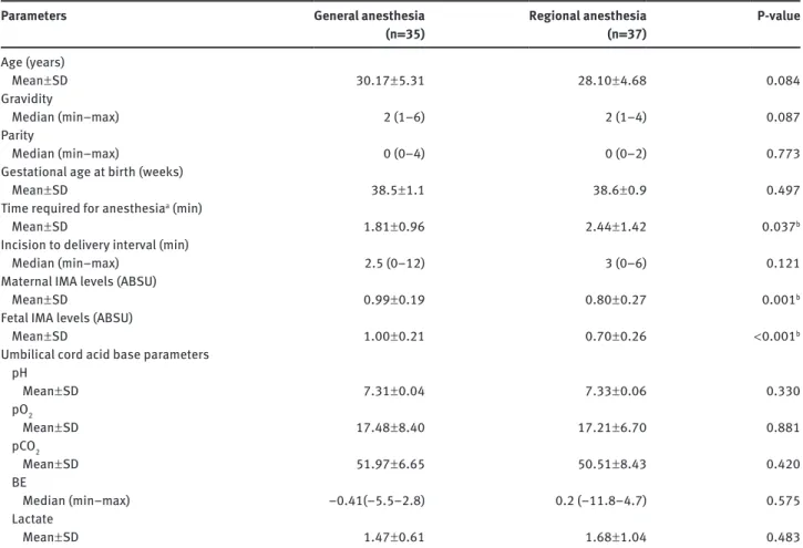
Table 1 Characteristics of general anesthesia and regional anesthesia groups.
Parameters General anesthesia
(n = 35) Regional anesthesia (n = 37) P-value Age (years) Mean ± SD 30.17 ± 5.31 28.10 ± 4.68 0.084 Gravidity Median (min–max) 2 (1–6) 2 (1–4) 0.087 Parity Median (min–max) 0 (0–4) 0 (0–2) 0.773
Gestational age at birth (weeks)
Mean ± SD 38.5 ± 1.1 38.6 ± 0.9 0.497
Time required for anesthesiaa (min)
Mean ± SD 1.81 ± 0.96 2.44 ± 1.42 0.037b
Incision to delivery interval (min)
Median (min–max) 2.5 (0–12) 3 (0–6) 0.121
Maternal IMA levels (ABSU)
Mean ± SD 0.99 ± 0.19 0.80 ± 0.27 0.001b
Fetal IMA levels (ABSU)
Mean ± SD 1.00 ± 0.21 0.70 ± 0.26 < 0.001b
Umbilical cord acid base parameters pH Mean ± SD 7.31 ± 0.04 7.33 ± 0.06 0.330 pO2 Mean ± SD 17.48 ± 8.40 17.21 ± 6.70 0.881 pCO2 Mean ± SD 51.97 ± 6.65 50.51 ± 8.43 0.420 BE Median (min–max) –0.41(–5.5–2.8) 0.2 (–11.8–4.7) 0.575 Lactate Mean ± SD 1.47 ± 0.61 1.68 ± 1.04 0.483
aInduction period in general anesthesia, time for regional anesthesia. bP < 0.05. SD = standard deviation, IMA = ischemia modified albumin,
ABSU = absorbance unit, pO2 = partial oxygen pressure, pCO2 = partial carbon dioxide pressure, BE = base excess.
A P-value < 0.05 was considered statistically significant. A power analysis was performed to estimate the number of patients needed in each group. It was assumed that 0.1 ABSU change (SD = 0.1) in serum IMA concentrations was clinically significant. Assuming a two-sided test with a probability of a type-I error of 0.05, a statis-tical power of 80%, 32 patients were required in each group.
Results
The mean age of the patients and the gestational age at
birth in the general and regional anesthesia groups are
given in Table 1. The median (min–max) gravidity and
parity of the patients were 2 (1–6) and 0 (0–4),
respec-tively (Table 1). When the groups were compared for
demographic characteristics (maternal age, gravidity,
parity, gestational age at birth) no significant difference
were found (P > 0.05, Table 1). Incision-to-delivery
inter-vals were similar in both groups (P > 0.05). The time for
induction was significantly shorter in general anesthesia
when compared with the time required for spinal
anes-thesia (Table 1). None of the umbilical artery acid base
parameters were abnormal in either group. No
signifi-cant difference was found when two groups were
com-pared for umbilical arter acid base parameters (Table 1).
Maternal (0.99 ± 0.19 vs. 0.80 ± 0.27) and fetal (1.00 ± 0.21
vs. 0.70 ± 0.26) IMA levels were significantly higher in the
general anesthesia group (Table 1). Fetal gender was male
in 57.1% (n = 20) of the newborns in general anesthesia and
50% (n = 18) in the spinal anesthesia group. Additionally,
fetal IMA levels were higher in male fetuses compared to
females (0.90 ± 0.26 vs. 0.77 ± 0.28; P = 0.039).
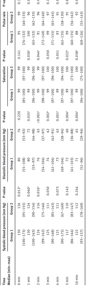
Systolic and diastolic blood pressure, SpO
2and pulse
rate values at 0, 1, 2, 3, 4, 5, and 10 min for both groups
are listed in Table 2. Both systolic and diastolic blood
pressures declined during the procedure. Neither the
changes in systolic or diastolic blood pressures differed in
the general and regional anesthesia groups (P = 0.875 and
P = 0.141, respectively). Systolic blood pressure was
signifi-cantly higher at 0, 1, and 2 min in the general anesthesia
Tab le 2 Blood pressu res, O 2 sat ur ation and p ulse r ates in gener al (Gro up 1) and region al anesthesi a (Gro up 2) gro ups. Time Medi an (min–max) Sy sto lic b lood pre ss ure (mm Hg) P-va lue Di as to lic b lood pre ss ure (mm Hg) P-va lue Sat ur ation P-va lue Pu lse r ate P-va lue Gro up 1 Gro up 2 Gro up 1 Gro up 2 Gro up 1 Gro up 2 Gro up 1 Gro up 2 0 min 130 126 0.012 a 80 76 0.220 99 99 0.141 95 99 0.344 (100–173) (95–155) (55–108) (53–92) (85–100) (97–100) (76–132) (40–133) 1 min 130 124 0.040 a 80 72 0.035 a 99 99 0.030 a 100 96 0.972 (100–163) (90–154) (53–97) (44–98) (96–100) (96–100) (65–132) (62–133) 2 min 124 116 0.016 a 76 63 < 0.001 a 99 99 0.044 a 91 96 0.454 (99–162) (75–146) (54–104) (37–97) (98–100) (97–100) (72–149) (64–140) 3 min 125 111 0.050 74 62 0.002 a 99 100 0.050 99 97 0.767 (90–168) (85–143) (42–104) (42–91) (97–100) (96–100) (71–155) (65–145) 4 min 121 111 0.073 73 63 0.001 a 99 100 0.051 93 91 0.989 (90–175) (67–169) (49–104) (38–94) (97–100) (94–100) (62–135) (58–130) 5 min 117 110 0.143 72 60 0.004 a 99 100 0.022 a 99 91 0.848 (86–164) (83–147) (41–103) (36–88) (97–100) (73–100) (60–135) (56–162) 10 min 122 112 0.244 71 65 0.004 a 99 100 0.038 a 88 92 0.918 (93–157) (78–169) (51–96) (29–94) (94–100) (75–100) (69–126) (55–143) aSt atistic al ly signific ant .
group. Diastolic blood pressures were significantly higher
in the general anesthesia group at all times (1, 2, 3, 4, 5,
and 10 min) except initiation of anesthesia (0 min).
SpO
2was stable during the procedure in both groups.
The changes in SpO
2values were similar between groups
(P = 0.841). Significantly higher values of SpO
2were found
at 1, 3, 5, and 10 min in the regional anesthesia group
when compared with the general anesthesia group
(P = 0.030, P = 0.050, P = 0.022, and P = 0.038, respectively).
The changes in pulse rate were similar between groups
(P = 0.077). The pulse rate did not change during the
pro-cedure, and no significant difference were found in the
pulse rate when the two groups were compared for 0, 1, 2,
3, 4, 5, and 10 min values.
According to the Spearman’s rank correlation
analy-ses, the baseline characteristics (age, gravidity, parity,
and gestational age at birth) were not correlated with
maternal IMA levels. However, fetal IMA levels were
posi-tively correlated with gravidity (r = 0.31; P = 0.008), and
parity (r = 0.25; P = 0.028). In addition, fetal birth weight
and fetal IMA levels were also found to be positively
corre-lated (r = 0.23, P = 0.045). An important finding was that as
time from incision to delivery lengthens, fetal IMA levels
increase (r = 0.29, P = 0.012). However, no correlation exists
between fetal umbilical cord acid base parameters and
fetal IMA levels (Table 3). In the general anesthesia group,
systolic blood pressure at 2 min was negatively correlated
with fetal IMA levels, while no correlation existed in the
regional anesthesia group.
The correlation analyses were repeated to clarify the
parameters recorded during anesthesia (systolic blood
pressure, diastolic blood pressure, SpO
2, and pulse rate)
that might elevate maternal and fetal IMA levels. The
results revealed that neither maternal nor fetal IMA levels
were correlated with SpO
2values (P > 0.05). Among blood
pressure values only 2 min systolic blood pressure was
negatively correlated with fetal IMA levels in the general
anesthesia group (r = 0.035; P = 0.037). However, none of
the blood pressure values were found to be correlated with
fetal IMA levels in the regional anesthesia group.
Discussion
This study is the first in the literature evaluating
mater-nal and fetal IMA levels in different anesthesia types
used for a cesarean section in uncomplicated term
ges-tations. The results showed significantly higher
mater-nal and fetal IMA levels in the general anesthesia group.
Moreover, in the general anesthesia group lower systolic
Table 3 The results of Spearman rank correlation analysis.
Parameters Maternal IMA Fetal IMA
Correlation
coefficient (r) P-value coefficient (r)Correlation P-value
Maternal age 0.076 0.526 0.157 0.187
Gestational age 0.134 0.262 –0.091 0.448
Gravidity 0.181 0.129 0.310 0.008a
Parity 0.090 0.453 0.259 0.028a
Fetal birth weight 0.311 0.008a 0.237 0.045a
pH 0.011 0.929 –0.044 0.715 pO2 –0.027 0.820 0.089 0.456 pCO2 –0.032 0.790 –0.007 0.951 BE –0.015 0.899 –0.109 0.367 Hb –0.032 0.787 0.064 0.596 Htc –0.030 0.805 0.066 0.582 Lactate –0.155 0.193 –0.150 0.209
Time required for anesthesiab –0.114 0.355 –0.131 0.285
Incision to delivery interval –0.118 0.327 –0.296 0.012a
aP < 0.05. b“Induction period” in general anesthesia, “time required” for regional anesthesia. IMA = ischemia modified albumin, pO 2 = partial
oxygen pressure, pCO2 = partial carbon dioxide pressure, BE = base excess, Hb = hemoglobin, Htc = hemotocrit.
blood pressure during delivery of the baby was found to
be correlated with increased fetal IMA levels. A previous
report [12, 14] documented that cord blood IMA levels of
neonates from complicated deliveries are significantly
higher than uncomplicated term deliveries. Complicated
delivery causes an almost 50% increase in fetal cord blood
IMA levels compared with the normal delivery group.
However, severe fetal hypoxia was related with a 300%
increase in IMA levels [12]. All these accumulating data
indicate that IMA can be a valuable marker in
perinatol-ogy in the future.
During abdominal entry in a cesarean section,
manip-ulation and traction of the anterior abdominal muscles
can be the explanation for higher maternal IMA levels
of women delivered by cesarean section when compared
with women who delivered vaginally [8]. As previously
reported by Troxler et al., ischemia in skeletal muscles
causes an increase in IMA [24]. Cesarean section is a
process needing external forces, and can also lead to
abdominal muscle injury to some extent. The operation
itself or anesthesia related factors can be the cause of
ele-vated maternal IMA levels. However, our results suggest
other factors rather than muscle injury contribute to
ele-vated maternal IMA levels. Mothers from the general
anes-thesia group had significantly higher IMA levels when
compared with the regional anesthesia group. A result
supported by studies on rat models where laparotomy by
transperitoneal anesthesia, on its own, did not obviously
change IMA levels pre- and postoperatively, in the sham
group [3]. The higher maternal IMA levels in the general
anesthesia group can be as a result of significantly lower
values of SpO
2in this group when compared with the
regional anesthesia group.
According to our study, one of the subjects of
impor-tance is the correlation of fetal weight with fetal IMA
levels. The results of our study documented a positive
correlation of fetal weight with fetal IMA levels.
Regard-ing these results, small for gestational age fetuses are
expected to have lower IMA levels. The clinical
impor-tance of this finding is that in cases of intrauterine growth
restriction, higher IMA levels might be detected as fetal
hypoxia is related to increased IMA levels. Hence, a
pre-vious study that compared umbilical cord blood IMA
levels of intrauterine growth restricted and appropriate
for gestational age fetuses, reported no significant
dif-ference in IMA levels [14]. Another major point found in
our study is the correlation between gravidity, parity and
IMA levels. This was reported by Iacovidou et al. [14] and
others [17, 23], previously. All the factors, such as fetal
weight, gravidity, and parity that might have an
influ-ence on IMA levels need to be clarified by large
popu-lation based studies to prevent misinterpretation of the
results.
Some authors suggest that oxidative stress in the fetal
circulation does not depend on the mode of delivery [10].
Others report higher fetal cord blood IMA levels in
neo-nates delivered by cesarean section compared with
vaginal deliveries, although arterial blood gas
analy-sis and Apgar scores were in the normal range [14]. This
study evaluated the maternal and fetal IMA levels during
different anesthesia types and our results showed that the
general anesthesia group had significantly higher blood
pressures at all times when compared with the regional
anesthesia group. Moreover, newborns from the general
anesthesia group had significantly higher levels of IMA
with normal fetal cord blood arterial blood gas values
and neonatal Apgar scores. Cesarean section under
anes-thesia might elevate IMA levels causing hypotension and
uterine hypoperfusion, which is a very similar mechanism
as in tourniquet and revascularization surgery reports.
Consequently, hypotension and blood pressure
altera-tions might not be the sole factors affecting fetal IMA
levels. Oxygenation might be another contributing factor
for elevated fetal IMA levels of fetuses delivered by
elec-tive cesarean section. Breathing room air under regional
anesthesia or 30% oxygen under general anesthesia is
usually adequate for maternal or fetal oxygenation [15].
In the presence of a hypoxic fetus, providing
supplemen-tary oxygen could lessen the severity of fetal hypoxia but
can also lead to reperfusion injury. All the previous
litera-ture documenting elevated IMA levels from complicated
deliveries might be due to reperfusion injury caused by
maternally high inspired oxygen fraction [15]. The clinical
importance and long-term consequences of fetuses with
normal umbilical cord acid base status and elevated IMA
levels is not yet clear.
In conclusion, cord blood IMA is a marker of transient
ischemia and the unknown long-term consequences of
neonates with high IMA level necessitates follow-up of
these children. Concerning our results, if general
anes-thesia is to be applied for elective cesarean sections then
blood pressure and maternal oxygenation should be
strictly controlled as systolic blood pressure before, and
during, the delivery of the baby is negatively correlated
with fetal IMA levels.
Received October 30, 2012. Accepted April 24, 2013. Previously pub-lished online June 8, 2013.
References
[1] Afolabi BB, Lesi FE. Regional versus general anaesthesia for caesarean section. Cochrane Database Syst Rev. 2012;10:CD004350.
[2] Apple FS, Quist HE, Otto AP, Mathews WE, Murakami MM. Release characteristics of cardiac biomarkers and ischemia-modified albumin as measured by the albumin cobalt-binding test after a marathon race. Clin Chem. 2002;48:1097–100. [3] Aran T, Guven S, Unsal MA, Alver A, Mentese A, Yulug E. Serum
ischemia-modified albumin as a novel marker of ovarian torsion: an experimental study. Eur J Obstet Gynecol Reprod Biol. 2010;150:72–5.
[4] Bar-Or D, Curtis G, Rao N, Bampos N, Lau E. Characterization of the Co(2+) and Ni(2+) binding amino-acid residues of the N-terminus of human albumin. An insight into the mechanism of a new assay for myocardial ischemia. Eur J Biochem. 2001;268:42–7.
[5] Bar-Or D, Winkler JV, Vanbenthuysen K, Harris L, Lau E, Hetzel FW. Reduced albumin-cobalt binding with transient myocardial ischemia after elective percutaneous transluminal coronary angioplasty: a preliminary comparison to creatine kinase-MB, myoglobin, and troponin I. Am Heart J. 2001;141: 985–91.
[6] Borderie D, Allanore Y, Meune C, Devaux JY, Ekindjian OG, Kahan A. High ischemia-modified albumin concentration reflects oxidative stress but not myocardial involvement in systemic sclerosis. Clin Chem. 2004;50:2190–3.
[7] Caglar GS, Oztas E, Karadag D, Pabuccu R, Demirtas S. Ischemia-modified albumin and cardiovascular risk markers in polycystic ovary syndrome with or without insulin resistance. Fertil Steril. 2011;95:310–3.
[8] Caglar GS, Tasci Y, Goktolga U, Oztas E, Pabuccu R, Ozdemir E, et al. Maternal and umbilical cord ischemia
modified albumin levels in nonreassuring fetal heart rate tracings regarding the mode of delivery. J Matern Fetal Neonatal Med. 2013;26:528–31.
[9] Can M, Demirtas S, Rolat O, Yıldız A. Evaluation of effects of ischemia on the albumin cobalt binding (ACB) assay in patients exposed to trauma. Emeg Med S. 2006;23:537–9. [10] Fogel I, Pinchuk I, Kupferminc MJ, Lichtenberg D, Fainaru O.
Oxidative stress in the fetal circulation does not depend on mode of delivery. Am J Obstet Gynecol. 2005;193:241–6. [11] Gottlieb MGV, Da Cruz IBM, Duarte MMF, Moresco RN,
Wiehe M, Schwanke CHA, et al. Associations among metabolic syndrome, ischemia, inflammatory, oxidatives, and lipids biomarkers. J Clin Endocrinol Metab. 2010;95:586–91. [12] Gugliucci A, Hermo R, Monroy C, Numaguchi M, Kimura S.
Ischemia-modified albumin levels in cord blood: a case-control study in uncomplicated and complicated deliveries. Clin Chim Acta. 2005;362:155–60.
[13] Guven S, Alver A, Mentese A, Ilhan FC, Calapoglu M, Unsal MA. The novel ischemia marker ‘ischemia-modified albumin’ is increased in normal pregnancies. Acta Obstet Gynecol Scand. 2009;88:479–82.
[14] Iacovidou N, Briana DD, Boutsikou M, Liosi S, Baka S, Boutsikou T, et al. Cord blood ischemia-modified albumin levels in normal and intrauterine growth restricted pregnancies. Mediators Inflamm. 2008;2008:523081. [15] Khaw KS, Ngan Kee WD. Fetal effects of maternal
supple-mentary oxygen during Caesarean section. Curr Opin Anaesthesiol. 2004;17:309–13.
[16] Lippi G, Brocco G, Salvagno GL, Montagnana M, Dima F, Guidi GC. High-workload endurance training may increase serum ischemia-modified albumin concentrations. Clin Chem Lab Med. 2005;43:741–4.
[17] Ness RB, Harris T, Cobb J, Flegal KM, Kelsey JL, Balanger A, et al. Number of pregnancies and the subsequent risk of cardiovascular disease. N Engl J Med. 1993;328:1528–33. [18] Özdemir S, Kıyıcı A, Balci O, Göktepe H, Çiçekler H,
Çelik Ç. Assessment of ischemia-modified albumin level in patients with recurrent pregnancy loss during the first trimester. Eur J Obstet Gynecol Reprod Biol. 2011;155: 209–12.
[19] Papageorghiou AT, Prefumo F, Leslie K, Gaze DC, Collinson PO, Thilaganathan B. Defective endovascular trophoblast invasion in the first trimester is associated with increased maternal serum ischemia-modified albumin. Hum Reprod. 2008;23:803–6.
[20] Piva SJ, Duarte MM, Da Cruz IB, Coelho AC, Moreira AP, Tonello R, et al. Ischemia-modified albumin as an oxidative stress biomarker in obesity. Clin Biochem. 2011;44: 345–7.
[21] Prefumo F, Gaze DC, Papageorghiou AT, Collinson PO, Thilaganathan B. First trimester maternal serum ischaemia-modified albumin: a marker of hypoxia-ischaemia-driven early trophoblast development. Hum Reprod. 2007;22:2029–32. [22] Seneş M, Kazan N, Coşkun O, Zengi O, Inan L, Yücel D.
Oxidative and nitrosative stress in acute ischaemic stroke. Ann Clin Biochem. 2007;44:43–7.
[23] Toescu V, Nuttall SL, Martin U, Kendall MJ, Dunne F. Oxidative stress and normal pregnancy. Clin Endocrinol (Oxf). 2002;57:609–13.
[24] Troxler M, Thompson D, Homer-Vanniasinkam S. Ischaemic skeletal muscle increases serum ischaemia modified albumin. Eur J Vasc Endovasc Surg. 2006;31:164–9.
The authors stated that there are no conflicts of interest regarding the publication of this article.