© 2019 by the Texas Heart ® Institute, Houston
Effectiveness of Artificial
Neochordae Implantation
in Tricuspid Valve Repair
Various techniques for treating tricuspid regurgitation have been described; however, be-cause of scarce data about the long-term outcomes of different repairs, the optimal tech-nique has not been established. We evaluated the effectiveness and durability of artificial neochordae implantation in the treatment of tricuspid regurgitation.
From 2009 through 2014, 507 patients underwent tricuspid valve repair at our institu-tion. Of those, 48 patients implanted with artificial neochordae were included in our study. The median age of the participants was 62 years (range, 4–77 yr) and 50% were women. Thirty patients (63%) were in New York Heart Association functional class III, and 11 (23%) were in class II. The cause of tricuspid regurgitation was functional in 33 patients (69%) and rheumatic in 15 (31%). In 46 patients, neochordae implantation was performed in ad-dition to Kay annuloplasty (n=13) or ring annuloplasty (n=33).
Forty-two patients were discharged from the hospital with absent or mild tricuspid re-gurgitation. The mean follow-up period was 44.3 ± 20.2 months. Follow-up echocardio-grams revealed that tricuspid regurgitation was absent, minimal, or mild in 38 patients (80.8%), moderate in 7, and severe in 2.
Our results indicate that the use of artificial neochordae implantation as an adjunct pro-cedure to annuloplasty leads to effective and durable repair in comparison with conven-tional techniques for treating tricuspid regurgitation. (Tex Heart Inst J 2019;46(2):100-6)
T
ricuspid regurgitation (TR) caused by primary lesions (organic disease) of the tricuspid valve (TV) is increasingly rare, particularly in western countries. However, rheumatic valve disease remains one of the most typical causes of primary TR in developing countries. In patients with this pathologic condition, TV motion is restricted by commissure fusion, chordae fusion, and thickening of the leaflets.1 Tricuspid regurgitation typically develops secondary to other valvular disease(most often, mitral valve [MV] disease) that affects the left side of the heart.2 The
optimal timing and procedure for the surgical management of secondary TR are not established.3,4 The correction of left-sided valvular disease without concomitant
correction of functional TR is associated with significant late morbidity and mortal-ity rates because of progressive right ventricular (RV) dysfunction and an increasing need for reoperation.5,6 Several annuloplasty techniques have been used to correct TV
dysfunction. Because severe dilation of the tricuspid annulus has been identified as the predominant lesion, these techniques have been aimed primarily at narrowing the orifice to achieve leaflet coaptation.3,7 However, recurrence rates of 15% to 30% have
been reported after tricuspid ring annuloplasty for the treatment of severe TR, and severe leaflet tethering is an independent predictor of TR recurrence after TV annu-loplasty.8,9 Implantation of neochordae made from polytetrafluoroethylene (PTFE),
which are widely used in MV repair, is an option for replacing the chordae tendineae in patients with TV prolapse. The purpose of this study was to evaluate the effective-ness and durability of this technique in the treatment of TR.
Patients and Methods
After our Institutional Ethics Committee’s approval of this retrospective study, we searched our hospital’s database for demographic, preoperative, intraoperative, and postoperative data on patients with TR. Of 507 who had undergone TV surgical repair at our hospital from January 2009 through December 2014, 48 also underwent implantation of expanded PTFE (e-PTFE) neochordae. These 48 patients had
under-Clinical
Investigation
Salih Salihi, MD H. Tarik Kiziltan, MD Ahmad Huraibat, MD Askin Ali Korkmaz, MD Ibrahim Kara, MD Mustafa Guden, MD
Key words: Cardiac
surgi-cal procedures/methods; chordae tendineae/surgery; disease-free survival; heart valve prosthesis implanta-tion; prognosis; suture tech-niques; treatment outcome; tricuspid valve/pathology/ physiopathology; tricuspid valve insufficiency/etiology/ surgery
From: Department of
Car-diovascular Surgery (Drs. Korkmaz and Salihi), Okan University Hospital, 34947 Istanbul; Department of Cardiovascular Surgery (Dr. Kiziltan), Ozel Adana Hospital, 01060 Adana; Department of Cardiology (Dr. Huraibat), Artvin State Hospital, 08000 Artvin; Department of Cardiovas-cular Surgery (Dr. Kara), Sakarya University, 54000 Sakarya; and Department of Cardiovascular Surgery (Dr. Guden), Medipol University, 34214 Istanbul; Turkey
Address for reprints:
Salih Salihi, MD, Department of Cardiovascular Surgery, Okan University Hospital, 34947 Istanbul, Turkey
gone transthoracic echocardiography (TTE) preopera-tively and before discharge from the hospital, as well as intraoperative transesophageal echocardiography (TEE) before and after repair.
Surgical Techniques
All patients underwent median sternotomy. All pro-cedures were performed with use of cardiopulmonary bypass (CPB) and moderate hypothermia. After an oblique right atriotomy was performed for optimal ex-posure, MV repair and concomitant procedures were completed through a transseptal approach. Subsequent-ly, the structure and function of the TV were evaluated by means of intraoperative saline injection leak-testing. We clamped the pulmonary artery while the RV was filled with saline solution. Secondary TV regurgitation was corrected by ring annuloplasty or bicuspidization (Kay annuloplasty). Our strategy for TV repair was to perform Kay annuloplasty in patients who had a tricus-pid annular diameter ≥40 mm and mild TR. If TR was moderate or severe, we performed ring annuloplasty. In performing tricuspid ring annuloplasty, we deter-mined the appropriate ring size from the surface area of the leaflet tissue attached to the chordae arising from the anterior papillary muscle. Tricuspid valve bicuspidi-zation is accomplished by plicating the annulus along the posterior leaflet.7 We used 5-0 e-PTFE sutures when
replacing the chordae tendineae. The e-PTFE neochor-dae were implanted by initially attaching the suture to the head of the papillary muscle and subsequently passing it through the free margin of the prolapsed TV leaflet, from the ventricular side to the atrial side. The length was adjusted by using the nonprolapsed leaflet as a guide. Finally, more than 10 knots were used to tie the suture. In patients with rheumatic TV disease, nerve hooks were used to analyze the TV morphology and to detect valvular lesions (Fig. 1).
Commissurotomy with division of fused chordae and resection of secondary chordae was frequently performed as the initial procedure. Primary chordae resection was
performed in some patients to enable leaflet mobiliza-tion. We used artificial neochordae in these patients to repair prolapsed segments that resulted from the pri-mary chordae resection. We evaluated TV competence after annuloplasty by filling the RV with saline solu-tion and observing leaflet coaptasolu-tion. We used TEE for our final evaluation of the repair after the patients were completely weaned from CPB. When TEE showed suboptimal results, a second cross-clamp was placed to achieve satisfactory repair.
All patients received anticoagulation for 3 months to maintain an international normalized ratio between 2 and 2.5. Lifelong anticoagulation was prescribed for patients who had atrial fibrillation (AF) or mechanical prostheses.
Statistical Analysis
Parametric data are presented as mean ± SD; nonpara-metric data are presented as median and range. Categor-ical variables are presented as number and percentage.
Results
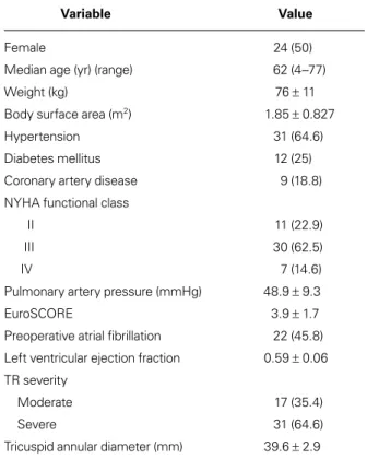
Table I documents the preoperative characteristics of the 48 patients. The median age was 62 years (range,
Fig. 1 Intraoperative photograph shows neochordae
implanta-tion on the anterior leaflet of a patient with rheumatic tricuspid valve disease.
TABLE I. Preoperative Characteristics of the 48 Patients
Variable Value
Female 24 (50)
Median age (yr) (range) 62 (4–77)
Weight (kg) 76 ± 11
Body surface area (m2) 1.85 ± 0.827
Hypertension 31 (64.6)
Diabetes mellitus 12 (25)
Coronary artery disease 9 (18.8)
NYHA functional class
II 11 (22.9)
III 30 (62.5)
IV 7 (14.6)
Pulmonary artery pressure (mmHg) 48.9 ± 9.3
EuroSCORE 3.9 ± 1.7
Preoperative atrial fibrillation 22 (45.8) Left ventricular ejection fraction 0.59 ± 0.06 TR severity
Moderate 17 (35.4)
Severe 31 (64.6)
Tricuspid annular diameter (mm) 39.6 ± 2.9 NYHA = New York Heart Association; TR = tricuspid regurgitation
Unless otherwise stated, data are presented as number and percentage or as mean ± SD.
4–77 yr), and 24 (50%) of the patients were women. The mean preoperative left ventricular ejection fraction was 0.59 ± 0.06.
Thirty patients (63%) were in New York Heart As-sociation (NYHA) functional class III, and 11 (23%) were in class II. Preoperatively, 22 patients had AF. Car-diovascular comorbidities included MV disease in 42 patients and secundum atrial septal defects in 5 (Fig. 2). Table II shows the intraoperative data.
Tricuspid Valve Disease and Repair
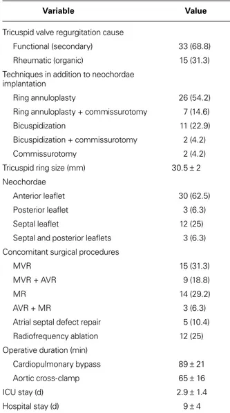
Table III lists the types of TV disease identified during surgery. Nineteen patients had anterior leaflet prolapse; 6 had involvement of both the posterior and septal leaf-lets. Commissural fusion was identified in 11 patients. Patients with annular dilation and prolapse under-went bicuspidization (n=13) or ring annuloplasty (n=33) in addition to neochordae implantation and other techniques (commissurotomy or resection of chordae tendineae). Figure 3 shows the distribution of the pros-thetic ring sizes.
In total, 103 chordae were replaced, as follows: 55 in the anterior leaflets of 30 patients, 33 in the septal leaflets of 12 patients, 3 in the posterior leaflets of 3 patients, and 12 in the septal and posterior leaflets of 3 patients.
Concomitant Procedures
Concomitant procedures were performed in 46 patients. These included 14 MV repairs, 15 MV replacements, 9 MV replacements and aortic valve replacements, 3 aortic valve replacements with mitral repair, and closure of atrial septal defects in 5 patients. Left atrial radio-frequency ablation was performed in 12 patients who had preoperative AF. Left atrial appendage ligation was performed in all patients with preoperative AF.
Follow-Up Findings
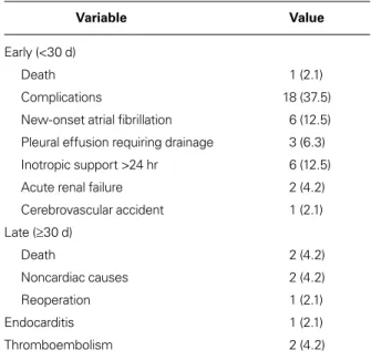
Table IV shows the early and late complications after TV repair. Early (<30-d) death occurred in one patient who had low cardiac output syndrome. The mean in-tensive care unit stay for all patients was 2.9 ± 1.4 days, and mean hospital stay, 9 ± 4 days (Table II). New-onset AF in 6 patients was medically resolved in all cases. Six patients needed inotropic support for longer than 24 hours. One patient had a cerebrovascular accident. Late follow-up data were obtained in 47 cases at an average of 44.3 ± 20.2 months postoperatively. One pa-tient died of noncardiac causes. One papa-tient who need-ed reoperation to treat infective endocarditis underwent mechanical valve replacement 14 months after MV and TV repair. 0 5 10 15 20 25 30 35 40 45
Secundum ASD Aortic valve
disease Mitral valvedisease
Pa
tien
ts
(n
)
Fig. 2 Graph shows the distribution of the patients’ preoperative
cardiovascular comorbidities. ASD = atrial septal defect
TABLE II. Intraoperative Data of the 48 Patients
Variable Value
Tricuspid valve regurgitation cause
Functional (secondary) 33 (68.8)
Rheumatic (organic) 15 (31.3)
Techniques in addition to neochordae implantation
Ring annuloplasty 26 (54.2)
Ring annuloplasty + commissurotomy 7 (14.6)
Bicuspidization 11 (22.9)
Bicuspidization + commissurotomy 2 (4.2)
Commissurotomy 2 (4.2)
Tricuspid ring size (mm) 30.5 ± 2
Neochordae
Anterior leaflet 30 (62.5)
Posterior leaflet 3 (6.3)
Septal leaflet 12 (25)
Septal and posterior leaflets 3 (6.3) Concomitant surgical procedures
MVR 15 (31.3)
MVR + AVR 9 (18.8)
MR 14 (29.2)
AVR + MR 3 (6.3)
Atrial septal defect repair 5 (10.4)
Radiofrequency ablation 12 (25)
Operative duration (min)
Cardiopulmonary bypass 89 ± 21
Aortic cross-clamp 65 ± 16
ICU stay (d) 2.9 ± 1.4
Hospital stay (d) 9 ± 4
AVR = aortic valve replacement; ICU = intensive care unit; MR = mitral repair; MVR = mitral valve replacement
Repair was satisfactory in all except 5 patients who had moderate TR when discharged from the hospi-tal. The TV repair was successful in 38 patients, who presented with absent or minimal or with mild TR. Seven patients (including 3 who had moderate TR at hospital discharge) presented with moderate TR, which was asymptomatic with medical therapy. Four of these patients had undergone Kay annuloplasty in addition to artificial neochordae implantation. We identified left
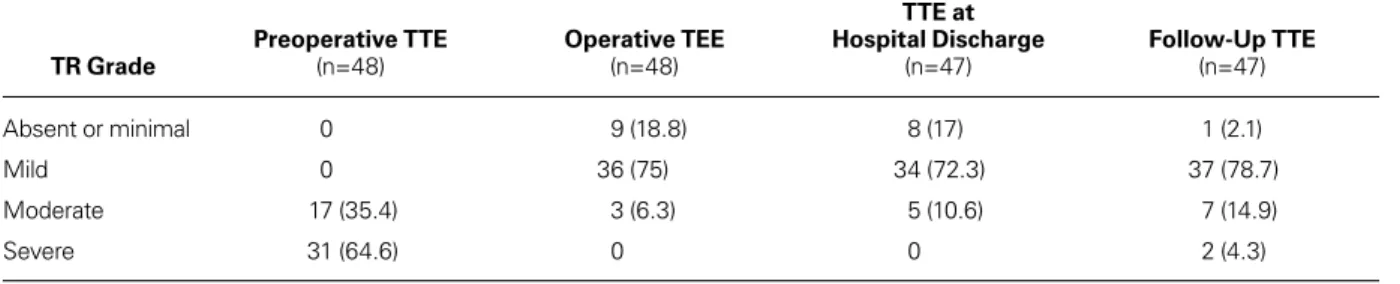
ventricular dysfunction in 4 of the 7 patients who had moderate TR during follow-up. Severe TR developed in 2 patients who had undergone concomitant MV replacement and had left ventricular dysfunction (ejec-tion frac(ejec-tion, ≤0.50). Table V and Figure 4 report the echocardiographic follow-up data of the patients.
Discussion
Functional TR, which results mainly from dilation of the tricuspid annulus secondary to RV enlargement, is the most prevalent type of TV insufficiency.4 Later in
the course of the disease, tethering of the tricuspid leaf-lets due to displacement of the papillary muscles may also contribute to TR.10 The principles of treatment for
secondary TR include correcting increased RV afterload (by treating left-sided heart disease and optimizing left ventricular function) and correcting tricuspid annular dilation and dysfunction, usually by TV annuloplasty.4
Traditionally, the primary goal of surgical treatment for secondary TR has been to correct tricuspid annular dilation by means of suture annuloplasty or ring an-nuloplasty. Most suture annuloplasty techniques are modified versions of Kay’s bicuspidization technique or De Vega annuloplasty, which consist of plicating the posterior and anterior annulus.7,11 In prosthetic ring
an-nuloplasty, the annulus is permanently fixed in a sys-tolic position, and the physiologic shape of the TV is restored.12 However, none of these techniques has
con-sistently eliminated functional TR.
The recurrence rate of substantial tricuspid insuf-ficiency after tricuspid annuloplasty ranges from 8% to 15% as early as one month after surgery.9,13 Dreyfus 0 2 4 6 8 10 12 28 30 32 33 Pa tien ts (n )
Prosthetic Ring Size (mm)
Fig. 3 Graph shows the distribution of annuloplasty ring sizes in
patients undergoing tricuspid valve repair.
TABLE III. Types of Tricuspid Valve Disease in the 48 Patients Variable Value Annular dilation 35 (72.9) Leaflet prolapse Anterior 19 (39.6) Posterior 3 (6.3) Septal 12 (25)
Septal and posterior 6 (12.5)
Leaflet tethering 12 (25) Chordal rupture 2 (4.2) Anterior 1 (2.1) Posterior 1 (2.1) Leaflet retraction 4 (8.3) Anterior 2 (4.2) Posterior 1 (2.1) Septal 1 (2.1) Chordal retraction Anterior 5 (10.4) Posterior 6 (12.5) Septal 6 (12.5)
Anterior and posterior 4 (8.3)
Commissural fusion 11 (22.9)
Data are presented as number and percentage.
TABLE IV. Early and Late Morbidity and Death in the 48 Patients
Variable Value
Early (<30 d)
Death 1 (2.1)
Complications 18 (37.5)
New-onset atrial fibrillation 6 (12.5) Pleural effusion requiring drainage 3 (6.3) Inotropic support >24 hr 6 (12.5)
Acute renal failure 2 (4.2)
Cerebrovascular accident 1 (2.1) Late (≥30 d) Death 2 (4.2) Noncardiac causes 2 (4.2) Reoperation 1 (2.1) Endocarditis 1 (2.1) Thromboembolism 2 (4.2)
and colleagues14 reported excellent outcomes, with only
a 2% recurrence rate after tricuspid ring annuloplasty for functional TR. However, this series did not include patients with severe TR or severe leaflet tethering, in whom reported recurrence rates have ranged from 15% to 30%.9,15 It has long been recognized that ring
an-nuloplasty is unlikely to successfully treat severe leaf-let tethering resulting in TR; several repair techniques have been suggested for these cases, including suture bicuspidization of the TV, and the clover technique.16,17
Aoyagi and colleagues18 reported a TR recurrence rate of
11.6%, a 10-year survival rate of 88.2%, and a freedom-from-reoperation rate of 97.6% after modified De Vega annuloplasty. However, when functional TR results from both severe annular dilation and leaflet tethering, annuloplasty alone is unlikely to be successful.8 In these
circumstances, several additional techniques have been proposed to achieve effective, durable repair. Augmenta-tion of the anterior leaflet with autologous pericardium has been used in patients with substantial tethering, and different procedures have been combined with annu-loplasty in the presence of prolapse or flail, including
chordae replacement, leaflet resection, chordal transpo-sition, and papillary muscle reimplantation.19-23
During the past 10 years, e-PTFE sutures have been used to replace chordae tendineae, especially in patients with MV prolapse. Reports of artificial chordae ten-dineae implantation for the treatment of TR regurgi-tation are sparse. Marin and co-authors24 reported the
case of a 72-year-old woman in NYHA class III who underwent successful TV repair with implantation of artificial neochordae; her preoperative echocardiograms revealed severe TV regurgitation caused by prolapse of the anterior leaflet (A1–A2) and annular dilation. Ar-tificial chordal implantation is also used in the repair of traumatic TR, which is associated with chordal dis-ruption, rupture of the anterior papillary muscle, leaf-let laceration, and leafleaf-let retraction.19-21 Tricuspid valve
malformations such as papillary muscle elongation and congenital absence of the papillary muscle can also be treated by artificial chordae implantation. Ito and col-leagues25 reported the use of artificial chordae to repair
isolated congenital TR in a 23-year-old woman. These reports have suggested that artificial chordal implanta-tion is a viable technique for the surgical repair of TR. To prevent TR recurrence, we use artificial neochor-dae implantation as an adjunctive procedure to ring or suture annuloplasty, neither of which has proved com-pletely effective in achieving TR repair. Since 2009, we have used artificial chordae to treat TR resulting from annular dilation associated with prolapse or severe teth-ering of multiple leaflets. If leaflet tethteth-ering persists after annuloplasty, we resect the native chordae of the leaflet and replace them with longer artificial neochor-dae. Lapenna and associates26 used the clover technique
to manage multiple prolapse and flail of the tricuspid leaflets in the presence of traumatic or degenerative TR and severe leaflet tethering secondary to advanced RV remodeling. In that series, TR was absent or mild in 55 patients (88.7%), moderate in 6 (9.6%), and severe in one (1.6%).26 In our institution, the size of the
prosthet-ic ring is chosen on the basis of the surface area of the leaflet tissue attached to the chordae arising from the anterior papillary muscle. In our study, prolapse of the
0 1 2 3 4 5 6 Moderate TR Severe TR Pa tien ts (n ) RheumaticFunctional
Fig. 4 Graph shows grading of recurrent tricuspid regurgitation
(TR) severity by means of transthoracic echocardiography in patients who had rheumatic and functional valvular disease during follow-up evaluation.
TABLE V. Results of Operation on Tricuspid Regurgitation Grade
TR Grade Preoperative TTE (n=48) Operative TEE (n=48)
TTE at Hospital Discharge (n=47) Follow-Up TTE (n=47) Absent or minimal 0 9 (18.8) 8 (17) 1 (2.1) Mild 0 36 (75) 34 (72.3) 37 (78.7) Moderate 17 (35.4) 3 (6.3) 5 (10.6) 7 (14.9) Severe 31 (64.6) 0 0 2 (4.3)
TEE = transesophageal echocardiography; TR = tricuspid regurgitation; TTE = transthoracic echocardiography Data are presented as number and percentage.
anterior or posterior leaflet was observed in 9 patients whose functional TR resulted from the restriction of the tricuspid annular motion by the ring. Ten other patients with functional TR presented with prolapse of one or multiple TV leaflets after Kay annuloplasty. In these complex cases, we used artificial neochordae implanta-tion as an adjunct to annuloplasty, to restore valvular competence and avoid post-repair leak and the need for valve replacement.
Surgical valve repair is now firmly established as the gold standard for valvular heart disease. Some research-ers have analyzed the outcomes of surgical treatment for functional TV regurgitation, but reports on patients with organic TV disease predominate in the medical literature.15,27-29 Surgical repair of rheumatic TV disease
often fails because of severe anatomic distortion of the valve apparatus. The results of this procedure are less satisfactory than those of functional TV repair because, in patients with rheumatic TV disease, the durability of repair is compromised by the active and rapidly progres-sive nature of the disease process. Bernal and colleagues30
reported that 38 patients (25.3%) with rheumatic val-vular heart disease needed TV reoperations after mitral and TV repair. Tang and colleagues31 obtained favorable
outcomes of rheumatic TV repair with use of autolo-gous pericardium; follow-up echocardiograms showed mild regurgitation in 8 patients (25.8%) and moderate regurgitation in one (3.2%). Among other modern tech-niques for repairing complex TV lesions is edge-to-edge valve plasty, which has been used as an effective adjunct procedure in patients with residual TR.32 Because TV
replacement is a high-risk procedure in patients with rheumatic heart disease33 and is associated with high
recurrence of TR, we chose to repair rheumatic TV by implanting artificial neochordae. This procedure was performed in 15 of our patients with rheumatic TV disease. Other techniques, including commissurotomy, leaflet mobilization, annuloplasty, and prosthetic ring implantation, were performed as needed. Primary chord resection was performed in those 15 patients in addition to secondary chord resection because of the thickened chordae. The consequent prolapse of the TV leaflets was corrected by implanting artificial neochordae. Fol-low-up echocardiograms showed no or mild TR in 9 of the 15 patients. Five patients had moderate TR and were treated medically. One patient had severe TR. Our results suggest that rheumatic pathology plays a role in determining the durability of TV repair.
Upon discharge from the hospital, only 5 of our sur-viving 47 patients had moderate TR; the remaining 42 had absent or minimal TR or mild TR. Follow-up TTE showed that TR was absent, minimal, or mild in 80.8% of patients. Seven patients (14.9%) had moderate TR, and 2 (4.3%) had severe TR. Our treatment approach might substantially increase the rate of successful repair in difficult cases, such as those described in our study.
However, careful intraoperative evaluation of the ana-tomic lesions underlying TR is necessary to decide if this technique can be applied with a reasonably high probability of success.
Study Limitations
The major limitations of this study are its retrospective design, the small number of patients included, and the short follow-up periods in some cases.
Conclusion
Our results indicate that neochordae implantation is a safe and durable technique in surgical repair of the TV, and that it leads to good outcomes in the management of TR. In carefully selected patients, organic rheumatic TV disease can be treated with neochordae implanta-tion adjunctively with other techniques. Although the preliminary results of our approach are encouraging, additional data from studies with longer follow-up pe-riods are necessary to confirm its effectiveness and to define its role as an alternative to TV replacement in selected patients.
References
1. Waller BF, Howard J, Fess S. Pathology of tricuspid valve ste-nosis and pure tricuspid regurgitation--part II. Clin Cardiol 1995;18(3):167-74.
2. Cohen SR, Sell JE, McIntosh CL, Clark RE. Tricuspid re-gurgitation in patients with acquired, chronic, pure mitral regurgitation. II. Nonoperative management, tricuspid valve annuloplasty, and tricuspid valve replacement. J Thorac Car-diovasc Surg 1987;94(4):488-97.
3. Braunwald NS, Ross J, Morrow AG. Conservative manage-ment of tricuspid regurgitation in patients undergoing mitral valve replacement. Circulation 1967;35(4 Suppl):I63-9. 4. De Bonis M, Taramasso M, Lapenna E, Alfieri O.
Manage-ment of tricuspid regurgitation. F1000Prime Rep 2014;6:58. 5. Matsuyama K, Matsumoto M, Sugita T, Nishizawa J, Tokuda
Y, Matsuo T. Predictors of residual tricuspid regurgitation after mitral valve surgery. Ann Thorac Surg 2003;75(6):1826-8.
6. Raja SG, Dreyfus GD. Surgery for functional tricuspid re-gurgitation: current techniques, outcomes and emerging con-cepts. Expert Rev Cardiovasc Ther 2009;7(1):73-84. 7. Kay JH, Maselli-Campagna G, Tsuji KK. Surgical treatment
of tricuspid insufficiency. Ann Surg 1965;162:53-8.
8. Fukuda S, Song JM, Gillinov AM, McCarthy PM, Daimon M, Kongsaerepong V, et al. Tricuspid valve tethering predicts residual tricuspid regurgitation after tricuspid annuloplasty. Circulation 2005;111(8):975-9.
9. McCarthy PM, Bhudia SK, Rajeswaran J, Hoercher KJ, Lytle BW, Cosgrove DM, Blackstone EH. Tricuspid valve repair: durability and risk factors for failure. J Thorac Cardiovasc Surg 2004;127(3):674-85.
10. Kim HK, Kim YJ, Park JS, Kim KH, Kim KB, Ahn H, et al. Determinants of the severity of functional tricuspid regurgita-tion. Am J Cardiol 2006;98(2):236-42.
11. De Vega NG. Selective, adjustable and permanent annulo-plasty. An original technic for the treatment of tricuspid insuf-ficiency [in Spanish]. Rev Esp Cardiol 1972;25(6):555-6.
12. Carpentier A, Deloche A, Hanania G, Forman J, Sellier P, Piwnica A, et al. Surgical management of acquired tricuspid valve disease. J Thorac Cardiovasc Surg 1974;67(1):53-65. 13. Navia JL, Nowicki ER, Blackstone EH, Brozzi NA, Nento
DE, Atik FA, et al. Surgical management of secondary tricus-pid valve regurgitation: annulus, commissure, or leaflet pro-cedure? J Thorac Cardiovasc Surg 2010;139(6):1473-82.e5. 14. Dreyfus GD, Corbi PJ, Chan KM, Bahrami T. Secondary
tri-cuspid regurgitation or dilatation: which should be the criteria for surgical repair? Ann Thorac Surg 2005;79(1):127-32. 15. Tang GH, David TE, Singh SK, Maganti MD, Armstrong
S, Borger MA. Tricuspid valve repair with an annuloplasty ring results in improved long-term outcomes. Circulation 2006;114(1 Suppl):I577-81.
16. De Bonis M, Lapenna E, La Canna G, Grimaldi A, Maisano F, Torracca L, et al. A novel technique for correction of se-vere tricuspid valve regurgitation due to complex lesions. Eur J Cardiothorac Surg 2004;25(5):760-5.
17. Ghanta RK, Chen R, Narayanasamy N, McGurk S, Lipsitz S, Chen FY, Cohn LH. Suture bicuspidization of the tricuspid valve versus ring annuloplasty for repair of functional tricus-pid regurgitation: midterm results of 237 consecutive patients. J Thorac Cardiovasc Surg 2007;133(1):117-26.
18. Aoyagi S, Tanaka K, Hara H, Kumate M, Oryoji A, Yasu-naga H, et al. Modified De Vega’s annuloplasty for functional tricuspid regurgitation--early and late results. Kurume Med J 1992;39(1):23-32.
19. Maisano F, Lorusso R, Sandrelli L, Torracca L, Coletti G, La Canna G, Alfieri O. Valve repair for traumatic tricuspid re-gurgitation. Eur J Cardiothorac Surg 1996;10(10):867-73. 20. Hachiro Y, Sugimoto S, Takagi N, Osawa H, Morishita K,
Abe T. Native valve salvage for post-traumatic tricuspid regur-gitation. J Heart Valve Dis 2001;10(2):276-8.
21. van Son JA, Danielson GK, Schaff HV, Miller FA Jr. Trau-matic tricuspid valve insufficiency. Experience in thirteen pa-tients. J Thorac Cardiovasc Surg 1994;108(5):893-8. 22. Dreyfus GD, Raja SG, John Chan KM. Tricuspid leaflet
aug-mentation to address severe tethering in functional tricuspid regurgitation. Eur J Cardiothorac Surg 2008;34(4):908-10. 23. Messika-Zeitoun, Thomson H, Bellamy M, Scott C,
Tri-bouilloy C, Dearani J, et al. Medical and surgical outcome of tricuspid regurgitation caused by flail leaflets. J Thorac Car-diovasc Surg 2004;128(2):296-302.
24. Marin D, Ramadan K, Hamilton C, Schuetz A. Tricuspid valve repair with artificial chordae in a 72-year-old woman. Thorac Cardiovasc Surg 2011;59(8):495-7.
25. Ito T, Katogi T, Aeba R, Fujii H, Goto T, Kawada S. Surgical repair of isolated congenital tricuspid regurgitation with arti-ficial chordae--a case of two year-follow up [in Japanese]. Jpn J Thorac Cardiovasc Surg 1998;46(12):1334-8.
26. Lapenna E, De Bonis M, Verzini A, La Canna G, Ferrara D, Calabrese MC, et al. The clover technique for the treatment of complex tricuspid valve insufficiency: midterm clinical and echocardiographic results in 66 patients. Eur J Cardiothorac Surg 2010;37(6):1297-303.
27. Grinda JM, Latremouille C, D’Attellis N, Berrebi A, Chauvaud S, Carpentier A, et al. Triple valve repair for young rheumatic patients. Eur J Cardiothorac Surg 2002;21(3):447-52.
28. Chaouch H, Kafsi N, Ben Ismail M. Indications and results of surgery of organic involvement of the tricuspid valve [in French]. Arch Mal Coeur Vaiss 1989;82(6):879-84. 29. Han QQ, Xu ZY, Zhang BR, Zou LJ, Hao JH, Huang SD.
Primary triple valve surgery for advanced rheumatic heart dis-ease in mainland China: a single-center experience with 871 clinical cases. Eur J Cardiothorac Surg 2007;31(5):845-50. 30. Bernal JM, Ponton A, Diaz B, Llorca J, Garcia I, Sarralde JA,
et al. Combined mitral and tricuspid valve repair in rheumatic valve disease: fewer reoperations with prosthetic ring annulo-plasty. Circulation 2010;121(17):1934-40.
31. Tang H, Xu Z, Zou L, Han L, Lu F, Lang X, Song Z. Valve re-pair with autologous pericardium for organic lesions in rheu-matic tricuspid valve disease. Ann Thorac Surg 2009;87(3): 726-30.
32. Lai YQ, Meng X, Bai T, Zhang C, Luo Y, Zhang ZG. Edge-to-edge tricuspid valve repair: an adjuvant technique for residual tricuspid regurgitation. Ann Thorac Surg 2006;81(6):2179-82.
33. Iscan ZH, Vural KM, Bahar I, Mavioglu L, Saritas A. What to expect after tricuspid valve replacement? Long-term results. Eur J Cardiothorac Surg 2007;32(2):296-300.