Yazışma Adresi/Address for Correspondence: Dr. Şehnaz Yılmaz, Çukurova University Faculty of Dentistry , Department of Endodontics, Adana, Turkey E-mail: [email protected]
Geliş tarihi/Received: 20.02.2016 Kabul tarihi/Accepted: 06.04.2016
ARAŞTIRMA/RESEARCH
Effectiveness of different irrigation systems on filling of simulated
lateral canals
Farklı irrigasyon sistemlerinin yapay yan kanalların doldurulması üzerindeki etkisi
Şehnaz Yılmaz
1, Ayşin Dumani
1, Ayfer Ateş
1, Kadriye Özdayı
11Çukurova University Faculty of Dentistry, Department of Endodontics, Adana, Turkey
Cukurova Medical Journal 2016;41(3):515-520.
Abstract Öz
Purpose: The purpose of this study was to evaluate the
effect of currently used sonic irrigation and activation systems on filling of simulated lateral canals in a closed system by radiographic analysis and tooth decalcification and clearing method.
Materials and Methods: Sixty freshly extracted human
maxillary anterior and mandibular premolar teeth with similar root length were selected for this study. The teeth were decoronated and lateral canals were created by inserting #15 reamer at 3, 6, 9 mm away from the apex, on the mesial and distal walls perpendicularly to the external surface. The root canals were instrumented and randomly assigned into three groups (n=20): group 1, Vibringe sonic irrigation; group 2, passive ultrasonic irrigation; and control group 3, conventional needle irrigation. The root canal were filled with AH plus and gutta-percha using the cold lateral compaction technique. Periapical radiographs were taken from each specimen and then decalcified for stereomicroscopic analysis.
Results: Significant difference was found at the middle
third between the control and the passive ultrasonic irrigation groups in the radiographic evaluation and between the control and the Vibringe group in the cleared specimens. However, the cleared specimens showed higher values than radiographic evaluation quantitatively.
Conclusion: Sonically or ultrasonically irrigation showed
significant differences on the filling of the simulated lateral canals at the middle third of the root canals. Ultrasonic activation of the irrigants represented better results in radiographic and cleared specimen evaluation.
Amaç: Bu çalışmanın amacı, güncel olarak kullanılan sonik
irrigasyon ve aktivasyon sistemlerinin kapalı yöntemle hazırlanmış yapay yan kanalların doldurulabilmesi üzerindeki etkisinin, radyografik analizle ve diş şeffaflaştırma yöntemiyle değerlendirilmesidir.
Gereç ve Yöntem: Bu çalışma için, kök uzunlukları
birbirine yakın, 60 adet yeni çekilmiş üst çene ön bölge ve alt çene küçükazı insan dişi seçildi. Dişlerin kronları uzaklaştırıldı ve kök ucunun 3, 6, 9 mm uzak noktalarında hem mezial hem de distal taraftan dişin dış yüzeyine dik olacak şekilde 15 nolu reamerla lateral kanallar oluşturuldu. Kök kanalları genişletildi ve rastgele olacak şekilde üç gruba (n=20) ayrıldı: grup 1, Vibringe sonik irrigasyon; grup 2, pasif ultrasonik irrigasyon; ve kontrol grubu, geleneksel iğne irrigasyonu. Kök kanalları AH Plus ve guta perka ile soğuk lateral sıkıştırma yöntemi ile dolduruldu. Her örnekten periapikal radyograf alındı ve stereomikroskop için dekalsifiye edildi.
Bulgular: Radyografik değerlendirmede, kontrol ve pasif
ultrasonik irrigasyon grupları arasında ve şeffaflaştırma yapılan örneklerin analizinde kontrol ve Vibringe grupları arasında orta üçlü seviyesinde istatistiksel olarak farklılık bulunmuştur. Bununla birlikte, şeffaflaştırma yapılan örnekler, radyografik incelemeye göre daha yüksek değerler göstermiştir.
Sonuç: Sonik veya ultrasonik irrigasyon kök kanallarının
orta üçlü seviyesinde oluşturulan yapay yan kanalların doldurulmasında istatistiksel olarak farklılık göstermiştir. İrrigantların ultrasonik olarak aktive edilmesi hem radyografik hem de şeffaflaştırılan örneklerin değerlendirilmesinde daha iyi sonuçlar göstermektedir.
Key words: irrigation, lateral compaction, root canal
INTRODUCTION
The complete sealing of the root canal system after a biomechanical procedure can achieve with effective removal of intracanal debris and sufficient filling. However, the complex anatomy of root canal systems has limited our ability to clean and disinfect it predictably. The debris accumulation in the uninstrumented fins, irregularities on the cross-section of the canal, apical delta and lateral canals may not allow for proper disinfection and may prevent the root canal filling from reaching these recesses1,2. Chemical debridement is paramount in
ensuring that canals are sufficiently cleaned before filling. Because, gutta-percha does not adhere to the dentinal walls, the sealer must fill the irregularities and the dentinal tubules of the root canal system. In order to achieve efficient debris and smear removal, irrigant is important as well as irrigation delivery system. Sodium hypochlorite (NaOCl), chlorhexidine (CHX), ethylenediaminetetraacetic acid (EDTA), MTAD, and alcohol are the most preferred irrigants by clinicians. In most of the clinical and in vitro studies, NaOCl, which have antimicrobial effect and tissue dissolution properties has been used in association with EDTA, which acts on the inorganic debris formed in instrumented root canals as a final irrigation protocol3, 4. On the other
hand, numerous devices have been proposed to increase the efficacy of irrigant delivery and improve canal cleanliness. Sonic devices have been shown to safely clean the canal system, including lateral canals, fins, and apical deltas, by energizing the root canal irrigants5 at a lower frequency (2–3 kHz) than
ultrasonic devices (25–40 kHz). When these systems are compared with conventional needle irrigation techniques, they have demonstrated better results in the removal of the smear layer from the canal walls6.
Lateral canals and apical ramifications are arguably difficult to reach, clean, disinfect, and fill during treatment. In this context, in vitro studies have aimed to evaluate the ability of filling lateral canals with different techniques7-9. Curiously, most of these
studies reported that no significant differences were observed for the efficacy of different techniques in forcing sealer into the lateral canal, even though thermoplasticized techniques obviously also tended to force gutta-percha in many specimens10. But, still
their possible clinical significance called the attention of clinicians and researchers as to how and
disinfect and fill.
The purpose of this study was to evaluate the effect of currently used sonic irrigation and activation systems on filling of simulated lateral canals in a closed system by radiographic analysis and tooth decalcification and clearing method.
MATERIALS AND METHODS
Preparation of the specimens
Sixty freshly extracted human maxillary anterior and mandibular premolar teeth with similar root length were selected for this study. This study was approved by the University of Cukurova Institutional Review Board. Teeth were kept in %10 formalin solution until they were used. Any visible calculus was removed ultrasonically. Presence of a single canal was verified radiographically by taking 3 angulated films and by direct exploration under the dental operating microscope.
The working length of each tooth was determined visually by subtracting 1 mm from the length at which the size 10 K–file tip extruded apically. The teeth were decoronated with a slow-speed diamond saw and their root length was standardized at 15 mm. Lateral canals were created by inserting #15 reamer at 3, 6, 9 mm away from the apex, on the mesial and distal walls perpendicularly to the external surface. To prevent the escape of irrigants from the root apex by simulating a clinical condition, a closed-canal design is used. All root surfaces were covered by nail polish for sealing of the lateral canals. The apices of the roots were sealed with hot glue. After setting, the tooth was inserted into a polyvinylsiloxane impression material-filled (Blue Moose, Parkell Inc, Farmington, CT) Eppendorf tube. The Eppendorf tube, in turn, was affixed to an experimental set up which permitted canal irrigation.
The root canals were instrumented using the RevoS rotary system (Micro-Mega, Besancon, France) according to the manufacturers’ instructions including SC1, SC2 and SU files. Irrigation performed with a 30G side venting needle using 5 ml 2.5% sodium hypochlorite between each instrument. The irrigation needles were introduced passively up to 1 mm from the working length. Upon completion of instrumentation, teeth were randomly divided into three groups. Final irrigation
517
5 ml 17%EDTA and 5 ml of 2.5% NaOCl according to the groups that described below:
Groups
Group 1 (n=20): Vibringe sonic activation: Sonic activation was delivered by using Vibringe that combines battery-driven vibrations (9000 cpm) with manually operated irrigation of the root canal. Vibringe was activated during the irrigation procedure and 30G side vented needle was inserted up to 1 mm from working length. Flow rate was approximately 5 ml/min.
Group 2 (n=20): Passive ultrasonic (PUI) activation:Ultrasonic activation was performed with a stainless steel ultrasonic file ISO 25. Final irrigation was performed with an in-and-out motion by a 30 G side vented needle inserted up to 1 mm from the working length. Flow rate was approximately 5 ml/30 seconds. The ultrasonic file was passively inserted up to 1 mm from working length and activated for 30 seconds for each 5 ml irrigant.
Group 3 (n=20): Control group: Final irrigation was performed with an in-and-out motion by a 30 G side vented needle inserted up to 1 mm from the working length. Flow rate was adjusted as 5 ml/min.
Root canal filling
The root canals were filled with AH plus sealer and 0.02 taper gutta-percha cones using the cold lateral compaction technique by one operator. Lateral compaction was accomplished using an ISO size 25 spreader and 25 gutta-percha accessory cones. The spreader initially reached to within 2 mm of the full working length and the process was repeated until accessory cones could not be inserted more than 2 mm into the canal.
Radiographic and photographic evaluation
of cleared specimens
Periapical digital radiographs (PSPIX Imaging Plates; Satelec SAS, France) of each specimen were taken using the Belmont 303-A Dental X-ray unit (Takara Belmont Corp., Osaka, Japan) after standardizing the positioning of the roots and the focus film distances. Specimens were decalcified in 5 % nitric acid for 36 hours, and the solution renewed every 8 hours. Samples were then
dehydrated ascending grades of ethyl alcohol and submerged in 99.9% methyl salicylate for clearing and re-hardening of dental tissues. The specimens were viewed at ×40 magnification using a stereomicroscope (Olympus SZ61, Tokyo, Japan) and digital images were captured with a digital camera (QImaging, Canada) that attached to the stereomicroscope. The radiographic and photographic images showing the root canal fillings in each specimen were imported into the Image J software program (Wayne Rasband, National Institutes of Health). The amount of filling material observed in each lateral canal at the level of 3, 6, 9 mm was calculated by the examiners. The data was expressed of the area filled (area in mm2 occupied
by the filling material inside the lateral canal in relation to entire area).
Statistical analysis
All analyses were performed using IBM SPSS Statistics Version 20.0 statistical software package. Continuous variables were summarized as median and minimum-maximum. The normality of distribution for the percentages of the filled area was confirmed with the Kolmogorov-Smirnov test. For comparison of two paired continuous variables (radiograph vs cleared specimens), Wilcoxon Signed Rank test was used. For non-normal distributed data, Kruskal Wallis test was used to compare three groups. Bonferroni adjusted Mann Whitney U test was used for multiple comparisons of groups. The statistical level of significance for all tests was considered to be 0.05.
RESULTS
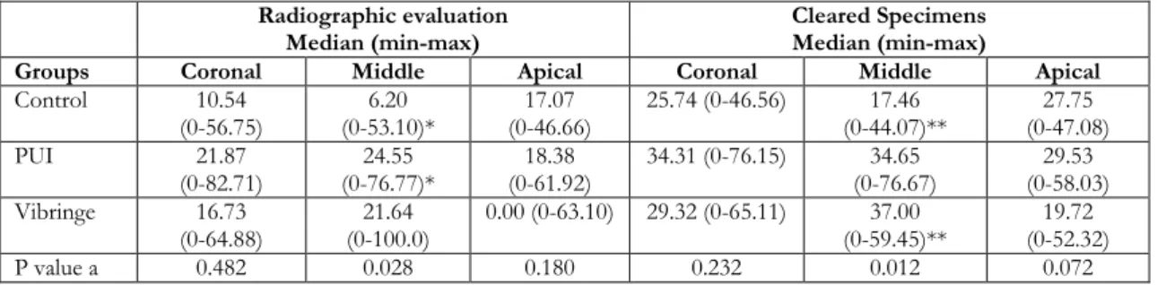
Table 1 show the ability of epoxy resin sealer to fill the simulated lateral canals (measured in linear percentages). Significant difference was found between the control and the PUI groups at the middle third in the radiographic evaluation (p<0.05). When considering cleared specimens measurements, Vibringe group showed significantly higher values than the control group at the middle third (p<0.05). In comparison between the radiograph (Fig 1) and cleared specimens (Fig 2), no significant difference was found among the groups at all thirds, however, the cleared specimens showed higher values than radiographic evaluation quantitatively.
Table 1. Radiographic and cleared specimens evaluation of filling material penetration into simulated lateral canals made in each third.
Radiographic evaluation
Median (min-max) Cleared Specimens Median (min-max)
Groups Coronal Middle Apical Coronal Middle Apical
Control 10.54 (0-56.75) (0-53.10)* 6.20 (0-46.66) 17.07 25.74 (0-46.56) (0-44.07)** 17.46 (0-47.08) 27.75 PUI 21.87 (0-82.71) (0-76.77)* 24.55 (0-61.92) 18.38 34.31 (0-76.15) (0-76.67) 34.65 (0-58.03) 29.53 Vibringe 16.73 (0-64.88) (0-100.0) 21.64 0.00 (0-63.10) 29.32 (0-65.11) (0-59.45)** 37.00 (0-52.32) 19.72 P value a 0.482 0.028 0.180 0.232 0.012 0.072
a Kruskal Wallis Test; * p=0.016, Mann Whitney U test with Bonferroni correction; ** p=0.033, Mann Whitney U test with Bonferroni
correction
Figure 1. Radiographic images of each group. (A) Control, (B) PUI, (C) Vibringe.
519
DISCUSSION
Removal of the smear layer from the root canal walls during instrumentation allows access of endodontic irrigants and filling materials into the dentinal tubules, lateral canals and intracanal irregularities11, 12. The filling capability of solid core
endodontic materials such as gutta-percha and resilon were investigated with various studies and showed good adaptation to the root canal system including the irregularities and filling in simulated lateral canals when these materials get warmed13.
However, if lateral condensation was used to fill the root canals, it was expected that the filling of the simulated lateral canals with root canal sealer. In this regard, this study designed to evaluate the effects of activation of NaOCl and EDTA with a sonic frequency on filling of simulated lateral canals. Therefore, the removal of smear layer appeared to improve the filling of the lateral canals did not investigate in this study.
Although a recent study has shown no correlation between sealer penetration and sealability of root-filling materials14, sealer penetration into the tubules
can be used as an indicator for smear layer removal. In addition, it can also be considered beneficial for preventing reinfection15 because of the sealer’s
antibacterial activity and blockade effect. It can also be effective at killing or entombing bacteria within the tubules16. Furthermore, the mechanical
interlocking of the sealer inside the tubules has been suggested to improve retention of the material17.
To be clinically relevant, in vitro studies should reproduce the clinical situation as much as possible. In order to prevent the extrusion of the irrigant through apically or laterally, a closed canal system was used in this study. Because, while testing the effect of irrigation and fluid dynamics, it should take into consideration the presence of the periradicular tissues surrounding the root surface, preventing passive extrusion of irrigant18. This physical
limitation explains the discrepancy found in the literature. Whereas some articles reported optimal results with positive pressure irrigation19,20, others
found its efficacy to be very limited21,22. In addition,
a reamer was used for creating the artificial lateral canals because of the specimen’s standardization. Thus, creating straight and smooth canals can be accepted as a limitation of this study which does not simulate clinical conditions. In vitro studies also evaluated decalcified and cleared specimens by
radiographic and visual analyses after filling of the root canals13,23. Almeida et al.23 observed that the
radiographic analysis did not detect lateral canal fillings in 8% of the specimens although it could be visualized in the decalcified and cleared teeth. In accordance with the mentioned studies, particular decrease of the lateral canal filling measurements in the radiographic analyses was seen in the present study. Even no significant differences were revealed between the results from the radiographic and visual analyses of the cleared specimens, it should be pointed out that radiographic analysis was performed digitally and the specimens had been previously subjected to the decalcification and clearing protocol, promoting better image quality. The results of this study indicated that after activation of irrigants with PUI caused significantly better quality of filling lateral canals at the middle third in the radiographic evaluation. But, these differences did not seen in the cleared specimen evaluation. However, in cleared specimen evaluation, the significant difference was seen between the control and the Vibringe group at the middle third. Thus, the results concluded that lateral condensation technique caused a difference only at the middle third of the root. This may be attributed to penetration of the spreader that used in the lateral condensation method. The penetration of the spreader to the 3 mm minus from the working length may cause filling of the lateral canals by pushing the sealer to the canal walls independently from the irrigation technique used.
In considering the overall of the data, PUI showed better results in both radiographic and cleared specimen evaluation. Rodig et al11 compared the
Vibringe, ultrasonics, and conventional needle irrigation in their ability to remove canal debris by using an artificial groove technique. In accordance with the present study, they found ultrasonics to be the most effective, followed by the Vibringe, and they both were significantly better than conventional needle irrigation. In addition, Gregorio et al24
reported that the PUI group demonstrated significantly more penetration of irrigant into lateral canals than syringe irrigation and sonic irrigation. In conclusion, sonically or ultrasonically irrigation showed significant differences on the filling of the simulated lateral canals at the middle third of the root canals. However, ultrasonic activation of the irrigants represented better results in radiographic and cleared specimen evaluation.
REFERENCES
1. Wu MK, Wesselink PR. A primary observation on the preparation and obturation in oval canals. Int Endod J. 2001;34:137–412.
2. De-Deus G, Gurgel-Filho ED, Magalhães KM, Coutinho-Filho T. A laboratory analysis of gutta-percha-filled area obtained using Thermafil, System B and lateral condensation. Int Endod J. 2006;39:378–83.
3. Baumgartner JC, Mader CL. A scanning electron microscopic evaluation of four root canal irrigation regimens. J Endod. 1987;13:147–57.
4. Calt S, Serper A. Time-dependent effects of EDTA on dentin structures. J Endod. 2002;28:17–9. 5. Ruddle CJ. Hydrodynamic disinfection: tsunami
endodontics. Dent Today. 2007;26:4–7.
6. Kuah HG, Lui JN, Tseng PSK, Chen NN. The effect of EDTA with and without ultra-sonics on removal of the smear layer. J Endod. 2009;35:393–6.
7. Venturi M. An ex vivo evaluation of a gutta-percha filling technique when used with two endodontic sealers: analysis of the filling of main and lateral canals. J Endod. 2008;34:1105–10.
8. DuLac KA, Nielsen CJ, Tomazic TJ, Ferrillo PJ Jr., Hatton JF. Comparison of the obturation of lateral canals by six techniques. J Endod. 1999;25:376–80. 9. Venturi M, Prati C, Capelli G, Falconi M, Breschi L.
A preliminary analysis of the morphology of lateral canals after root canal filling using a tooth-clearing technique. Int Endod J. 2003;36:54–63.
10. Ricucci D, Siqueira JF Jr. Fate of the tissue in lateral canals and apical ramifications in response to pathologic conditions and treatment procedures. J Endod. 2010;36:1–15.
11. Rödig T, Bozkurt M,Konietschke F, Hülsmann M. Comparison of the Vibringe system with syringe and passive ultrasonic irrigation in removing debris from simulated root canal irregularities. J Endod. 2010;36:1410–3.
12. Passarinho-Neto JG, Marchesan MA, Ferreira RB, Silva RG, Silva-Sousa YT, Sousa- Neto MD. In vitro evaluation of endodontic debris removal as obtained by rotary instrumentation coupled with ultrasonic irrigation. Aust Endod J. 2006;32:123–8.
13. Sant'Anna-Junior A, Guerreiro-Tanomaru JM, Martelo RB, Silva GF, Tanomaru Filho M. Filling of
simulated lateral canals with gutta-percha or thermoplastic polymer by warm vertical compaction. Braz Oral Res. 2015;29:1-6.
14. De-Deus G, Brandao MC, Leal F, Reis C, Souza EM, Luna AS et al. Lack of correlation between sealer penetration into dentinal tubules and sealibility in nonbonded root fillings. Int Endod J. 2012;45:642– 51.
15. Kokkas AB, Boutsioukis AC, Vassiliadis LP, Stavrianos CK. The influence of the smear layer on dentinal tubule penetration depth by three different root canal sealers: an in vitro study. J Endod. 2004;30:100–2.
16. Heling I, Chandler NP. The antimicrobial effect within dentinal tubules of four root canal sealers. J Endod. 1996;22:257–9.
17. White RR, Goldman M, Lin PS. The influence of the smeared layer upon dentinal tubule penetration by plastic filling materials. J Endod. 1984;10:558–62. 18. Migun NP, Shnip AI. Model of film flow in a
dead-end conic capillary. J Eng Phys Thermophys. 2002;75:1422–8.
19. Khademi A, Yazdizadeh M, Feizianfard M. Determination of the minimum instrumentation size for penetration of irrigants to the apical third of root canal systems. J Endod. 2006;32:417–20.
20. Grandini S, Balleri P, Ferrari M. Evaluation of Glyde File Prep in combination with sodium hypochlorite as a root canal irrigant. J Endod. 2002;28:300–3. 21. Usman N, Baumgartner JC, Marshall JG. Influence
of instrument size on root canal debridement. J Endod. 2004;30:110–2.
22. O’Connell MS, Morgan LA, Beeler WJ, Baumgartner JC. A comparative study of smear layer removal using different salts of EDTA. J Endod. 2000;26:739–43.
23. Almeida JFA, Gomes BPFA, Ferraz CCR, Souza-Filho FJ, Zaia AA. Filling of artificial lateral canals and microleakage and flow of five endodontic sealers. Int End J. 2007;40:692–9.
24. Gregorio C, Estevez R, Cisneros R, Paranjpe A,
Cohenca N. Efficacy of different irrigation and activation systems on the penetration of sodium hypochlorite into simulated lateral canals and up to working length: an in vitro study. J Endod. 2010;36:1216–2