1122
M
acrophages play central roles in the development of atherosclerosis through modulation of cholesterol homeostasis, the immune-inflammatory response, and plaque cellularity.1 Macrophage activation and survival are crucialdeterminants of atherosclerotic lesion development.2 In
addi-tion, macrophages contribute to the integration of immune and metabolic responses, and their dysfunction contributes to chronic metabolic disorders, such as obesity, type 2 diabetes mellitus, and cardiovascular disease.3
The c-Jun NH2-terminal kinases (JNK) belong to the stress-activated protein kinase family, which are stress-activated by a vari-ety of environmental (radiation, osmotic, and redox stress) and metabolic stresses, cytokines, and growth factors.4,5 JNK plays
an important role in inflammatory signaling, and its activation is crucial for programmed cell death.6 In mammals, the JNK
protein kinases are encoded by 3 genes: Jnk1, Jnk2 and Jnk3, which transcribe several alternatively spliced isoforms.7 Jnk1
and Jnk2 genes are expressed ubiquitously, whereas the Jnk3 gene is restricted to the brain, cardiac smooth muscle, pan-creatic islets, and testis.4 The targeted disruption of the Jnk1
or Jnk2 genes revealed that they compensate for each other’s activity and are functionally redundant,8 but each isoform also
exhibits distinct roles.9 For example, activation of CD8+ T cells
is impaired in Jnk1 knockout mice but enhanced in Jnk2 null mice.10 Loss of Jnk1, but not Jnk2, suppresses obesity and
improves insulin sensitivity in mice.11 JNK1, but not JNK2,
© 2016 American Heart Association, Inc.
Arterioscler Thromb Vasc Biol is available at http://atvb.ahajournals.org DOI: 10.1161/ATVBAHA.116.307580
Objective—The c-Jun NH2-terminal kinases (JNK) are regulated by a wide variety of cellular stresses and have been implicated in apoptotic signaling. Macrophages express 2 JNK isoforms, JNK1 and JNK2, which may have different effects on cell survival and atherosclerosis.
Approach and Results—To dissect the effect of macrophage JNK1 and JNK2 on early atherosclerosis, Ldlr−/− mice were
reconstituted with wild-type, Jnk1−/−, and Jnk2−/− hematopoietic cells and fed a high cholesterol diet. Jnk1−/−→Ldlr−/− mice
have larger atherosclerotic lesions with more macrophages and fewer apoptotic cells than mice transplanted with wild-type or Jnk2−/− cells. Moreover, genetic ablation of JNK to a single allele (Jnk1+/−/Jnk2−/− or Jnk1−/−/Jnk2+/−) in marrow
of Ldlr−/− recipients further increased atherosclerosis compared with Jnk1−/−→Ldlr−/− and wild-type→Ldlr−/− mice. In
mouse macrophages, anisomycin-mediated JNK signaling antagonized Akt activity, and loss of Jnk1 gene obliterated this effect. Similarly, pharmacological inhibition of JNK1, but not JNK2, markedly reduced the antagonizing effect of JNK on Akt activity. Prolonged JNK signaling in the setting of endoplasmic reticulum stress gradually extinguished Akt and Bad activity in wild-type cells with markedly less effects in Jnk1−/− macrophages, which were also more resistant to
apoptosis. Consequently, anisomycin increased and JNK1 inhibitors suppressed endoplasmic reticulum stress–mediated apoptosis in macrophages. We also found that genetic and pharmacological inhibition of phosphatase and tensin homolog abolished the JNK-mediated effects on Akt activity, indicating that phosphatase and tensin homolog mediates crosstalk between these pathways.
Conclusions—Loss of Jnk1, but not Jnk2, in macrophages protects them from apoptosis, increasing cell survival, and this accelerates early atherosclerosis. (Arterioscler Thromb Vasc Biol. 2016;36:1122-1131. DOI: 10.1161/ATVBAHA. 116.307580.)
Key Words: apoptosis ◼ atherosclerosis ◼ endoplasmic reticulum stress ◼ macrophages ◼ MAP kinase signaling system
Received on: February 9, 2015; final version accepted on: April 4, 2016.
From the Departments of Medicine (V.R.B., M.Y., L.D., Y.Z., J.M.M., M.F.L.) and Pharmacology (M.F.L.), Vanderbilt University Medical Center, Nashville, TN; Department of Molecular Biology and Genetics, Bilkent University, Ankara, Turkey (E.E.); Department of Medicine, Oregon Health & Science University, Portland, OR (S.F.); and Department of Genetics & Complex Diseases & Sabri Ulker Center, Harvard School of Public Health, Boston, MA (G.S.H.).
The online-only Data Supplement is available with this article at http://atvb.ahajournals.org/lookup/suppl/doi:10.1161/ATVBAHA.116.307580/-/DC1.
Correspondence to Vladimir R. Babaev, PhD, or MacRae F. Linton, MD, Vanderbilt University School of Medicine, 2220 Pierce Ave, Nashville, TN 37232. E-mail [email protected] or [email protected]
Macrophage Apoptosis and Increases Atherosclerosis
in Low-Density Lipoprotein Receptor Null Mice
Vladimir R. Babaev, Michele Yeung, Ebru Erbay, Lei Ding, Youmin Zhang, James M. May,
Sergio Fazio, Gökhan S. Hotamisligil, MacRae F. Linton
by guest on June 27, 2018 http://atvb.ahajournals.org/ Downloaded from by guest on June 27, 2018 http://atvb.ahajournals.org/ Downloaded from by guest on June 27, 2018 http://atvb.ahajournals.org/ Downloaded from by guest on June 27, 2018 http://atvb.ahajournals.org/ Downloaded from by guest on June 27, 2018 http://atvb.ahajournals.org/ Downloaded from by guest on June 27, 2018 http://atvb.ahajournals.org/ Downloaded from by guest on June 27, 2018 http://atvb.ahajournals.org/ Downloaded from by guest on June 27, 2018 http://atvb.ahajournals.org/ Downloaded from by guest on June 27, 2018 http://atvb.ahajournals.org/ Downloaded from by guest on June 27, 2018 http://atvb.ahajournals.org/ Downloaded from
activation plays an important role in the pathogenesis of insulin resistance.12–14 Examination of cell types involved in metabolic
functions of JNK illustrated contributions from many stromal cell types, including neuronal cells, adipocytes, and hepato-cytes.14,15 Several studies also demonstrated the involvement
of macrophage JNK activity at varying degrees in obesity and insulin resistance.8,12,14 Ricci et al16 have shown that apoE null
(apoE−/−) mice lacking Jnk2 (apoE−/−/Jnk2−/− mice) develop
less atherosclerosis than apoE−/− or apoE−/−/Jnk1−/− mice. The
effect of loss of Jnk2 on atherosclerosis was attributed to reduced scavenger receptor A expression and foam cell forma-tion by macrophages.16 However, the role of macrophage JNK
isoforms on apoptosis in the setting of atherosclerosis was not assessed, and additional studies are needed to evaluate the role of individual macrophage JNK isoforms in atherogenesis.5
JNK signaling has been implicated in apoptosis in response to a variety of stress stimuli.4,6 Although both JNK1 and JNK2
are involved in apoptotic signaling, only JNK1 is considered to be essential for apoptosis.17 Murine embryonic fibroblasts
lacking Jnk1, but not Jnk2, have reduced c-Jun phosphoryla-tion and ultraviolet-induced cell death.18 Loss of both Jnk1 and
Jnk2 in murine embryonic fibroblasts produces a defect in death signaling and protects them from apoptosis.19 Interestingly, the
role of JNK in apoptosis depends on the activity of other cel-lular signaling pathways, including the prosurvival phosphati-dylinositol-3-kinase (PI3K/Akt).20,21 Aikin et al22 were the first
to report cross talk between the PI3K/Akt and JNK pathways that protects islet cells from apoptosis. In addition, Sunayama et al23 have shown that JNK signaling antagonizes Akt activity
in mammalian cells making them more susceptible to apop-tosis. Similarly, JNK inhibition significantly suppresses pan-creatic β-cell death24 and decreases macrophage apoptosis.25
Interestingly, phosphatase and tensin homolog (PTEN) may play a key role in the cross talk between the PI3K/Akt and JNK pathways, and PTEN deficiency impairs negative feed-back regulation of PI3K in cancer cells.26 However, the precise
role of JNK signaling in apoptosis depends on the cell type and the nature of the death stimulus.6,17 It is unclear whether JNK
antagonizes Akt activity in mouse macrophages or whether this cross talk is mediated via PTEN with consequent suppres-sion of cell survival that affects atherogenesis.
Here, we used genetic loss-of-function and pharmacologi-cal inhibition approaches to investigate the effect of JNK1 and JNK2 on Akt signaling in mouse macrophages and ath-erogenesis. Our data demonstrate the critical role of JNK1 signaling in macrophage apoptosis and development of early atherosclerosis.
Materials and Methods
Materials and Methods are available in the online-only Data Supplement.
Results
JNK Deficiency in Hematopoietic Cells Increases Early-Stage Atherosclerotic Lesions
To examine the effect of hematopoietic cell Jnk1 and Jnk2 deficiency on atherosclerosis, 22-week-old male Ldlr−/− mice
were lethally irradiated and transplanted with male wild-type (WT; n=14), Jnk1−/− (n=11), or Jnk2−/− (n=13) bone marrow.
After 4 weeks on a normal chow diet, mice were fed with the Western diet for another 8 weeks. No significant differences between the recipient groups were detected in body weight, serum total cholesterol, and triglyceride levels on the chow and the Western diets (Table, A). Size exclusion chromatography of serum revealed an accumulation of cholesterol in very low-density lipoprotein, low-low-density lipoprotein, and intermedi-ate-density lipoproteins fractions in Ldlr−−/− recipients with no
differences between control and experimental groups in either experiment (data not shown). Mice reconstituted with WT,
Jnk1−/−, and Jnk2−/− marrow had similar levels of blood
glu-cose (133.7±5.3, 139±6.8, and 137±6.7 mg/dL, respectively),
Nonstandard Abbreviations and Acronyms
ER endoplasmic reticulum
JNK c-Jun NH2-terminal kinases
PTEN phosphatase and tensin homolog
WT wild-type
Table. BW, TC, and TG Levels in Male Ldlr−/− Mice Reconstituted With WT, Jnk1−/−, Jnk2−/−, Jnk1+/−/Jnk2−/−, and Jnk1−/−/Jnk2+/− Hematopoietic Cells on Chow and High-Fat Diets
Type of Bone Marrow Reconstituted
Chow Diet High-Fat Diet
BW, g TC, mg/dL TG, mg/dL BW, g TC, mg/dL TG, mg/dL A WT (n=14) 29.3±0.6 242±11 125±4 32.3±0.7 1063±49 453±40 Jnk1−/− (n=11) 27.8±0.5 245±16 137±5 29.8±0.9 1030±74 472±79 Jnk2−/− (n=13); P values 28.0±0.8; 0.21 213±13; 0.20 135±6; 0.15 29.9±0.4; 0.75 1073±84; 0.44 465±59; 0.90 B WT (n=10) 28.0±0.7 208±4 116±6 30.8±1.2 974±98 322±21 Jnk1−/− (n=10) 26.9±0.6 216±8 121±5 30.3±2.2 982±62 362±39 Jnk1+/−/2−/−(n=13) 27.3±0.9 218±9 118±3 29.7±1.1 955±52 334±13 Jnk1−/−/2+/−(n=12); P values 27.2±0.8; 0.66 213±5; 0.75 121±5; 0.12 29.3±0.7; 0.33 966±48; 0.99 345±24; 0.72
Values are in mg/dL (mean±SEM). The number of recipient mice in each group is indicated by n. The differences are not statistically significant between the groups by 1-way ANOVA. BW indicates body weight; TC, total serum cholesterol; TG, triglyceride; and WT, wild-type.
by guest on June 27, 2018
http://atvb.ahajournals.org/
erythrocytes (9.7±0.8, 9.9±1.1, and 9.7±0.9×10⁶/μL),
plate-lets (649±66, 679±73, and 613±61×103/μL), and white blood
cells (7.8±06, 9.1±0.7, and 7.5±0.45×10⁶/mL). In contrast, the
extent of atherosclerotic lesions in aortic sinus of the Jnk1−/−→
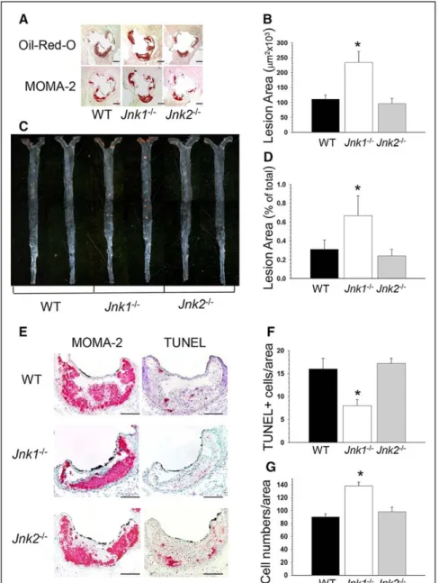
Ldlr−/− mice was markedly increased (Figure 1A and 1B)
com-pared with mice reconstituted with WT or Jnk2−/− marrow cells
(Figure 1B; 241.6±38.1 versus 110.8±13.4 and 95.8±17.6×10³
μm2, respectively). Similarly, Jnk1−/−→Ldlr−/− mice had
sig-nificantly increased size of atherosclerotic lesions in the distal aorta compared with WT→ Ldlr−/− and Jnk2−/−→ Ldlr−/− mice
(Figure 1C and 1D; 0.67%±0.22% versus 0.31%±0.10% and 0.24%±0.07%, respectively).
Next, examination of the cellular composition of ath-erosclerotic lesions in the aortic sinus of recipients showed that the proportion of smooth muscle, T, and B cells in ath-erosclerotic lesions did not differ significantly between the 3 groups (data not shown). The lesions predominantly consisted of macrophage-derived foam cells, and Jnk1−/−→Ldlr−/− mice
had significantly bigger lesion area stained with antibody
to MOMA-2 versus WT→Ldlr−/− and Jnk2−/−→Ldlr−/− mice
(Figure 1A; 167.1±29.4 versus 82.4±10.3 and 76.4±4.6×10³
μm2, respectively). The analysis of serial aortic sections
stained with MOMA-2 and terminal deoxynucleotidyl trans-ferase-mediated dUTP nick-end labeling (TUNEL) revealed that Jnk1−/−→Ldlr−/− mice contained significantly fewer
num-bers of apoptotic cells in macrophage-rich areas of lesions than WT→Ldlr−/− and Jnk2−/−→Ldlr−/− mice (Figure 1E and
1F). Double staining of macrophages with MOMA-2 and cell nuclei with DAPI revealed increased (153%) numbers of nuclei per macrophage lesion area in Jnk1−/−→Ldlr−/− mice
compared with lesions of WT→Ldlr−/− and Jnk2−/−→Ldlr−/−
mice (Figure 1G). Together, the data indicate that the lack of
Jnk1 in hematopoietic cells increases the burden of early ath-erosclerotic lesions in the absence of changes in plasma lipid or glucose levels. The dramatic increase of macrophage num-bers together with reduced apoptosis in atherosclerotic lesions of Jnk1−/−→Ldlr−/− mice also suggested changes in viability of
JNK1−/−macrophages in vivo.
Figure 1. Loss of Jnk1 in hematopoietic
cells increases atherosclerosis. A and C,
Detection of atherosclerotic lesions in the aortic sinus and aortas pinned out en face in wild-type (WT)→Ldlr−/−, Jnk1−/−→Ldlr−/−,
and Jnk2−/−→Ldlr−/− mice. Serial sections
of the aortic sinus were stained with Oil Red O to detect neutral lipids or with the MOMA-2 antibody followed by biotinylated goat antirat IgG as the secondary antibody, avidin–biotin complex labeled with alkaline phosphatase, and Fast Red TR/naphthol AS-NX substrate to reveal macrophages. Aortas were pinned out and stained with Sudan IV. Scale bar, 200 μm; a pin size, 10 μm. B and D, The extent of atherosclerotic lesions in the proximal and distal aorta of Ldlr−/− mice reconstituted with WT (◼),
Jnk1−/− (□), or Jnk2−/− (◼) bone marrow. Note:
atherosclerotic lesions are bigger in Jnk1−/−
→Ldlr−/− than in WT→Ldlr−/− and Jnk2−/−→L
dlr−/− mice. Graphs represent atherosclerotic
lesion area (mean±SEM) of the recipient
Ldlr−/− mice (*P<0.05 compared with control
group, WT→Ldlr−/− mice, by Kruskal–Wallis
1-way ANOVA on ranks, Dunn method).
E, Detection of macrophages by staining
with anti–MOMA-2 antibodies and apop-totic cells by terminal deoxynucleotidyl transferase-mediated dUTP nick-end label-ing (TUNEL) in serial sections of the aortic sinus. Scale bar, 50 μm. F and G, Percent of TUNEL+ cells (F) and DAPI-stained nucleus
numbers in MOMA-2+ area (G) in
athero-sclerotic lesions of WT→Ldlr−/−, Jnk1−/−→
Ldlr−/−, and Jnk2−/−→Ldlr−/− mice (*P<0.05
compared with the control group by 1-way ANOVA on ranks).
by guest on June 27, 2018
http://atvb.ahajournals.org/
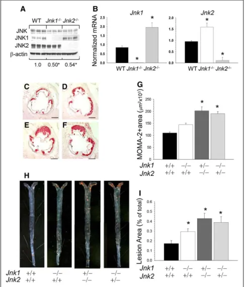
Genetic Ablation to a Single JNK Allele Further Increases Atherosclerosis
Peritoneal macrophages isolated from Jnk1−/−→Ldlr−/− and Jnk
2−/−→Ldlr−/− mice exhibited a significant decrease in JNK
pro-tein content compared with WT cells (Figure 2A; 0.49±0.03 and 0.54±0.03 versus 1.0±0.01; P<0.05 by 1-way ANOVA) and JNK kinase activity (Figure I in the online-only Data Supplement). They also had minimal residual expression of the knocked out isoform with compensatory increased expression of the other isoform (Figure 2B), indicating that maintaining total JNK activity is a vital for macrophages. Hence, to exam-ine the effect of further genetic suppression of JNK signal-ing on atherosclerosis, we generated mice expresssignal-ing a ssignal-ingle allele of Jnk1 or Jnk2 in hematopoietic cells. Because the com-plete absence of both Jnk1 and Jnk2 causes early embryonic lethality, we intercrossed Jnk1+/−/Jnk2+/− mice and collected
fetal liver cells. Then, 17-week-old male Ldlr−/− mice were
lethally irradiated and reconstituted with male WT (n=10),
Jnk1−/− (n=10), Jnk1+/−/Jnk2−/− (n=13), and Jnk1−/−/Jnk2+/−
(n=12) fetal liver cells. Four weeks after transplantation, these mice were challenged with the Western diet for 8 weeks. Again, there were no differences between the recipient groups in body weight and plasma lipid levels either on the chow or the Western diets (Table, B). Macrophages isolated from
mice with a single JNK allele exhibited further decrease in JNK protein content compared with Jnk1−/− and WT cells
(Figure II in the online-only Data Supplement). Remarkably, both Jnk1+/−/Jnk2−/−→Ldlr−/− and Jnk1−/−/Jnk2+/−→Ldlr−/−
mice developed larger atherosclerotic lesions with increased macrophage MOMA-2–positive area in the proximal aorta (Figure 2E and 2F) than Jnk1−/−→Ldlr−/− and WT−/−→Ldlr−/−
mice (Figure 2C, 2F, and 2G; 183% and 172% versus 131% and 100%, respectively). Similarly, the analysis of aorta en face demonstrated that these Jnk1+/−/Jnk2−/−→Ldlr−/− and
Jnk1−/−/Jnk2+/−→Ldlr−/− mice had larger atherosclerotic lesions
compared with Jnk1−/−→Ldlr−/− and WT−/−→Ldlr−/− mice
(Figure 2H and 2I; 248% and 225% versus 171% and 100%). Thus, genetic ablation of JNK to a single allele in hematopoi-etic cells resulted in further increases of atherosclerosis. JNK1 Signaling Antagonizes Akt
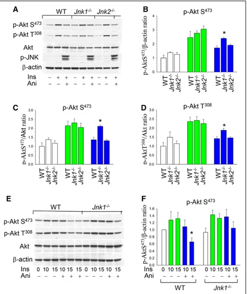
Activity in Macrophages
Next, we investigated the mechanism(s) responsible for the increased macrophage numbers in atherosclerotic lesions of
Jnk1−/−→Ldlr−/− mice by focusing on Akt signaling, which is
crucial for cell survival.20 In macrophages, Akt is constitutively
activated, and inhibition of Akt signaling induces apoptosis.27,28
In addition, a recent report demonstrated that JNK activity
Figure 2. Genetic suppression of c-Jun
NH2-terminal kinase (JNK) signaling to a Jnk
single allele further increases atheroscle-rosis. A, JNK protein contents in wild-type
(WT), Jnk1−/−, and Jnk2−/− macrophages (n=3
per group); proteins were isolated, and JNK protein contents were analyzed by Western blot; the ratio of JNK/β-actin is presented compared with WT cells (*P<0.05 by 1-way
ANOVA analysis). B, Jnk1 or Jnk2 gene
expression levels in peritoneal macrophages from mice reconstituted with WT (◼), Jnk1−/−
(□), or Jnk2−/− (◼) fetal liver cells (FLC);
mRNA levels were analyzed by real-time polymerase chain reaction. Graphs represent data (mean±SEM) with the same number (n=3) of mice per group (*P<0.05 by 1-way
ANOVA). C–F, Detection of macrophages in
the aortic sinus lesions of mice reconstituted with WT (C), Jnk1−/− (D), Jnk1+/−/Jnk2−/− (E),
or Jnk1−/−/Jnk2+/− (F) FLC. Sections were
stained with MOMA-2; Scale bar, 50 μm.
G, The extent of macrophage lesion area
in the proximal aorta of mice reconstituted with WT (◼), Jnk1−/− (□), Jnk1+/−/Jnk2−/− (◼),
or Jnk1−/−/Jnk2+/− (◼) FLC (*P<0.05 by 1-way
ANOVA, multiple comparisons vs control group; Tukey test). H, Atherosclerotic lesions
in pinned out en face aorta of mice reconsti-tuted with WT, Jnk1−/−, Jnk1+/−/Jnk2−/− or Jnk
1−/−/Jnk2+/− FLC. A pin size, 10 μm.
I, The extent of the atherosclerotic lesion
area in Ldlr−/− mice reconstituted with WT,
Jnk1, or Jnk1+/−/Jnk2−/− or Jnk1−/−/Jnk2+/−
FLC (*P<0.05 by Kruskal–Wallis 1-way
ANOVA on ranks, Dunn method, vs control group; WT→Ldlr−/− mice).
by guest on June 27, 2018
http://atvb.ahajournals.org/
antagonizes Akt signaling in some types of cells.23 To
exam-ine whether JNK affects p-Akt in mouse macrophages, WT,
Jnk1−/−, and Jnk2−/− peritoneal macrophages were treated with
insulin alone or together with anisomycin, a known activator of JNK signaling.23 Insulin significantly (2–3-fold) activated
phosphorylation of both Akt sites (p-AktS473 and T308) in all
types of cells (Figure 3A), whereas anisomycin suppressed Akt signaling activity in WT and Jnk2−/− macrophages, respectively,
with no changes in total Akt or β-actin content (Figure 3A). Importantly, Jnk1−/− macrophages showed significantly less
effect of JNK signaling on Akt activity than WT or Jnk2−/− cells
(Figure 3B). The analysis of p-AktS473/Akt and p-Akt T308/Akt
ratio in the same blot indicated a similar protective effect of
Jnk1 deficiency compared with WT or Jnk2−/− cells (Figure 3C
and 3D). Direct comparison of WT and Jnk1−/− macrophages
treated with insulin and anisomycin demonstrated a statistically significant inhibitory effect of JNK signaling in the p-Akt/β-actin ratio of WT but not of Jnk1−/− macrophages (Figure 3E
and 3F). Thus, JNK1 is the isoform primarily responsible for JNK-mediated inhibition of Akt signaling in macrophages.
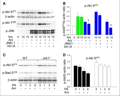
We also examined whether pharmacological inhibition of JNK can prevent the negative effects of JNK signaling on p-Akt. WT peritoneal macrophages were incubated with a mixture of insulin and anisomycin alone or in the presence of
a JNK inhibitor. There was a 39% reduction of p-Akt levels in WT cells treated with anisomycin and a cell-permeable pep-tide inhibitor of JNK1, JNKI1, which preserved p-Akt levels in macrophages (Figure 4A and 4B). In contrast, treatment with a cell-permeable inhibitor IX, selective for JNK2 and JNK3 with little or no activity against JNK1, had no protective effects on Akt activity (Figure 4A and 4B). Taken together, these data indicate that genetic ablation and pharmacological inhibition of JNK1, but not JNK2, eliminate the suppressive effects of JNK signaling on Akt activity.
JNK1 Deficiency Protects Macrophages From Apoptosis
JNK signaling has proapoptotic or antiapoptotic functions, depending on the cell type, nature of the death stimulus, dura-tion of its activadura-tion, and the activity of other signaling path-ways.17 Taking into consideration the critical role of Akt in
cell survival,20 we suggested that sustained JNK activation
(1–6 hours) may promote apoptosis by exhausting antiapop-totic Akt signaling and by subsequently reducing Bad S136
phosphorylation, which normally serves to inhibit apoptosis in macrophages.27,29 To test this hypothesis, we examined
the effect of anisomycin on Akt signaling in WT and Jnk1−/−
macrophages treated with palmitic acid, a stress-mediated
Figure 3. c-Jun NH2-terminal kinase (JNK)
signaling antagonizes p-Akt activity, and loss of JNK1 obliterated this effect. A,
Wild-type (WT), Jnk1−/−, and Jnk2−/− peritoneal
macrophages were preincubated in serum-free media for 24 hours and then untreated or treated with insulin (100 nmol/L) alone or together with anisomycin (10 μg/mL) for 15 minutes. Macrophage proteins were extracted, resolved by electrophoresis (50 μg), and analyzed by Western blot. B–D, Ratio of p-AktS473/b-actin, p-AktS473/ Akt, and p-Akt T308/Akt in untreated (white color) or treated with insulin (green color) or insulin plus anisomycin (blue color). Graphs represent data (mean±SEM) of 3 experi-ments (*P<0.05 by 1-way ANOVA on rank
compared with control WT cells treated with insulin together with anisomycin).
E and F, WT and Jnk1−/− macrophages were
treated with insulin alone (green color) or together with anisomycin (blue color) for 10 and 15 minutes. Graphs represent data (mean±SEM) of 3 experiments (*P<0.05 by
1-way ANOVA on rank compared with WT cells treated with insulin).
by guest on June 27, 2018
http://atvb.ahajournals.org/
lipotoxic factor inducing endoplasmic reticulum (ER) stress and apoptosis.30 The increased JNK signaling gradually
sup-pressed p-Akt S473 in WT cells, whereas Jnk1−/− macrophages
had higher p-Akt S473 levels and were more resistant to p-Akt
suppression (Figure 4C and 4D). Similarly, the treatment progressively reduced p-Bad S136 levels in WT macrophages,
but there was less attenuation of p-Bad S136 in Jnk1−/− cells
(Figure 4C). Thus, compared with WT cells, Jnk1−/−
macro-phages were able to preserve higher levels of Akt and Bad phosphorylation, which are important protective and anti-apoptotic factors under conditions of ER stress.31
In addition, to define the role of JNK signaling in macro-phage apoptosis, WT, Jnk1−/−, and Jnk2−/− macrophages were
treated with bovine serum albumin or palmitic acid. Treatment with bovine serum albumin generated only a few apoptotic TUNEL-positive (TUNEL+) cells with no differences between
cell types, whereas palmitic acid increased TUNEL+ cells
4-fold in WT and Jnk2−/− macrophages but not in Jnk1−/− cells
(Figure 5A and 5B). The addition of anisomycin markedly (3-fold) increased the percentage of TUNEL+ cells to a
simi-lar degree in WT and Jnk2−/− cells, whereas apoptosis was
sig-nificantly reduced (57% of WT cells) in Jnk1−/− macrophages
(Figure 5C). In contrast, the selective inhibitor JNKI1 sig-nificantly (2-fold) reduced apoptosis in all types of cells, but
Jnk1−/− macrophages had less apoptosis than WT and Jnk2−/−
cells (Figure 5D). When WT macrophages were treated with the specific inhibitors of JNK, JNKI1, and SP600125, they dem-onstrated similar levels of apoptosis (Figure 5E). Importantly, when cells were loaded with human oxidized or acetylated LDL in combination with an ACAT (acetyl-coenzyme A acet-yltransferase) inhibitor, Jnk1−/− macrophages generated
signifi-cantly less apoptosis than WT and Jnk2−/− cells (Figure 5F). In
addition, macrophages expressing a single Akt isoform (Figure III in the online-only Data Supplement), Akt1 (Akt2−/−/Akt3−/−),
or Akt3 (Akt1−/−/Akt2−/−) palmitic acid–bovine serum albumin
treatment induced a stepwise increase in apoptosis that was especially high in Akt1−/−/Akt2−/− cells compared with WT cells.
However, suppression of JNK signaling by the JNK inhibitor, SP600125, completely reversed the effect on cell survival with no differences between the groups (Figure IV in the online-only Data Supplement). Taken together, our data indicate that JNK1 signaling regulates ER stress–mediated apoptosis in mouse macrophages and Jnk1−/− macrophages displayed clear
resistance to apoptosis induced by different stimuli. PTEN Suppression Impairs Effects of JNK Signaling on Akt Activity
Recently, Vivanco et al26 have shown that JNK regulates
p-Akt via PTEN, and Pten null mouse embryonic fibroblasts exhibit an impaired negative feedback loop. To test whether PTEN plays a critical role in regulating this pathway in mouse macrophages, WT and Pten−/− cells were treated with insulin
alone or together with anisomycin. In contrast to WT cells, which showed increased p-Akt S473 in response to insulin and
reduced p-Akt S473 after treatment with anisomycin, Pten−/−
macrophages had markedly increased basal p-Akt, which was not suppressed in response to anisomycin (Figure 6A and 6B). Similarly, treatment with bpV(pic), a potent PTEN inhibitor, with an IC50 ≈10- to 100-fold lower than for other tyrosine phosphatases,32 decreased the inhibitory effects of JNK on
p-Akt (Figure 6C and 6D). Taken together, these results indi-cate that both genetic ablation and pharmacological inhibition of PTEN effectively eradicated JNK-mediated inhibition of Akt phosphorylation in mouse macrophages.
Discussion
Numerous studies have linked macrophage or hematopoietic JNK1 activity to insulin resistance and abnormal glucose homeostasis in obesity.12,33–35 These studies targeting individual
Figure 4. c-Jun NH2-terminal kinase-1
(JNK1) inhibitor, JNKI1, preserves Akt signaling, and Jnk1−/− macrophages are
more resistant to endoplasmic reticulum stress than wild-type (WT) cells. A and B,
WT peritoneal macrophages were prein-cubated in serum-free media for 24 hours and then treated with insulin alone (green color) or together with anisomycin (Ani; blue color) without or with the specific JNK inhibitor 1, JNKI1 (3 μmol/L) or specific JNK2 and JNK3 inhibitor, inhibitor IX (50 nmol/L), for indicated time. Macrophage proteins were extracted, resolved (60 μg per well), and analyzed by Western blot with noted antibodies. Graphs represent data (mean±SEM) of experiments with 4 mice per group (*P<0.05 compared with control
WT cells treated with insulin for 15 minutes by 1-way ANOVA on ranks). C and D, WT
(◼) and Jnk1−/− (□) peritoneal macrophages
were untreated or treated with 0.5 mmol/L palmitic acid–bovine serum albumin and Ani (10 μg/mL) for the indicated time. Graphs represent data (mean±SEM) of 3 experiments.
by guest on June 27, 2018
http://atvb.ahajournals.org/
JNK isoforms have produced varying degrees of effect in dif-ferent models, perhaps because of interactions between iso-forms and redundancies.8 In fact, a recent report using Jnk1
and Jnk2-combined deletion has shown that macrophage JNK promotes the establishment of obesity-induced insulin resis-tance and pancreatic islet dysfunction.12 These findings suggest
that macrophage JNK signaling may be crucial in other patho-logical conditions and warrants detailed studies of individual isoforms in cardiovascular disease models. Here, we examined the effect of Jnk1 or Jnk2 deficiency in hematopoietic cells on early stages of atherosclerosis using the Ldlr-deficiency model. Mice reconstituted with Jnk1−/− hematopoietic cells
had significantly bigger atherosclerotic lesions compared with mice transplanted with WT or Jnk2−/− marrow with no
differ-ences in serum lipids. Genetic ablation to a single Jnk allele (either Jnk1+/−/Jnk2−/− or Jnk1−/−/Jnk2+/−) in hematopoietic
cells further increased atherosclerosis compared with Jnk1−/−
→Ldlr−/− mice. We also found that JNK signaling antagonizes
Akt activity in mouse macrophages acting mainly through JNK1. Therefore, Jnk1−/− macrophages had less suppression
of p-Akt in response to sustained ER stress and were protected from apoptosis. On the basis of these data, we conclude that this resistance to apoptotic stimuli in Jnk1 null macrophages increases lesion burden at the early stages of atherogenesis.
JNK signaling is overexpressed and activated in atheroscle-rotic lesions of cholesterol-fed rabbits.36 Considering the role
of JNK in inflammatory and metabolic responses, it is plau-sible that this stress-mediated JNK activation may affect mac-rophage viability and atherosclerosis. In fact, Ricci et al16 were
the first to report the involvement of JNK2 in atherosclerosis showing that Jnk2−/−/apoE−/− mice developed less
atheroscle-rosis compared with control apoE−/− and Jnk1−/−/apoE−/− mice.
They analyzed a later stage of atherosclerosis with more severe lesions induced by a high cholesterol (1.25%) diet for
Figure 5. Jnk1−/− macrophages are protected from apoptosis and anisomycin (Ani) increases, whereas JNK inhibition suppresses
endo-plasmic reticulum–mediated apoptosis. A, Detection of apoptosis in wild-type (WT), Jnk1−/−, and Jnk2−/− macrophages treated with bovine
serum albumin (BSA; control) and 0.5 mmol/L palmitic acid (PA)–BSA for 24 hours by terminal deoxynucleotidyl transferase-mediated dUTP nick-end labeling (TUNEL) assay. Note: TUNEL-positive cells (red), nuclei counterstained with Mayer hematoxylin. B, Percent of TUNEL+
WT, Jnk1−/−, and Jnk2−/− macrophages treated with BSA or PA-BSA (*P<0.05 by 1-way ANOVA on rank compared with WT cells treated
with PA-BSA). C and D, Percent of TUNEL+ WT, Jnk1−/−, and Jnk2−/− macrophages treated with BSA or PA-BSA together with Ani (10 mg/
mL) or the JNK inhibitor, JNKI1 (3 μmol/L), for 24 hours (*P<0.05 by 1-way ANOVA on rank compared with WT cells treated with PA+Ani or PA+SP600125 [SP]). E, Percent of TUNEL+ cells in WT macrophages untreated or treated with 0.5 mmol/L PA-BSA alone or together with
JNK inhibitors, SP (100 nmol/L) or JNKI1 (3 μmol/L) for 24 hours (*P<0.05 by 1-way ANOVA on rank compared with untreated WT cells). F, Percent of TUNEL+ in WT, Jnk1−/−, and Jnk2−/− macrophages untreated (control) or treated with human acetylated low-density lipoprotein
(AcLDL; 100 μg/mL) in the presence of the ACAT (acetyl-coenzyme A acetyltransferase) inhibitor CP-113,818 (2 μg/mL) or human oxidized LDL (OxLDL; 100 μg/mL) for 48 hours (*P<0.05 compared with control WT cells treated with AcLDL by 1-way ANOVA on rank).
by guest on June 27, 2018
http://atvb.ahajournals.org/
14 weeks in total body JNK isoform deficiency in the apoE-deficienct model on a hybrid C57BL6/129SV background, whereas in the current study, we explored early-stage athero-sclerosis using Ldlr−/− mice on C57BL/6 background
recon-stituted with hematopoietic cells null for JNK isoforms and fed with the Western diet (containing 21% milk fat and 0.15% cholesterol) for 8 weeks. The variation in genetic background of mice, stage-specific lesion burden, and Jnk deficiency in specific compartments are all important determinants of cho-lesterol absorption37 and susceptibility to atherosclerosis,38 and
they may underlie the apparent differences in our results. In the current study, we observed a higher lesion burden in early atherosclerosis as a result of deficiency of Jnk1, but not Jnk2, in hematopoietic cells in the Ldlr null mice. Similar results were also obtained when combined deletion models (either Jnk1+/−/Jnk2−/− or Jnk1−/−/Jnk2+/−) were used as donors
to produce hematopoietic JNK deficiency. These results may point to several possibilities. For example, it is possible that total JNK activity may be a more important determinant of the effect on macrophage apoptosis and atherogenesis than separate JNK isoforms. In the future, it would be highly informative to examine interactions between JNK isoforms in supporting total JNK activity in vivo. In this sense, our data are consistent with a recent report,39 indicating that loss of apoptosis
signal-regu-lating kinase 1, which is upstream of JNK in certain contexts, in apoE null mice significantly reduced apoptosis and increased atherosclerosis by forming lesions enriched with macrophages. Because of the complexity of signaling upstream of JNK, mul-tiple mechanisms may affect atherogenesis in a differential manner. For example, lack of mitogen-activated protein kinase phosphotase-1 protects apoE null mice from atherosclerosis,40
whereas genetic deletion of Jnk1 reduces apoptosis in endothe-lial cells at atheroprone sites of the artery and thus diminishes
atherosclerosis.41 Similarly, the administration of anisomycin
via osmotic minipump increased apoptosis and decreased the macrophage content in atherosclerotic lesions of rabbits.42 In
this scenario, prevention of macrophage death is likely a domi-nant feature of Jnk deficiency, at least during early stages of atherosclerosis, supporting the growth of vascular lesions enriched in macrophages. If this is the case, careful consider-ation of JNK’s role in atherosclerosis and how it could be best used for therapeutic intervention would be well warranted. It is, however, equally likely that Jnk1 deficiency and early pres-ervation of macrophage death may yield favorable functional outcomes by ensuring plaque stability and preventing rupture, the predominant cause of morbidity and mortality caused by atherosclerosis.43 In fact, this would be quite reminiscent of the
role of certain ER stress responses that are also related to mac-rophage death.2 For example, C/EBP homologous protein
defi-ciency can prevent macrophage death and support the stability of vascular lesions and prevent rupture.44 Finally, it is possible
that isolated examination of hematopoietic JNK activity only may have limitations and may not reflect the complete role of JNK in the pathogenesis of atherosclerosis. Future studies should dissect these possibilities in additional models.
Next, to identify the mechanism(s) responsible for the actions of JNK signaling in macrophages, we focused on the fact that Jnk1−/−→Ldlr−/− mice had a dramatic decrease in apoptosis
and increased numbers of macrophages in their atherosclerotic lesions compared with lesions of WT→Ldlr−/− and Jnk2−/−→L
dlr−/− mice. These results suggested that Jnk1 deficiency changes
the balance between survival and proapoptotic signaling in mac-rophages at least in the setting they are examined. Indeed, our in vitro studies demonstrated that JNK signaling directly antago-nizes Akt activity in mouse macrophages. This effect occurs within a short time (3–15 minutes) and may be beneficial for Figure 6. Genetic and pharmacological
inhi-bition of phosphatase and tensin homolog (PTEN) eradicates anisomycin-mediated suppression of p-Akt in macrophages.
A and B, Akt signaling in wild-type (WT) and
Pten−/− macrophages treated with insulin
and anisomycin. Cells were preincubated with serum-free media for 16 hours and then untreated or treated with insulin alone (green color) or together with anisomycin (blue color) for the indicated time. Graphs represent data (mean±SEM) of 3 experi-ments (*P<0.05 between untreated and
treated cells by 1-way ANOVA). C and D,
PTEN inhibitor bpV(pig) preserves p-Akt signaling in WT peritoneal macrophages treated with anisomycin. Cells were preincu-bated in serum-free media for 24 hours and treated with insulin alone (green color) or with anisomycin (blue color) with or without bpV(pig) (0.1 μmol/L) for 15 minutes. Graphs represent data (mean±SEM) of 2 experi-ments (*P<0.05 by 1-way ANOVA on rank
compared with cells treated with insulin).
by guest on June 27, 2018
http://atvb.ahajournals.org/
inflammatory and stress responses by diverting energy sources from the synthetic Akt pathway.3 In contrast, prolonged or
sus-tained JNK activation suppresses Akt signaling and induces cell apoptosis.6 Interestingly, this antagonizing effect is
medi-ated mainly through JNK1, but not JNK2, and genetic ablation or pharmacological inhibition of JNK1 completely obliterated this effect. These data are consistent with the previous reports, indicating that JNK signaling acts as a negative feedback loop that attenuates insulin action and insulin-induced PI3K activa-tion.7,12,23,45–47 Together, our data indicate that JNK1 signaling
antagonizes and suppresses Akt activity in mouse macrophages. It is important to note that bone marrow transplantation may change every component of hematopoietic system in mice, including monocyte-macrophages, T and B cells, and platelets. Several studies have shown that JNK is required for effector T-cell function.48 JNK2 is important for T-cell
activa-tion and apoptosis of immature thymocytes49 and plays a role
in control of CD8+ T-cell expansion in vivo, whereas JNK1
is involved in survival of activated T cells during immune responses.50 Moreover, JNK1 is essential for platelet secretion
and thrombus formation.51 Therefore, we cannot exclude that
these changes may also affect atherogenesis.
It is known that sustained JNK signaling restrains Akt activity, the major prosurvival signaling pathway that opposes apoptosis,20 suggesting a potential mechanism for impaired
macrophage viability. In our experiments, sustained JNK sig-naling under conditions of ER stress gradually extinguished Akt and Bad (S136) activity in WT cells, whereas Jnk1−/−
macro-phages were much less affected (Figure 4C and 4D). Compared with WT cells, Jnk1−/− macrophages were also protected from
apoptosis initiated by different stimuli. Moreover, JNK1 inhi-bition distinctly decreased ER stress–mediated apoptosis in macrophages. These results are consistent with the concept that chronically activated JNK1 signaling is crucial in type 2 diabe-tes mellitus and obesity.8,11,47,52 JNK-mediated phosphorylation
of insulin receptor substrates 1 and 2 disrupts Akt signaling33,46
possibly by releasing Bad for translocation to the mitochondria23
or association with Bcl-2/Bcl-xL and initiation of apoptosis. In addition, we examined whether macrophages use a natural brake of Akt signaling, PTEN, to suppress p-Akt. Given that PTEN has been reported to cooperate with JNK53 to couple the
PI3K/Akt and JNK signaling pathways,26 we examined whether
PTEN mediates cross talk between these pathways in mouse macrophages. Our results demonstrate that genetic and phar-macological inhibition of PTEN virtually eradicates the JNK-mediated effect on p-Akt in macrophages. Thus, JNK signaling may also act via PTEN to antagonize Akt activity and suppress macrophage survival. Macrophage-derived foam cells are the predominant cell type of early atherosclerotic lesions, and loss of macrophages through increased apoptosis may reduce the size of early atherosclerotic lesions.54 Together, these data
dem-onstrate that Jnk1 deficiency significantly increases macrophage survival, and this leads to cell accumulation in early-stage ath-erosclerotic lesions. Importantly, JNK and PTEN signaling in macrophages can be altered pharmacologically with the use of their ligands or inhibitors, supporting these pathways as new potential therapeutic targets for the prevention of atherosclerosis and allowing for functional studies in a stage-specific manner.
Sources of Funding
This work was supported, in part, by National Institutes of Health grants HL105375, HL116263, DK50435, DK52539, and DK59637 (Lipid, Lipoprotein and Atherosclerosis Core of the Vanderbilt Mouse Metabolic Phenotype Centers).
Disclosures
None.
References
1. Moore KJ, Sheedy FJ, Fisher EA. Macrophages in atherosclerosis: a dynamic balance. Nat Rev Immunol. 2013;13:709–721. doi: 10.1038/ nri3520.
2. Tabas I, Ron D. Integrating the mechanisms of apoptosis induced by endo-plasmic reticulum stress. Nat Cell Biol. 2011;13:184–190. doi: 10.1038/ ncb0311-184.
3. Hotamisligil GS. Inflammation and metabolic disorders. Nature. 2006;444:860–867. doi: 10.1038/nature05485.
4. Davis RJ. Signal transduction by the JNK group of MAP kinases. Cell. 2000;103:239–252.
5. Vallerie SN, Hotamisligil GS. The role of JNK proteins in metabolism. Sci
Transl Med. 2010;2:60rv5. doi: 10.1126/scitranslmed.3001007. 6. Dhanasekaran DN, Reddy EP. JNK signaling in apoptosis. Oncogene.
2008;27:6245–6251. doi: 10.1038/onc.2008.301.
7. Waetzig V, Czeloth K, Hidding U, Mielke K, Kanzow M, Brecht S, Goetz M, Lucius R, Herdegen T, Hanisch UK. c-Jun N-terminal kinases (JNKs) mediate pro-inflammatory actions of microglia. Glia. 2005;50:235–246. doi: 10.1002/glia.20173.
8. Tuncman G, Hirosumi J, Solinas G, Chang L, Karin M, Hotamisligil GS. Functional in vivo interactions between JNK1 and JNK2 isoforms in obe-sity and insulin resistance. Proc Natl Acad Sci U S A. 2006;103:10741– 10746. doi: 10.1073/pnas.0603509103.
9. Karin M, Gallagher E. From JNK to pay dirt: jun kinases, their biochem-istry, physiology and clinical importance. IUBMB Life. 2005;57:283–295. doi: 10.1080/15216540500097111.
10. Conze D, Krahl T, Kennedy N, Weiss L, Lumsden J, Hess P, Flavell RA, Le Gros G, Davis RJ, Rincón M. c-Jun NH(2)-terminal kinase (JNK)1 and JNK2 have distinct roles in CD8(+) T cell activation. J Exp Med. 2002;195:811–823.
11. Hirosumi J, Tuncman G, Chang L, Görgün CZ, Uysal KT, Maeda K, Karin M, Hotamisligil GS. A central role for JNK in obesity and insulin resis-tance. Nature. 2002;420:333–336. doi: 10.1038/nature01137.
12. Han MS, Jung DY, Morel C, Lakhani SA, Kim JK, Flavell RA, Davis RJ. JNK expression by macrophages promotes obesity-induced insulin resistance and inflammation. Science. 2013;339:218–222. doi: 10.1126/science.1227568. 13. Samuel Varman T, Shulman Gerald I. Mechanisms for insulin resistance:
common threads and missing links. Cell. 2012;148:852–871.
14. Vernia S, Cavanagh-Kyros J, Garcia-Haro L, Sabio G, Barrett T, Jung DY, Kim JK, Xu J, Shulha HP, Garber M, Gao G, Davis RJ. The PPARα-FGF21 hormone axis contributes to metabolic regulation by the hepatic JNK signaling pathway. Cell Metab. 2014;20:512–525. doi: 10.1016/j. cmet.2014.06.010.
15. Sabio G, Das M, Mora A, Zhang Z, Jun JY, Ko HJ, Barrett T, Kim JK, Davis RJ. A stress signaling pathway in adipose tissue regulates hepatic insulin resistance. Science. 2008;322:1539–1543. doi: 10.1126/science.1160794. 16. Ricci R, Sumara G, Sumara I, et al. Requirement of JNK2 for scaven-ger receptor A-mediated foam cell formation in atherogenesis. Science. 2004;306:1558–1561. doi: 10.1126/science.1101909.
17. Liu J, Lin A. Role of JNK activation in apoptosis: a double-edged sword.
Cell Res. 2005;15:36–42. doi: 10.1038/sj.cr.7290262.
18. Liu J, Minemoto Y, Lin A. c-Jun N-terminal protein kinase 1 (JNK1), but not JNK2, is essential for tumor necrosis factor alpha-induced c-Jun kinase activation and apoptosis. Mol Cell Biol. 2004;24:10844–10856. doi: 10.1128/MCB.24.24.10844-10856.2004.
19. Tournier C, Hess P, Yang DD, Xu J, Turner TK, Nimnual A, Bar-Sagi D, Jones SN, Flavell RA, Davis RJ. Requirement of JNK for stress-induced activation of the cytochrome c-mediated death pathway. Science. 2000;288:870–874.
20. Duronio V. The life of a cell: apoptosis regulation by the PI3K/PKB path-way. Biochem J. 2008;415:333–344. doi: 10.1042/BJ20081056. 21. Manning BD, Cantley LC. AKT/PKB signaling: navigating downstream.
Cell. 2007;129:1261–1274. doi: 10.1016/j.cell.2007.06.009.
by guest on June 27, 2018
http://atvb.ahajournals.org/
22. Aikin R, Maysinger D, Rosenberg L. Cross-talk between phosphatidylino-sitol 3-kinase/AKT and c-jun NH2-terminal kinase mediates survival of isolated human islets. Endocrinology. 2004;145:4522–4531. doi: 10.1210/ en.2004-0488.
23. Sunayama J, Tsuruta F, Masuyama N, Gotoh Y. JNK antagonizes Akt-mediated survival signals by phosphorylating 14-3-3. J Cell Biol. 2005;170:295–304. doi: 10.1083/jcb.200409117.
24. Bonny C, Oberson A, Negri S, Sauser C, Schorderet DF. Cell-permeable peptide inhibitors of JNK: novel blockers of beta-cell death. Diabetes. 2001;50:77–82.
25. Du H, Sun X, Guma M, Luo J, Ouyang H, Zhang X, Zeng J, Quach J, Nguyen DH, Shaw PX, Karin M, Zhang K. JNK inhibition reduces apop-tosis and neovascularization in a murine model of age-related macu-lar degeneration. Proc Natl Acad Sci U S A. 2013;110:2377–2382. doi: 10.1073/pnas.1221729110.
26. Vivanco I, Palaskas N, Tran C, Finn SP, Getz G, Kennedy NJ, Jiao J, Rose J, Xie W, Loda M, Golub T, Mellinghoff IK, Davis RJ, Wu H, Sawyers CL. Identification of the JNK signaling pathway as a functional target of the tumor suppressor PTEN. Cancer Cell. 2007;11:555–569. doi: 10.1016/j. ccr.2007.04.021.
27. Babaev VR, Chew JD, Ding L, Davis S, Breyer MD, Breyer RM, Oates JA, Fazio S, Linton MF. Macrophage EP4 deficiency increases apoptosis and suppresses early atherosclerosis. Cell Metab. 2008;8:492–501. doi: 10.1016/j.cmet.2008.09.005.
28. Liu H, Perlman H, Pagliari LJ, Pope RM. Constitutively activated Akt-1 is vital for the survival of human monocyte-differentiated macrophages. Role of Mcl-1, independent of nuclear factor (NF)-kappaB, Bad, or cas-pase activation. J Exp Med. 2001;194:113–126.
29. Danial NN. BAD: undertaker by night, candyman by day. Oncogene. 2008;27(suppl 1):S53–S70. doi: 10.1038/onc.2009.44.
30. Borradaile NM, Han X, Harp JD, Gale SE, Ory DS, Schaffer JE. Disruption of endoplasmic reticulum structure and integrity in lipotoxic cell death. J
Lipid Res. 2006;47:2726–2737. doi: 10.1194/jlr.M600299-JLR200. 31. Datta SR, Ranger AM, Lin MZ, Sturgill JF, Ma YC, Cowan CW, Dikkes
P, Korsmeyer SJ, Greenberg ME. Survival factor-mediated BAD phos-phorylation raises the mitochondrial threshold for apoptosis. Dev Cell. 2002;3:631–643.
32. Schmid AC, Byrne RD, Vilar R, Woscholski R. Bisperoxovanadium com-pounds are potent PTEN inhibitors. FEBS Lett. 2004;566:35–38. doi: 10.1016/j.febslet.2004.03.102.
33. Aguirre V, Werner ED, Giraud J, Lee YH, Shoelson SE, White MF. Phosphorylation of ser307 in insulin receptor substrate-1 blocks interac-tions with the insulin receptor and inhibits insulin action. J Biol Chem. 2002;277:1531–1537.
34. Solinas G, Vilcu C, Neels JG, Bandyopadhyay GK, Luo JL, Naugler W, Grivennikov S, Wynshaw-Boris A, Scadeng M, Olefsky JM, Karin M. JNK1 in hematopoietically derived cells contributes to diet-induced inflammation and insulin resistance without affecting obesity. Cell Metab. 2007;6:386–397. doi: 10.1016/j.cmet.2007.09.011.
35. Vallerie SN, Furuhashi M, Fucho R, Hotamisligil GS. A predominant role for parenchymal c-Jun amino terminal kinase (JNK) in the regulation of systemic insulin sensitivity. PLoS One. 2008;3:e3151. doi: 10.1371/jour-nal.pone.0003151.
36. Metzler B, Hu Y, Dietrich H, Xu Q. Increased expression and activation of stress-activated protein kinases/c-Jun NH(2)-terminal protein kinases in atherosclerotic lesions coincide with p53. Am J Pathol. 2000;156:1875– 1886. doi: 10.1016/S0002-9440(10)65061-4.
37. Jolley CD, Dietschy JM, Turley SD. Genetic differences in cholesterol absorption in 129/Sv and C57BL/6 mice: effect on cholesterol responsive-ness. Am J Physiol. 1999;276(5 pt 1):G1117–G1124.
38. Dansky HM, Charlton SA, Sikes JL, Heath SC, Simantov R, Levin LF, Shu P, Moore KJ, Breslow JL, Smith JD. Genetic background determines
the extent of atherosclerosis in ApoE-deficient mice. Arterioscler Thromb
Vasc Biol. 1999;19:1960–1968.
39. Yamada S, Ding Y, Tanimoto A, Wang KY, Guo X, Li Z, Tasaki T, Nabesima A, Murata Y, Shimajiri S, Kohno K, Ichijo H, Sasaguri Y. Apoptosis signal-regulating kinase 1 deficiency accelerates hyperlipid-emia-induced atheromatous plaques via suppression of macrophage apop-tosis. Arterioscler Thromb Vasc Biol. 2011;31:1555–1564. doi: 10.1161/ ATVBAHA.111.227140.
40. Shen J, Chandrasekharan UM, Ashraf MZ, Long E, Morton RE, Liu Y, Smith JD, DiCorleto PE. Lack of mitogen-activated protein kinase phos-phatase-1 protects ApoE-null mice against atherosclerosis. Circ Res. 2010;106:902–910. doi: 10.1161/CIRCRESAHA.109.198069.
41. Amini N, Boyle JJ, Moers B, Warboys CM, Malik TH, Zakkar M, Francis SE, Mason JC, Haskard DO, Evans PC. Requirement of JNK1 for endo-thelial cell injury in atherogenesis. Atherosclerosis. 2014;235:613–618. doi: 10.1016/j.atherosclerosis.2014.05.950.
42. Croons V, Martinet W, Herman AG, Timmermans JP, De Meyer GR. The protein synthesis inhibitor anisomycin induces macrophage apoptosis in rabbit atherosclerotic plaques through p38 mitogen-activated protein kinase.
J Pharmacol Exp Ther. 2009;329:856–864. doi: 10.1124/jpet.108.149948. 43. Burke AP, Kolodgie FD, Farb A, Weber DK, Malcom GT, Smialek J,
Virmani R. Healed plaque ruptures and sudden coronary death: evidence that subclinical rupture has a role in plaque progression. Circulation. 2001;103:934–940.
44. Thorp E, Li G, Seimon TA, Kuriakose G, Ron D, Tabas I. Reduced apopto-sis and plaque necroapopto-sis in advanced atherosclerotic lesions of Apoe-/- and Ldlr-/- mice lacking CHOP. Cell Metab. 2009;9:474–481. doi: 10.1016/j. cmet.2009.03.003.
45. Gual P, Le Marchand-Brustel Y, Tanti JF. Positive and negative regula-tion of insulin signaling through IRS-1 phosphorylaregula-tion. Biochimie. 2005;87:99–109. doi: 10.1016/j.biochi.2004.10.019.
46. Lee YH, Giraud J, Davis RJ, White MF. c-Jun N-terminal kinase (JNK) mediates feedback inhibition of the insulin signaling cascade. J Biol
Chem. 2003;278:2896–2902. doi: 10.1074/jbc.M208359200.
47. Solinas G, Naugler W, Galimi F, Lee MS, Karin M. Saturated fatty acids inhibit induction of insulin gene transcription by JNK-mediated phos-phorylation of insulin-receptor substrates. Proc Natl Acad Sci U S A. 2006;103:16454–16459. doi: 10.1073/pnas.0607626103.
48. Dong C, Yang DD, Tournier C, Whitmarsh AJ, Xu J, Davis RJ, Flavell RA. JNK is required for effector T-cell function but not for T-cell activation.
Nature. 2000;405:91–94. doi: 10.1038/35011091.
49. Sabapathy K, Hu Y, Kallunki T, Schreiber M, David JP, Jochum W, Wagner EF, Karin M. JNK2 is required for efficient T-cell activation and apoptosis but not for normal lymphocyte development. Curr Biol. 1999;9:116–125. 50. Arbour N, Naniche D, Homann D, Davis RJ, Flavell RA, Oldstone MB.
c-Jun NH(2)-terminal kinase (JNK)1 and JNK2 signaling pathways have divergent roles in CD8(+) T cell-mediated antiviral immunity. J Exp Med. 2002;195:801–810.
51. Adam F, Kauskot A, Nurden P, Sulpice E, Hoylaerts MF, Davis RJ, Rosa JP, Bryckaert M. Platelet JNK1 is involved in secretion and thrombus forma-tion. Blood. 2010;115:4083–4092. doi: 10.1182/blood-2009-07-233932. 52. Weston CR, Davis RJ. The JNK signal transduction pathway. Curr Opin
Cell Biol. 2007;19:142–149. doi: 10.1016/j.ceb.2007.02.001.
53. Hübner A, Mulholland DJ, Standen CL, Karasarides M, Cavanagh-Kyros J, Barrett T, Chi H, Greiner DL, Tournier C, Sawyers CL, Flavell RA, Wu H, Davis RJ. JNK and PTEN cooperatively control the development of invasive adenocarcinoma of the prostate. Proc Natl Acad Sci U S A. 2012;109:12046–12051. doi: 10.1073/pnas.1209660109.
54. Seimon T, Tabas I. Mechanisms and consequences of macrophage apop-tosis in atherosclerosis. J Lipid Res. 2009;50(suppl):S382–S387. doi: 10.1194/jlr.R800032-JLR200.
• c-Jun NH2-terminal kinase-1 (JNK1) signaling antagonizes prosurvival Akt activity in mouse macrophages.
• Jnk1 null macrophages were less affected by the stress factors and more protected from apoptosis than wild-type and Jnk2 null macrophages. • Loss of Jnk1, but not Jnk2, in hematopoietic cells significantly increases early atherosclerosis.
• Genetic ablation of JNK to a single allele (Jnk1+/−/Jnk2−/− or Jnk1−/−/Jnk2+/−) in bone marrow recipients further increased atherosclerosis
compared with mice reconstituted with wild-type or Jnk1 null bone marrow.
Highlights
by guest on June 27, 2018
http://atvb.ahajournals.org/
Sergio Fazio, Gökhan S. Hotamisligil and MacRae F. Linton
Vladimir R. Babaev, Michele Yeung, Ebru Erbay, Lei Ding, Youmin Zhang, James M. May,
Atherosclerosis in Low-Density Lipoprotein Receptor Null Mice
Print ISSN: 1079-5642. Online ISSN: 1524-4636
Copyright © 2016 American Heart Association, Inc. All rights reserved. Greenville Avenue, Dallas, TX 75231
is published by the American Heart Association, 7272 Arteriosclerosis, Thrombosis, and Vascular Biology
doi: 10.1161/ATVBAHA.116.307580
2016;36:1122-1131; originally published online April 21, 2016;
Arterioscler Thromb Vasc Biol.
http://atvb.ahajournals.org/content/36/6/1122
World Wide Web at:
The online version of this article, along with updated information and services, is located on the
http://atvb.ahajournals.org/content/suppl/2016/04/20/ATVBAHA.116.307580.DC2 http://atvb.ahajournals.org/content/suppl/2016/04/20/ATVBAHA.116.307580.DC1
Data Supplement (unedited) at:
http://atvb.ahajournals.org//subscriptions/ at:
is online Arteriosclerosis, Thrombosis, and Vascular Biology
Information about subscribing to
Subscriptions:
http://www.lww.com/reprints
Information about reprints can be found online at:
Reprints:
document. Question and Answer
Permissions and Rights page under Services. Further information about this process is available in the
which permission is being requested is located, click Request Permissions in the middle column of the Web Copyright Clearance Center, not the Editorial Office. Once the online version of the published article for
can be obtained via RightsLink, a service of the Arteriosclerosis, Thrombosis, and Vascular Biology
in
Requests for permissions to reproduce figures, tables, or portions of articles originally published
Permissions:
by guest on June 27, 2018
TPG
+ + + - - -
WT 1 2 WT 1 2
b
-actin
P32 c-Jun
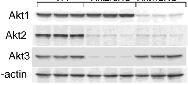
Figure SII. Dose-dependent suppression of JNK protein contents in wild type,
Jnk1
-/-and single Jnk allele macrophages.
A,B. JNK protein contents in WT, Jnk1
-/-, Jnk1
+/-/Jnk2
-/-and Jnk1
-/-/Jnk2
+/-macrophages and the ratio of JNK/
b
-actin is presented compared to WT cells.
Macrophage proteins were extracted, resolved (40μg/well) and analyzed by
Western blot.
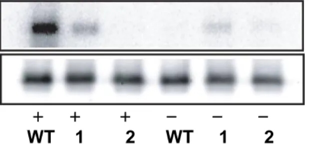
Figure SI. c-Jun kinase assay in WT, Jnk1
-/-and Jnk2
-/-macrophages treated
with thapsigargin.
Peritoneal macrophages from WT, Jnk1
-/-(1) and Jnk2
-/-(2) mice were isolated and
two days later, untreated or treated with 1mM thapsigargin (TPG) for 6 hours. Then
proteins were extracted, resolved (50μg/well) and analyzed by Western blot.
Jnk2
+/+ -/- +/- -/-
+/+ +/+ -/- +/-
Jnk1
c-Jun
JNK
0.0 0.2 0.4 0.6 0.8 1.0 1.2A
JNK/
b
-a
ctin
ratio
Jnk2
+/+ -/- +/- -/-
+/+ +/+ -/- +/-
Jnk1
B
0 5 10 15 20