Australian and New Zealand Journal of Obstetrics and Gynaecology 2006; 46: 384–387
384 © 2006 The Authors
Journal compilation © 2006 The Royal Australian and New Zealand College of Obstetricians and Gynaecologists Blackwell Publishing Asia
Original Article
X-inactivation and recurrent spontaneous abortion
Extremely skewed X-chromosome inactivation patterns in women with
recurrent spontaneous abortion
Sevgi BAGISLAR,
1* Isik USTUNER,
2* Bora CENGIZ,
2Feride SOYLEMEZ,
2Cemaliye Boylu
AKYERLI,
1Serdar CEYLANER,
3Gulay CEYLANER,
3Aynur ACAR
4and Tayfun OZCELIK
11Department of Molecular Biology and Genetics, Faculty of Science, Bilkent University, Ankara, Turkey, 2Department of Obstetrics and Gynaecology, Ankara University School of Medicine, Ankara, Turkey, 3Genetics Section, Zekai Tahir Burak Women’s Hospital, Ankara, Turkey,
4Department of Medical Biology, Selcuk University Meram Medical Faculty, Konya, Turkey
Abstract
Background: The role of extremely skewed X-chromosome inactivation (XCI) has been questioned in the pathogenesis
of recurrent spontaneous abortion (RSA) but the results obtained were conflicting.
Aims: We therefore investigated the XCI patterns in peripheral blood DNA obtained from 80 patients who had RSA
and 160 age-matched controls.
Methods: Pregnancy history, age, karyotype, and disease information was collected from all subjects. The methylation
status of a highly polymorphic cytosine-adenine-guanine repeat in the androgen-receptor (AR) gene was determined by use of methylation-sensitive restriction enzyme HpaII and polymerase chain reaction.
Results: Skewed XCI (> 85% skewing) was observed in 13 of the 62 patients informative for the AR polymorphism
(20.9%), and eight of the 124 informative controls (6.4%) (P = 0.0069; χ2
test). More importantly, extremely skewed XCI, defined as > 90% inactivation of one allele, was present in 11 (17.7%) patients, and in only two controls (P = 0.0002; χ2
test).
Conclusions: These results support the interpretation that disturbances in XCI mosaicism may be involved in the
pathogenesis of RSA.
Key words: androgen receptor, mosaicism, mutation, recurrent abortion; X-chromosome inactivation.
Introduction
Recurrent spontaneous abortion (RSA) is an important medical problem that affects 1–2% of couples wishing to have children. It is defined as loss of three or more consecutive pregnancies prior to 20 week of gestation.1
The aetiologic factors that have been implicated in RSA include anatomical abnormalities (15%), infectious agents (1–2%), hormonal imbalances (20%), immunologic factors (20%) and genetic disorders (2–5%).2,3
However, a large proportion of couples (37–79%) receive no explanation for their pregnancy losses. Based on the observation that an Xq28 deletion is associated with a high spontaneous abortion rate and familial skewed X-chromosome inactivation (XCI) in a large family,4
male lethal X-linked mutations that cause skewed XCI in female carriers was proposed as an aetiologic factor in RSA.
X-chromosome inactivation is a physiologic event that takes place during early embryonic development and results in the transcriptional silencing of one of the two X-chromosomes in females.5
The inactive X can be either paternally or maternally derived in different cells of the same individual, and once the decision as to which X to inactivate is made in a
particular cell, all the clonal descendants of that cell abide by the decision. Therefore, females who are heterozygous for X-linked genes are mosaics of two cell types, with either the paternal or the maternal alleles active. The choice of which X to inactivate is generally a random event.6
However, under extraordinary circumstances, exclusive or almost exclusive inactivation of one X-chromosome which leads to skewed XCI may be observed.7,8
According to the ‘male lethal X-linked mutations as a cause of recurrent abortions’ model,4
This manuscript was presented in European Human
Genetics Conference, 12–15 June 2004, Munich, Germany as a poster presentation.
*Sevgi Bagislar and Isik Ustuner are equally contributing first authors.
Correspondence: Professor Tayfun Ozcelik, Department of Molecular Biology and Genetics, B-242, Bilkent University, Bilkent, Ankara 06800, Turkey.
Email: [email protected] DOI: 10.1111/j.1479-828X.2006.00622.x Received 09 January 2006; accepted 19 April 2006.
X-inactivation and recurrent spontaneous abortion
© 2006 The Authors 385
Journal compilation © 2006 The Royal Australian and New Zealand College of Obstetricians and Gynaecologists; 46: 384–387
X-chromosomal mutations that are cell lethal or associated with cell-growth disadvantage could be tolerated in females as a result of the XCI process. This means that in those cells in which the mutant X-chromosome is the active X, cell death eventually occurs during development, and the XCI pattern becomes skewed. However, at the phenotypic level, the carrier female lives and exhibits some (if not no) symptoms. However, in males, as there is only one X-chromosome that is always active, the putative cell lethal mutation becomes a male lethal trait, and thus contributes to the aetiology of RSA.
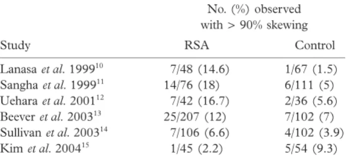
In the studies conducted to test the hypothesis that skewed XCI may be involved in the pathogenesis of RSA, both supporting9 –13
and refuting14,15
results have been obtained (Table 1). We therefore investigated the peripheral blood XCI patterns of controls and females who experience recurrent pregnancy loss to delineate the molecular basis of RSA in the Turkish population.
Materials and methods
Patients
Eighty Caucasian women diagnosed with RSA, and 160 apparently healthy female controls, were genotyped to deter-mine whether women with a history of RSA have a greater frequency of extremely skewed XCI than control subjects. The ethics review board of the participating institutions approved the study protocol. Pregnancy history, age, and disease information, accompanied by informed consent, was obtained from all subjects. We included those cases with three or more clinically recognised spontaneous abortions of unknown cause. A positive urine pregnancy test or serum βhCG and evidence of the loss of conception products accompanied by vaginal bleeding, or ultrasonographic findings of fetal demise with subsequent reduction in serum βhCG, were used to confirm spontaneous abortion. While ectopic or molar pregnancies were excluded, only those abortions that have occurred before week 20 of gestation were included in the study. Clinical evaluation of the cases excluded anatomic, cytogenetic, infectious, immunologic, hormonal, and social/exposure as causes of spontaneous abortion.2,3
Also abnormal hystero-salpingogram, antiphospholipid antibodies, or connective tissue disorder were included in the exclusion criteria. While all of
the RSA cases were karyotyped using peripheral blood samples, this was not done in the control group because of the lack of an indication for cytogenetic studies. Gravida 2, para 2 women with no history of pregnancy loss comprised the control group. The mean age of the cases and controls were 30.4 ± 5 (mean ± SD; range = 22–42 years), and 32.4 ± 4.9 years (range = 21– 47 years), respectively.
X-chromosome inactivation study
DNA was extracted from 10 mL venous blood samples of patients and controls using Nucleospin DNA isolation kit (Macherey-Nagel, Dueren, Germany). Genotyping of a polymorphic site in the androgen receptor gene (AR) was performed and quantified to assess the XCI patterns as described.16
The degree of skewing was estimated by an assay based on a methylation-sensitive HpaII restriction site located near the AR gene. This site is methylated on the inactive X, and unmethylated on the active X-chromosomes. When the genomic DNA is cleaved with HpaII prior to polymerase chain reaction (PCR), only the methylated AR allele, which represents the inactive X-chromosome, is amplified. A trinucleotide cytosine-adenine-guanine (CAG) repeat polymorphism located within the amplified region is used to distinguish between the two alleles. For each patient and control two separate PCRs, with or without HpaII treatment, were performed using the same set of primers. Male DNA with cytogenetically verified 46XY karyotype was used as control for complete digestion. All of the PCR products, before and after digestion, were separated by employing two independent detection methods. One is electrophoretic separation of the alleles in 4% MetaPhor agarose (FMC Bioproducts, Rockland, ME, USA) and ethidium bromide staining. The other is separation of radio-active PCR products (α-[33
P]-dCTP) (PerkinElmer, Wellesley, MA, USA) on 8% sequencing gels. Densitometric analysis of the alleles was performed using the Multi-Analyst software version 1.1 (Bio-Rad Laboratories Inc., Hercules, CA, USA). The results of the densitometric analyses included normali-sation of the ratios based on the non-digested samples by dividing the allele ratio of the digested sample by the ratio of the non-digested sample from the same specimen. The use of this ratio corrects for preferential amplification of one allele, which often occurs for the shorter microsatellite allele. The results from control and test groups were compared by the χ2
test with Yates’ correction.
Results
X-chromosome inactivation status was found to be informative in 77.5% of both the cases (62/80) and the controls (124/160). Only those individuals whose alleles resolve adequately for densitometric analysis were included in the study. Extremely skewed XCI was present in 11 (17.7%) cases and in two (1.6%) controls (P = 0.0002) (Table 2). Extremely skewed XCI, defined as > 90% inactivation of one allele, is observed in ∼1–7% of females aged 25 or younger, and in ∼2–16% of women aged 60 years or older.17,18
When XCI values between 85 and 89% Table 1 Studies of skewed X-chromosome inactivation and
recurrent spontaneous abortion (RSA)
Study No. (%) observed with > 90% skewing RSA Control Lanasa et al. 199910 7/48 (14.6) 1/67 (1.5) Sangha et al. 199911 14/76 (18) 6/111 (5) Uehara et al. 200112 7/42 (16.7) 2/36 (5.6) Beever et al. 200313 25/207 (12) 7/102 (7) Sullivan et al. 200314 7/106 (6.6) 4/102 (3.9) Kim et al. 200415 1/45 (2.2) 5/54 (9.3)
S. Bagislar et al.
386 © 2006 The Authors
Journal compilation © 2006 The Royal Australian and New Zealand College of Obstetricians and Gynaecologists; 46: 384–387
were also considered, skewed XCI was observed in 13 of the 62 (20.9%) informative cases, and eight of the 124 (6.4%) controls (P = 0.0069). These results indicate that extremely skewed XCI is more frequent in RSA cases than in controls, and raises the important question as to what causes this observation.
Discussion
Skewed XCI could originate from primary (bias in the initial choice) or secondary (selection following random XCI) causes.8
Two different hypotheses have been proposed to explain the aetiology of skewed XCI in women who experience RSA: (i) male lethal X-linked mutations4,19
and (ii) a reduction of the size of the follicular pool13
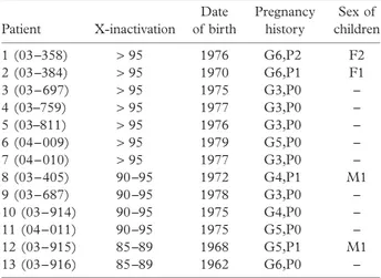
associated with skewed XCI and trisomic pregnancies. A gender bias towards female offspring among liveborn children and maternal inheritance of skewed X-inactivation was documented in conjunction with the first hypothesis. However, a significant excess of boys among live births was observed in females with skewed XCI who were ascertained on the basis of trisomic pregnancies. Among our 11 patients with extreme skewing (> 90%), only three patients had live births and the observed female : male ratio is 3 : 1 ( Table 3). Deviation from the expected female : male ratio of 1 : 1 raises the possibility that male lethal X-linked mutations could be an aetiologic factor in our RSA cases, at least in the group of patients with extreme skewing. In addition to deleterious X-linked mutations, X-autosome translocations, normal ageing, twining, confined placental mosaicism, reduction of the size of the follicular pool, monoclonal expansion of peripheral blood cells, and environmental insults, such as chemotherapy, are examples of secondary causes. Some of these factors are unlikely to be involved in our patients: for example only those patients with a normal karyotype were included in the study, excluding the potential contribution of X-autosome translocations. Twining, because of negative history, and ageing, because of the mean age of diagnosis at 30.4 ± 5 years, are also highly unlikely. A history or present diagnosis of haematologic malignancies, which may be associated with monoclonal expansion of peripheral blood cells, or exposure to environ-mental insults, such as chemotherapy, was not documented in any study subject. With respect to the primary causes of skewed XCI, we do not think dysfunction of gene(s) that regulates XCI20
could be implicated in the aetiology of RSA as dosage compensation for X-linked genes is not physiolog-ically required in XY cells and therefore is not expected to
be lethal in male fetuses. Our findings also leave us with the question of why some studies did not show a statistically significant association between RSAs and skewed XCI patterns.14,15
It is important to note that a heterogeneous group of aetiologic factors is known to contribute to the development of RSAs,2,3
and that small sample size15
may influence the outcome of statistical analyses.
We plan to direct our future efforts towards the ascertain-ment of extended pedigrees to assess the parental origin of the inactive X-chromosome. In addition, detailed high-resolution genomic analysis by newer molecular techniques, such as comparative genomic hybridisation microarray analysis,21
could prove to be very valuable at least in a subgroup of patients who may harbour X-chromosomal submicroscopic deletions.
Acknowledgements
We thank Iclal Buyukdevrim-Ozcelik for critical reading of the manuscript. Supported by grants from the Scientific and Technical Research Foundation of Turkey TÜBITAK– SBAG 3334, International Centre for Genetic Engineering and Biotechnology – ICGEB-CRP/TUR04-01, and Bilkent University Research Fund (to Dr Ozcelik).
References
1 Wilcox AJ, Weinberg CR, O’Connor JF et al. Incidence of early loss of pregnancy. N Engl J Med 1988; 319: 189 –194. 2 Stephenson MD. Frequency of factors associated with habitual
abortion in 197 couples. Fertil Steril 1996; 66: 24 –29. 3 Branch DW. Management of recurrent early pregnancy loss.
2001; ACOG Practice Bulletin No. 24. Washington: American College of Obstetricians and Gynecologists.
4 Pegoraro E, Whitaker J, Mowery-Rushton P, Surti U, Lanasa M, Hoffman EP. Familial skewed X inactivation: A molecular trait associated with high spontaneous-abortion rate maps to Xq28. Am J Hum Genet 1997; 61: 160 –170.
Table 2 Proportion of the cases and controls with skewed X-chromosome inactivation
Group
No. (%) observed with 85 – 89% skewing > 90% skewing Total RSA patients (n = 62) 2 (3.2) 11 (17.7) 13 (20.9) Control females (n = 124) 6 (4.8) 2 (1.6) 8 (6.4) For comparison by χ2
, P = 0.0002. RSA, recurrent spontaneous abortion.
Table 3 Characteristics of the cases with extremely skewed X-chromosome inactivation Patient X-inactivation Date of birth Pregnancy history Sex of children 1 (03 –358) > 95 1976 G6,P2 F2 2 (03 –384) > 95 1970 G6,P1 F1 3 (03 – 697) > 95 1975 G3,P0 – 4 (03–759) > 95 1977 G3,P0 – 5 (03–811) > 95 1976 G3,P0 – 6 (04 – 009) > 95 1979 G5,P0 – 7 (04 – 010) > 95 1977 G3,P0 – 8 (03 – 405) 90 –95 1972 G4,P1 M1 9 (03 – 687) 90 –95 1978 G3,P0 – 10 (03 – 914) 90 –95 1975 G4,P0 – 11 (04 – 011) 90 –95 1975 G5,P0 – 12 (03 – 915) 85 –89 1968 G5,P1 M1 13 (03 – 916) 85 –89 1962 G6,P0 –
X-inactivation and recurrent spontaneous abortion
© 2006 The Authors 387
Journal compilation © 2006 The Royal Australian and New Zealand College of Obstetricians and Gynaecologists; 46: 384–387 5 Lyon MF. Gene action in the X-chromosome of the mouse
(Mus musculus). Nature 1961; 190: 372–373.
6 Migeon BR. X chromosome inactivation: theme and variations. Cytogenet Genome Res 2002; 99: 8 –16.
7 Lyon MF. X-chromosome inactivation and human genetic disease. Acta Paediatr (Suppl.) 2002; 91: 107–112.
8 Brown CJ. Skewed X-chromosome inactivation: Cause or consequence? J Natl Cancer Inst 1999; 91: 304 –305. 9 Lanasa MC, Hogge WA, Hoffman EP. The X-chromosome
and recurrent spontaneous abortion: The significance of trans-manifesting carriers. Am J Hum Genet 1999; 64: 934 – 938. 10 Lanasa MC, Hogge WA, Kubick C, Blancato J, Hoffman EP.
Highly skewed X-chromosome inactivation is associated with idiopathic recurrent spontaneous abortion. Am J Hum Genet 1999; 65: 252–254.
11 Shanga KK, Stephenson MD, Brown CJ, Robinson WP. Extremely skewed X-chromosome inactivation is increased in women with recurrent spontaneous abortion. Am J Hum Genet 1999; 65: 913 – 917.
12 Uehara S, Hashiyada M, Sato K, Sato Y, Fujimori K, Okamura K. Preferential X-chromosome inactivation in women with idiopathic recurrent pregnancy loss. Fertil Steril 2001; 76: 908 – 914.
13 Beever CL, Stephenson MD, Penaherrera MS et al. Skewed X-chromosome inactivation is associated with trisomy in women ascertained on the basis of recurrent spontaneous abortion or chromosomally abnormal pregnancies. Am J Hum Genet 2003; 72: 399 – 407.
14 Sullivan AE, Lewis T, Stephenson M et al. Pregnancy outcome in recurrent miscarriage patients with skewed X chromosome inactivation. Obstet Gynecol 2003; 101: 1236 –1242.
15 Kim JW, Park SY, Kim YM, Kim JM, Han JY, Ryu HM. X-chromosome inactivation patterns in Korean women with idiopathic recurrent spontaneous abortion. J Korean Med Sci 2004; 19: 258 –262.
16 Allen RC, Zoghbi HY, Moseley AB, Rosenblatt HM, Belmont JW. Methylation of HpaII and HhaI sites near the polymorphic CAG repeat in the human androgen-receptor gene correlates with X-chromosome inactivation. Am J Hum Genet 1992; 51: 1229 –1239.
17 Buller RE, Sood AK, Lallas T, Buekers T, Skilling JS. Association between nonrandom X-chromosome inactivation and BRCA1 mutation in germline DNA of patients with ovarian cancer. J. Natl Cancer Inst 1999; 91: 339 –346. 18 Sharp A, Robinson D, Jacobs P. Age- and tissue-specific variation
of X-chromosome inactivation ratios in normal women. Hum Genet 2000; 107: 343 –349.
19 Lanasa MC, Hogge WA, Kubik CJ et al. A novel X chromosome-linked genetic cause of recurrent spontaneous abortion. Am J Obstet Gynecol 2001; 185: 563 –568.
20 Plenge RM, Hendrich BD, Schwartz C et al. A promoter mutation in the XIST gene in two unrelated families with skewed X-chromosome inactivation. Nat Genet 1997; 17: 353 –356. 21 Larrabee PB, Johnson KL, Pestova E et al. Microarray analysis
of cell-free fetal DNA in amniotic fluid: a prenatal molecular karyotype. Am J Hum Genet 2004; 75: 485 – 491.