https://doi.org/10.1007/s00590-018-2356-y
ORIGINAL ARTICLE • HIP - FRAC TUR ES
The effect on outcomes of the application of circumferential cerclage
cable following intramedullary nailing in reverse intertrochanteric
femoral fractures
Ahmet Imerci1,2 · Nevres Hurriyet Aydogan1 · Kursad Tosun3
Received: 15 September 2018 / Accepted: 3 December 2018 / Published online: 6 December 2018 © Springer-Verlag France SAS, part of Springer Nature 2018
Abstract
Introduction The aim of this study was to evaluate the effect on union results of the application of cerclage cable around the lateral femoral wall (LFW) in postoperative fracture displacement.
Patients and methods A retrospective evaluation was made of all the cases of reverse intertrochanteric fractures over a 7-year period in a trauma center. The study included 69 fractures treated with intramedullary nailing. The age of the patients, postoperative reduction quality and complications such as mechanical failure and non-union (1.4%) were obtained from the medical records. Changes in the neck-shaft angle, the amount of LFW displacement and telescoping of proximal femoral nail antirotation blades and lag screws were measured on anteroposterior and lateral radiographs. The functional evaluation of the Harris Hip Score was recorded at the final follow-up examination.
Results The mean age of the patients was 56.31 years (range 18–93 years). Closed reduction was applied in 37 (53.7%) cases, open reduction in 10 (14.5%) and with cerclage cable in 22 (31.8%). Mechanical failure developed in 8 (11.5%) cases where cerclage cable was not used. In 2 of these 8 cases, open reduction was successful. In the patients where cerclage cable was applied, no mechanical failure developed and no case underwent revision surgery. In the cases where cerclage cable was not used, a significantly higher rate of telescoping was found. (p = 0.001). The application of cerclage cable was seen to significantly reduce the amount of LFW displacement (2.23 mm vs 8.86 mm) and shorten the time to partial weight bearing (p = 0.000).
Conclusion In reverse intertrochanteric fractures, the application of circumferential cerclage cable with cephalomedullary nailing contributes to primary stability and accelerates mobilization. Therefore, it can be considered a good option. It reduces the risk of failure internal fixation for intertrochanteric fracture.
Keywords Intertrochanteric fracture · Cable · Intramedullary nailing
Introduction
Reverse intertrochanteric fractures are less common than other (AO/OTA 31-A1 and A2) intertrochanteric fractures and are estimated to constitute approximately 2% of all hip fractures [1]. These fractures are classified in the Ortho-paedic Trauma Association system as AO/OTA 31-A3. There are two known deforming forces on these fractures. The first is that due to shear forces formed of the iliopsoas and adductor muscles, there is a tendency for shaft medi-alization and shortening in the fracture area. The second is that the deforming force of the hip abductors which tends to move the proximal fragment to varus and external rota-tion [2]. Due to these high tensile and compressive forces working in different directions, it is difficult to prevent
* Ahmet Imerci
[email protected] Nevres Hurriyet Aydogan [email protected] Kursad Tosun
1 Department of Orthopaedics and Traumatology, Faculty of Medicine, Mugla Sitki Kocman University, Mugla, Turkey
2 Karamehmet Mah. 19 sokak No:5 C blok daire:10, Mentese, Mugla, Turkey
fracture displacement and provide recovery. Furthermore, calcar support is lost because of the fractured large pos-teromedial fragment. Thus, there is a tendency of implant failure with axial loading. Therefore, the optimal device for stabilization of these fractures would be an implant that resists the tendency for shaft medialization, rotation and varus [3]. Intramedullary nails can more reliably resist the relatively high axial forces across the medial calcar that are typically borne by the implant in an reverse fracture and owing to this feature it allows to early partial weight bearing [1–3].
In addition, a specific feature of 31-A3 fractures is the extension to the lateral femoral wall (LFW) [4, 5]. The LFW represents a region from the vastus corner of the tro-chanter extending distally along the lateral femoral cortex, which prevents the sliding of the proximal fragment [6, 7]. Although seen more often in A3.3 fractures in particular, in fractures which extend to the trochanter major, there may be free proximal fracture fragments in the anterior or posterior and free posteromedial fragment [8].
These fractures where the integrity of the lateral wall is impaired are repaired much better with intramedullary nailing than with dynamic hip screw. In addition, sufficient fixation may not be able to be provided with nailing alone because of the fracture of the LFW and the associated free fragments and therefore the fracture may remain displaced [8–10]. The use of circumferential cable provides bone contact and may be useful in avoiding postoperative lat-eral migration and angulation due to pulling of abductor arm to proximal fragment [11]. There are several studies in literature which state that the application of circumfer-ential cerclage cable as support to intramedullary nailing provides better functional results in trochanteric region fractures [11–13]. However, to the best of our knowledge, there is no study in literature which has compared the use of cerclage cable additional to nailing, specifically in reverse intertrochanteric fractures.
The aim of this study was to evaluate the effects of cir-cumferential cerclage cable in addition to intramedullary nailing for reverse intertrochanteric fractures.
Patients and methods
A retrospective evaluation was made of consecutive patients who presented at the Our Hospital with a reverse intertrochanteric fracture (31-A3) over a 7-year period. A total of 10 patients were excluded from the study because of pathological fractures, multiple trauma, mortality in the early postoperative period, a lack of regular follow-up examinations, and cephalomedullary nailing was not used.
Surgical technique and the physical therapy protocol
At half an hour before the start of the operation, antibiotic prophylaxis (cefazolin sodium 1 g/day IV) was applied to all patients and in the postoperative period, treatment of low molecular weighted heparin (enoxaparin sodium 40 mg/0.4 mL/day) was administered. All the operations were conducted under spinal or general anesthesia. All the operations were applied in a single center with the participa-tion of a surgical team of surgeons with at least 5 years of experience in hip surgery. Patients were positioned and rig-idly fixed in the lateral decubitus position on a conventional surgery table. The fixation device was checked to determine fluoroscopic visualization. Reduction in the fracture was provided by manual longitudinal traction and internal rota-tion. The quality of the reduction was confirmed using an image intensifier positioned in a lateral mode to take images for both anteroposterior and lateral views. The lateral view was obtained by flexing the hip to nearly 90° and abducting to 45°. In fractures where undisplaced reduction could not be provided in complex or LFW and posteromedial fragmented fractures, an open surgical approach was used. Circumfer-ential cerclage cable (1 or more) was applied to maintain fracture reduction or to fix the free lateral wall fragment and posteromedial fragment, usually when the clamp was removed after the nailing in patients with a free proximal fragment or with displaced trochanteric fragment or postero-medial fragment. Postoperatively, standard medical care was applied to all the patients. With the assistance of a physi-otherapist in the postoperative period, partial weight-bearing mobilization was encouraged, depending on patient toler-ance, biomechanical structure stability and bone quality. Toe touch with double crutches is allowed at first postoperative day. The patients were followed up regularly in the trauma clinic at 4, 6, 8 and 12 weeks 6 and 12 months. Non-union or delayed unions were controlled radiographically at 4 weeks intervals after 12 weeks.
At the 1-year follow-up examination, functional evalua-tion was made with the Harris Hip Score (HHS). Throughout follow-up, changes in the neck-shaft angle, time to fracture union, time to partial weight-bearing and mechanical com-plications such as hip screw migration, cutout, shaft fracture and non-union were reported in the radiological evaluations. Fracture union was defined as the visualization of osseous bridging in at least 3 cortices on anteroposterior and lateral radiographs and painless full weight bearing [14]. LFW displacement, reduction quality and telescoping of the lag screw and blade were also measured in all 69 patients.
Radiographic measurements
Measurements were taken using a measuring device in a digital environment on the radiographs taken on the post-operative 1st day and at 1 year. The measurements were standardized and evaluated by 2 experienced orthopedic surgeons. To prevent bias, the radiographic evaluations were completed before the evaluation of the functional results. Proportional correction was made of the meas-ured screw size with the known screw size to eliminate radiological magnifications and differences in the meas-urements arising from the extremity position [9].
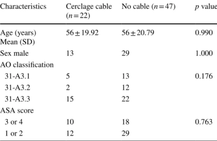
On postoperative day 1, plain anteroposterior and lat-eral radiographs were taken. The quality of the fracture reduction and the placement of the implant were analyzed. Reduction quality was classified as good, acceptable or poor [15]. LFW displacement was evaluated on the radio-graph as the perpendicular distance from the apex of the lateral femoral fragment to the lateral edge of the nail (Fig. 1).
According to the Cleveland–Bosworth Quadrants, center–center and inferior–center quadrants were accepted as optimal and the remaining quadrants were accepted as suboptimal [16]. Telescoping was defined as the distance along the blade from the lateral border of the nail to the blade end (Fig. 1). Proximal fragment sliding was con-sidered as the difference in telescoping between the post-operative 1st day radiograph and the 1st year radiograph.
Statistical analysis
Differences between the two techniques in respect of HHS, telescoping, neck-shaft angle changes, time to union and partial weight bearing were assessed using two-sample t tests. In the tables, data were expressed as mean ± standard deviation values. Chi-square tests were applied to evaluate the associations between the two techniques and quality of reduction, AO classification, ASA scores and implant posi-tions in the studied patient cohorts. All statistical testing was performed with software R (www.r-proje ct.org) [17].
Results
Of the 69 cases with reverse intertrochanteric fracture, cer-clage cable was used in 22 (31.9%). Closed reduction was applied in 37 (53.7%) cases, open reduction in 10 (14.5%) and open reduction with cerclage cable in 22 (31.9%). Of the patients with open reduction, support with cerclage cable was applied in 68.7%. The demographic data of age, gen-der, ASA score and fracture pattern of the fractures treated with cerclage and without cerclage are presented in Table 1. There were no statistical difference in terms of age, gender, ASA score and fracture pattern variables. (t test, p = 0.990, Chi-square test, p = 1.000, Fisher’s exact test, p = 0.176 and Chi-square test, p = 0.763, respectively).
The mean HHS was 84 ± 8.33 in the patients treated with cerclage cable and 79 ± 11.62 in those without cerclage cable. No statistically significant difference was deter-mined (t test p = 0.085). The time to union was deterdeter-mined as mean 17 ± 4.92 weeks in the cerclage cable group and 17 ± 4.64 weeks in the group without cerclage cable, with no statistically significant difference (p = 0.569) (Fig. 2). No statistically significant difference was determined between the two groups in respect of implant placement or reduction quality (Fisher’s exact test, p = 0.440 and Chi-square test,
Fig. 1 Measurement of LFW displacement (a). Telescoping (b)
Table 1 Comparison of baseline characteristics among the groups
Characteristics Cerclage cable
(n = 22) No cable (n = 47) p value Age (years) Mean (SD) 56 ± 19.92 56 ± 20.79 0.990 Sex male 13 29 1.000 AO classification 31-A3.1 5 13 0.176 31-A3.2 2 12 31-A3.3 15 22 ASA score 3 or 4 10 18 0.763 1 or 2 12 29
p = 1.000). Partial weight-bearing time was significantly
shorter in the cable group. (p = 0.0001) (Table 2).
Revision surgery was not required in any patient where cerclage cable was used. In 4 patients where cerclage cable was not used, arthroplasty was applied due to neck screw or blade cutout. In these three patients, the initial position of the implant was optimal, one was suboptimal. Erosion devel-oped in the acetabulum in 1 patient because of protuberance of the neck screw, and total hip arthroplasty was applied. Revision surgery was necessary in 1 patient due to symp-tomatic non-union and in 2 patients due to severe lateral migration of the neck screw. Seven of these eight patients underwent open reduction without cable. One patient under-went closed reduction. The amount of telescoping showing shortening of the femoral neck was found to be statistically significantly high in the group without cerclage cable (t test,
p = 0.001) (Table 2). The LFW displacement was determined as 2 ± 2.74 mm in the group without cerclage cable and 8 ± 6.35 mm (t test, p = 0.001) in the group where cerclage cable was used.
Discussion
Our results show that cable fixation appears to maintain good reduction, anatomic or close to anatomic reduction (Figs. 2, 3). This is often not technically possible with closed reduction alone. We believe that the good reduction not only eases the nailing procedure but also makes the entire fixation more stable, as most of the weight-bearing forces can be transferred through the aligned osseous fragments.
In 31-A3 fractures, the implant to be used must be opti-mal to provide anatomic reduction, to maintain the reduction until union and to allow early weight bearing. Following
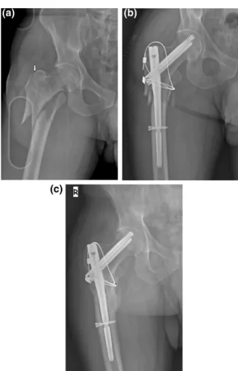
Fig. 2 Extension of the main fracture to the greater trochanter (white arrow), creating a free LFW fragment (a) and the LFW was reduced and fixed with two cerclage cable along the lateral cortex and tro-chanter major (b). Six-month postoperative radiograph showing the stable implant with union (c)
Table 2 Comparison of the operating factors and the radiological and functional results of the fractures treated with and without cerclage cable
Characteristics Cerclage cable (n = 22) No cable (n = 47) p value
HHS 84 ± 8.33 79 ± 11.62 0.085
Union time (weeks) (SD) 17 ± 4.92 17 ± 4.64 0.569 Partial weight-bearing (days) mean (SD) 4 ± 1.95 7 ± 3.67 0.000 Quality of reduction Good 18 31 0.440 Acceptable 3 11 Poor 1 5 Implant position Optimal 16 35 1.000 Suboptimal 6 12
Nail type 10 pfna, 12 InterTan 23 pfna, 24 InterTan 0.991 Nail length long % 7 (31.8%) 13 (27.6%) 0.994 Neck-shaft angle change (degrees) (SD) 3 ± 3.21 5 ± 5.45 0.080 Telescoping [mm (SD)] 3 ± 2.80 6 ± 6.68 0.001 LDD (mm) (SD) 2 ± 2.74 8 ± 6.35 0.000
fixation of these fractures with dynamic hip screw, compli-cations such as implant failure and functional impairment are often seen [18]. At the same time, uncontrolled collapse of the proximal fragment is permitted following medializa-tion of the distal femoral fragment [2, 19]. Therefore, tro-chanteric stabilization plates have been shown to be effec-tive in these types of fractures in biomechanical and clinical studies [20–22]. The combination of dynamic hip screw and trochanteric stabilization plate allows for partial fixation and reduction in the trochanter major and LFW fragments. How-ever, there is no such possibility with nailing [21, 22].
Proximal femoral nailing is the optimum treatment choice in unstable fractures, especially in reverse 31-A3 fractures with LFW fracture [3, 14, 23]. Boopalan et al. [4] reported
that as intramedullary nails functioned as support in 31-A1 and A2 intertrochanteric fractures, there was a limited effect of instability created by an unstable LFW. However, it is not clear whether or not there is any contribution to healing and stability of the support function provided by intramedul-lary nailing applied in these fractures which have sustained fractured and displaced LFW during the injury [4, 9, 20]. Therefore, there are no clear guidelines for the additional treatment of laterally displaced and free LFW fragments. The findings of the current study indicated that in contrast to what was assumed, there was no harmful effect of the application of circumferential cerclage cable such as infec-tion, lateral thigh pain or delayed union.
Yun et al. [10] used supporting mini locking plates for fix-ation of the trochanteric major fragment in pertrochanteric fractures or for the repair of the LFW during intramedullary hip nailing. The use of these plates has been reported to be simple and allows multi-directional use. In the current study, cerclage cable can be used after nailing for the fixation of the trochanter major and repair of the LFW. Its advantages in simple, rapid and stable manipulation, as well as its ability to fulfill load bearing requirement early, can reduce complica-tions related to long-term bed rest.
There have been reports in orthopaedic literature related to the addition of minimally invasive cerclage cable in sub-trochanteric fractures exposed to forces with a similar effect as in reverse intertrochanteric fractures [24]. Cerclage cable is advocated in unsuccessful closed reduction interventions. With the assistance of a clamp, reduction combinations have been used in oblique and spiral fractures [12, 13, 25]. Fol-lowing the use of cerclage cable, re-operation rates and reduction quality have been reported to be better and the amount of fracture displacement is less [12, 13]. It was also reported in another study of AO 31-A3.3 fractures that the presence of displaced LFW after closed reduction had no negative effect on the radiological healing of pertrochanteric fractures [9]. In our study, after the use of cerclage cable, better LFW reduction was seen to have been obtained.
The change in neck-shaft angle was not determined to have any effect on subsequent development of varus angula-tion. Furthermore, no statistically significant difference was determined between the two groups treated with or without cerclage cable in respect of the functional scores.
Although there have been low rates of open reduction reported in previously published studies, it was necessary to apply open reduction to almost half of the cases in the cur-rent series to obtain better stability and in the majority, ana-tomic or nearly anaana-tomic reduction was achieved [26–28]. Despite correct technical application during nail placement, because of the difficulty in reduction in medialization of the distal fragment and abduction of the proximal fragment with the effect of the abductor arm, open reduction must be applied in some patients to determine the correct nail entry
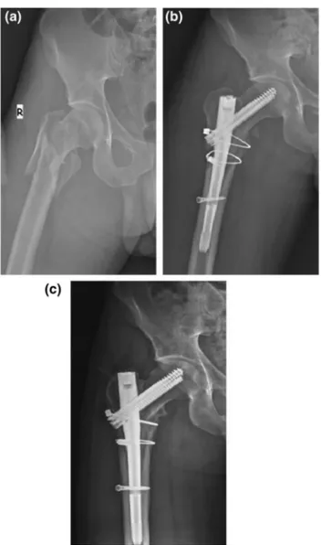
Fig. 3 Plain radiograph showing AO 31-A3.3 intertrochanteric femur
fracture with four part comminution (a). Initial postoperative radio-graph demonstrating anatomic reduction of LFW and posteromedial fragment by application of two circumferential cable (b). One-year postoperative radiograph showing a stable implant with union (c)
point and obtain anatomic or close to anatomic reduction [11, 29, 30]. The use of cerclage cable can be recommended after open reduction to obtain good primary stability and to help decrease abduction of the proximal fragment (LFW and trochanter major).
When weight bearing is applied early, to protect the reduction and reduce complication rates, it is important to provide weight transfer with the alignment of bone frag-ments. In our study, as there was stable reduction, the time to partial weight bearing was seen to be shorter than in with-out cable group. In a biomechanical study, it was reported that the wrapping of additional cerclage cable provided important posteromedial support and could reduce the risk of osteosynthesis failure in complex fractures [31]. Revi-sion surgery was necessary because of a symptomatic non-union in a cableless patient with poor posteromedial support (Fig. 4). No patient treated with cable in the current study required re-operation because of mechanical failure. It was also observed that good fracture repositioning was provided by the cerclage cable reducing the lateral fracture gap and provided to good posteromedial support. Other surgical
factors such as the type and length of nail and the implant position were not found to be associated with complications.
Gao et al. [32] reported that the use of cerclage cable was a useful surgical technique for the treatment of intertrochan-teric femoral fractures that could not be reduced with closed maneuvers in young patients, because the cable facilitated reaming and nailing, reduced intraoperative complications and helped to protect the reduction of the fracture. However, Ban et al. [11] applied cerclage cable additional to nailing in 35 patients with reverse fractures and reported that mobiliza-tion was accelerated in approximately three-quarters of the cases and reduction quality was increased. In our study also, no difference in reduction quality was determined between those treated with or without cerclage cable. A study by Fogagnolo et al. [33] included only 5 AO 31 A3 fractures, and fewer than half were permitted immediate weight bear-ing. Wei et al. [29] reported that intraoperative LFW recon-struction additional to cephalomedullary nailing for LFW integrity accelerated early mobilization. In the current study, it was also observed that the use of cerclage cable shortened the time to partial weight bearing and accelerated mobi-lization. Studer et al. [8] determined that the treatment of reverse pertrochanteric femoral fractures in particular with closed reduction and internal fixation with cephalomedullary nailing caused a serious loss in mobility of the displaced greater trochanteric fragment. In another study, cerclage cable applied to the LFW in reverse oblique fractures was determined to increase stability [26]. Another biomechani-cal study reported that the application of an auxiliary lock-ing plate additional to naillock-ing in intertrochanteric fractures reduced interfragmentary movement, increased the rigidity of osteosynthesis and reduced the load on the intramedullary nail [34]. Therefore, the reduced time to early weight bear-ing in the current study can be associated with the provision of better stability.
In our study, to prevent excessive sliding of the head and neck fragment, the application of circumferential cerclage cable additional to nailing of LFW fracture was seen to be beneficial. The rates of telescoping, which show head-neck collapse, were greater in the patients where cerclage cable was not applied. Revision with arthroplasty was applied to 2 cases because of serious lateral screw migration.
Biber et al. [35] noticed that in some cases of a series, nail gliding was blocked by cortical support below the sleeve of the lag screw on the lateral side. By removing this bone block, fracture impaction is provided along the femoral shaft axis, and with this procedure, known as “lateral notching,” the risk of implant fracture has been reported to have been reduced. Lateral notching for dynamization or removal of the distal locking screw was not applied to any of the cur-rent study patients. Tomás-Hernández et al. [36] associated nail breakage primarily with fracture gaps of > 5 mm. In the current study, the reason for nail breakage in 1 case applied
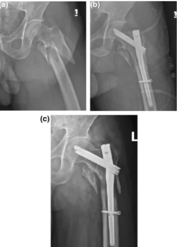
Fig. 4 a AP Radiograph of the left hip showing a AO/OTA 31-A3.3
fracture b postoperative 1 day and c at 11 months, a case of mechani-cal insufficient due to the non-union
with nailing without cable was considered due to a larger medial cortical gap (Fig. 4a–c).
There is a belief that non-union is caused as a result of impaired fracture vascularity and blood support of the bone related to the use of cerclage cable [37]. This has been asso-ciated with over-dissection of the periosteal and soft tissue. If minimally invasive techniques are applied, the deteriora-tion in soft tissue and bone vascularity is reduced [11]. In an animal model without fractures, vascular support was seen to be protected following the use of cerclage cable [38]. In the current study, there was seen to be no negative effect from the use of cerclage cable. Complete bone union was achieved in all the cases treated with cable. Kennedy et al. [25] reported no harmful effect on the time to union. In the this study, cutout developed in 4 patients who were not treated open reduction without cerclage, and revision was made with arthroplasty.
Limitations of the current study can be said to be that the study was retrospective, and as the fracture reduction in all cases was applied without the use of a fracture table, this could have affected the surgical approach rates.
Conclusion
Taking everything into consideration, the provision of sta-ble fixation is important to reduce the risk of re-operation and obtain early mobilization. Although it cannot be recom-mended for routine use, the addition of cerclage cable for selected patients under optimal conditions, especially those with 31-A3.3 fractures with free, displaced LFW fracture, can be considered to increase the chance of surgical success and help to protect the reduction. This approach is an easy, safe and effective treatment for intertrochanteric fracture.
Compliance with ethical standards
Conflict of interest The authors declare that they have no competing interests.
References
1. Haidukewych GJ, Israel TA, Berry DJ (2001) Reverse obliquity fractures of the intertrochanteric region of the femur. J Bone Joint Surg Am 83-A:643–650
2. Sadowski C, Lubbeke A, Saudan M, Riand N, Stern R, Hoffmeyer P (2002) Treatment of reverse oblique and transverse intertro-chanteric fractures with use of an intramedullary nail or a 958 screw-plate: a prospective, randomized study. J Bone Joint Surg 84:372–381
3. Kuzyk PR, Lobo J, Whelan D, Zdero R, McKee MD, Schemitsch EH (2009) Biomechanical evaluation of extramedullary versus intramedullary fixation for reverse obliquity intertrochanteric fractures. J Orthop Trauma 23(1):31–38
4. Boopalan PR, Oh JK, Kim TY, Oh CW, Cho JW, Shon WY (2012) Incidence and radiologic outcome of intraoperative lateral wall fractures in OTA 31A1 and A2 fractures treated with cephalomed-ullary nailing. J Orthop Trauma 26(11):638–642
5. Palm H, Jacobsen S, Sonne-Holm S, Gebuhr P, Hip Fracture Study Group (2007) Integrity of the lateral femoral wall in intertrochan-teric hip fractures: an important predictor of a reoperation. J Bone Joint Surg Am 89(3):470–475
6. Ma Z, Chang SM (2014) Letter to the editor: where is the lateral femoral wall? Int Orthop 38(12):2645–2646
7. Hsu CE, Chiu YC, Tsai SH, Lin TC, Lee MH, Huang KC (2015) Trochanter stabilising plate improves treatment outcomes in AO/ OTA 31-A2 intertrochanteric fractures with critical thin femoral lateral walls. Injury 46(6):1047–1053
8. Studer P, Suhm N, Wang Q, Rosenthal R, Saleh HA, Jakob M (2015) Displaced trochanteric fragments lead to poor functional outcome in pertrochanteric fractures treated by cephalomedullary nails. Injury 46(12):2384–2388
9. Kim Y, Bahk WJ, Yoon YC, Cho JW, Shon WY, Oh CW, Oh JK (2015) Radiologic healing of lateral femoral wall fragments after intramedullary nail fixation for A3.3 intertrochanteric fractures. Arch Orthop Trauma Surg 135(10):1349–1356
10. Yun HH, Lee YI, Kim KH, Yun S (2015) Use of auxiliary locking plates for the treatment of unstable pertrochanteric femur frac-tures. Orthopedic 38(5):305–309
11. Ban I, Birkelund L, Palm H, Brix M, Troelsen A (2012) Cir-cumferential wires as a supplement to intramedullary nailing in unstable trochanteric hip fractures: 4 reoperations in 60 patients followed for 1 year. Acta Orthop 83(3):240–243
12. Hoskins W, Bingham R, Joseph S, Liew D, Love D, Bucknill A, Oppy A, Griffin X (2015) Subtrochanteric fracture: the effect of cerclage wire on fracture reduction and outcome. Injury 46(10):1992–1995
13. Tomás J, Teixidor J, Batalla L, Pacha D, Cortina J (2013) Sub-trochanteric fractures: treatment with cerclage wire and long intramedullary nail. J Orthop Trauma 27(7):e157–e160
14. Park SY, Yang KH, Yoo JH, Yoon HK, Park HW (2008) The treatment of reverse obliquity intertrochanteric fractures with the intramedullary hip nail. J Trauma 65(4):852–857
15. Vidyadhara S, Rao SK (2007) One and two femoral neck screws with intramedullary nails for unstable trochanteric fractures of femur in the elderly–randomised clinical trial. Injury 38:806–814 16. Cleveland M, Bosworth DM, Thompson FR, Wılson HJ Jr,
Ishızuka T (1959) A ten-year analysis of intertrochanteric frac-tures of the femur. J Bone Joint Surg Am 41-A:1399–1408 17. R Core Team (2016) R: a language and environment for
statisti-cal computing. R Foundation for Statististatisti-cal Computing, Vienna, Austria. ISBN 3-900051-07-0. http://www.R-proje ct.org/
18. Min WK, Kim SY, Kim TK, Lee KB, Cho MR, Ha YC, Koo KH (2007) Proximal femoral nail for the treatment of reverse obliquity intertrochanteric fractures compared with gamma nail. J Trauma 63(5):1054–1060
19. Matre K, Havelin LI, Gjertsen JE, Vinje T, Espehaug B, Fevang JM (2013) Sliding hip screw versus IM nail in reverse oblique tro-chanteric and subtrotro-chanteric fractures. A study of 2716 patients in the Norwegian Hip Fracture Register. Injury 44(6):735–742 20. Gupta RK, Sangwan K, Kamboj P, Punia SS, Walecha P (2010)
Unstable trochanteric fractures: the role of lateral wall reconstruc-tion. Int Orthop 34(1):125–129
21. Babst R, Renner N, Biedermann M, Rosso R, Heberer M, Harder F, Regazzoni P (1998) Clinical results using the trochanter sta-bilizing plate (TSP): the modular extension of the dynamic hip screw (DHS) for internal fixation of selected unstable intertro-chanteric fractures. J Orthop Trauma 12(6):392–399
22. Langford J, Pillai G, Ugliailoro AD, Yang E (2011) Periopera-tive lateral trochanteric wall fractures: sliding hip screw versus
percutaneous compression plate for intertrochanteric hip fractures. J Orthop Trauma 25(4):191–195
23. Nherera L, Trueman P, Horner A, Watson T, Johnstone AJ (2018) Comparison of a twin interlocking derotation and compression screw cephalomedullary nail (InterTAN) with a single screw dero-tation cephalomedullary nail (proximal femoral nail antirodero-tation): a systematic review and meta-analysis for intertrochanteric frac-tures. J Orthop Surg Res 2(13):46
24. Afsari A, Liporace F, Lindvall E, Infante A Jr, Sagi HC, Haiduke-wych GJ (2009) Clamp-assisted reduction of high subtrocanteric fractures of the femur. J Bone Joint Surg (Am) 91(8):1913–1918 25. Kennedy MT, Mitra A, Hierlihy TG, Harty JA, Reidy D, Dolan M
(2011) Subtrochanteric hip fractures treated with cerclage cables and long cephalomedullary nails: a review of 17 consecutive cases over 2 years. Injury 42(11):1317–1321
26. Honkonen SE, Vihtonen K, Jarvinen MJ (2004) Second generation cephalo medullary nails in the treatment of reverse obliquity inter-trochanteric fractures of the proximal femur. Injury 35:179–183 27. Al-yassari G, Langstaff RJ, Jones JW, Al- Lami M (2002) The
AO/ASIF proximal femoral nail (PFN) for the treatment of unsta-ble trochanteric femoral fracture. Injury 33(5):395–399
28. Ozkan K, Eceviz E, Unay K, Tasyikan L, Akman B, Eren A (2011) Treatment of reverse oblique trochanteric femoral fractures with proximal femoral nail. Int Orthop 35(4):595–598
29. Wei J, Qin DA, Guo XS (2015) Curative effect analysis on proxi-mal fermoral nail antirotation for the treatment of femoral inter-trochanteric fracture and integrity of lateral inter-trochanteric wall. Zhongguo Gu Shang 28(6):572–575
30. Angelini A, Battiato C (2016) Combination of low-contact cer-clage wiring and osteosynthesis in the treatment of femoral frac-tures. Eur J Orthop Surg Traumatol 26(4):397–406
31. Müller T, Topp T, Kühne CA, Gebhart G, Ruchholtz S, Zettl R (2011) The benefit of wire cerclage stabilisation of the medial hinge in intramedullary nailing for the treatment of subtro-chanteric femoral fractures: a biomechanical study. Int Orthop 35(8):1237–1243
32. Gao YS, Guo YJ, Yu XG, Chen Y, Chen C, Lu NJ (2018) A novel cerclage wiring technique in intertrochanteric femoral fractures treated by intramedullary nails in young adults. BMC Musculo-skelet Disord 6(19):359
33. Fogagnolo F, Kfuri M Jr, Paccola CA (2004) Intramedullary fixation of pertrochanteric hip fractures with the short AO-ASIF proximal femoral nail. Arch Orthop Trauma Surg 124(1):31–37 34. Eberle S, Gabel J, Hungerer S, Hoffmann S, Pätzold R, Augat P,
Bühren V (2012) Auxiliary locking plate improves fracture stabil-ity and healing in intertrochanteric fractures fixated by intramed-ullary nail. Clin Biomech (Bristol, Avon) 27(10):1006–1010 35. Biber R, Bail HJ, Stedtfeld HW (2013) Lateral cortical
notch-ing in specific cases of delayed unions or nonunions after inter-trochanteric and reversed fractures. Arch Orthop Trauma Surg 133:495–501
36. Tomás-Hernández J, Núñez-Camarena J, Teixidor-Serra J, Guerra-Farfan E, Selga J, Antonio Porcel J, Andrés-Peiró JV, Molero V (2018) Salvage for intramedullary nailing breakage after operative treatment of trochanteric fractures. Injury 49(Suppl 2):S44–S50 37. Perren SM, Fernandez Dell’Oca A, Lenz M, Windolf M (2011)
Cerclage, evolution and potential of a Cinderella technology. An overview with reference to periprosthetic fractures. Acta Chir Orthop Traumatol Cech 78(3):190–199
38. Apivatthakakul T, Phaliphot J, Leuvitoonvechkit S (2013) Percu-taneous cerclage wiring, does it disrupt femoral blood supply? A cadaveric injection study. Injury 44(2):168–174