CORNEA
Application of autologous serum eye drops after pterygium surgery:
a prospective study
Sabahattin Sul1&Safak Korkmaz2&Goksu Alacamli1&Pelin Ozyol1&Erhan Ozyol1 Received: 24 February 2018 / Revised: 25 June 2018 / Accepted: 12 July 2018 / Published online: 18 July 2018 # Springer-Verlag GmbH Germany, part of Springer Nature 2018
Abstract
Purpose The study aims to determine the effect of 50% autologous serum drops (ASD) on corneal healing and patient comfort following pterygium surgery.
Methods Fifty eyes of 50 patients who underwent pterygium excision combined with autologous conjunctival graft were included in this prospective randomized study: in 25 eyes, 50% ASD. In the remaining 25 eyes, conventional artificial tears (CAT) were applied postoperatively until corneal epithelium had completely epithelialized. Corneal epithelium healing speed, visual analog scale (VAS) for postoperative pain assessment, conjunctival inflammation, and recurrences were evaluated. Patients were followed up for 6 months.
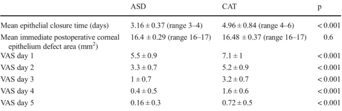
Results Mean corneal epithelium closure time was 3.16 ± 0.37 days (range 3 and 4 days) in ASD group and 4.96 ± 0.84 days in CAT group (range 4 and 6 days), and the difference was statistically significant (p < 0.001). VAS scores were significantly lower in ASD group than CAT group in the first 5 days after surgery. In 9 of 50 eyes, moderate conjunctival inflammation continued 1 month: 4 (16%) in ASD group and 5 (20%) in CAT group (p = 0.713). In total, pterygium recurrence was seen in 5 (10%) eyes: 2 eyes (8%) in ASD group and 3 eyes (12%) in CAT group (p = 0.637).
Conclusion ASD accelerated corneal epithelial healing following pterygium surgery. ASD group had lesser pain that was seem to be related with accelerated corneal epithelial healing.
Keywords Pterygium surgery . Corneal epithelial defect . Patient discomfort . Autologous serum drops
Introduction
Pterygium is a common ocular surface disorder characterized by a fibrovascular growth of the bulbar conjunctiva onto the cornea. The main treatment of pterygium is surgical excision. Surgery is usually performed under subconjunctival anesthesia [1]. Patients would experience pain and ocular discomfort after elimination of the effect of anesthesia (general or subconjunctival).
Non-steroidal anti-inflammatory drugs, topical anesthetics, and opioids have been used to relieve pain and discomfort after surgery [2–5]. Delayed epithelial healing was also asso-ciated with postoperative pain and discomfort [6]. Soft contact
lenses were used to accelerate corneal re-epithelization after ocular surgery [7].
Autologous serum has been used widely to treat dry eye disease and persistent epithelial defects [8,9]. Furthermore, autologous serum drops have been used to promote epithelial healing following penetrating keratoplasty, vitreoretinal sur-gery, and refractive surgery [10–12].
In the literature, there is no data that evaluates the effect of autologous serum after pterygium surgery on acceleration of epithelial healing and decreasing the patient pain and discom-fort after surgery. In this study, we aimed to determine the effect of autologous serum on epithelial healing and patient discomfort after pterygium surgery.
Materials and methods
Fifty eyes of 50 patients who underwent pterygium excision combined with conjunctival autograft were included in this prospective study. Written informed consent was obtained * Sabahattin Sul
1
Department of Ophthalmology, Muğla Sıtkı Koçman University, 48000 Muğla, Turkey
from all patients. The study was approved by the Ethics Committee of Muğla Sıtkı Koçman University Faculty of Medicine (registration no. 0806201711/1), and the tenets of Declaration of Helsinki were followed throughout the study.
Recorded information included preoperative and postopera-tive medical and ocular history, snellen visual acuity, applanation tonometry, slit-lamp examination, and fundoscopy. Anesthesia of the conjunctiva and cornea was induced by topical application of 0.5% alcaine and supplemented by 0.5 mL of jetocaine injected subconjunctivally with a 25-gauge needle, which was inserted into the body of the pteryg-ium. The head of the pterygium was dissected from the cornea with crescent knife. The body of the pterygium, including the adjacent Tenon’s capsule, and a 0.5- to 1-mm strip of border free of pterygium were excised from the surrounding conjunc-tiva with Westcott scissors, leaving a bare sclera. Minimal cautery was applied to bleeding vessels. Of the jetocaine, 0.5 mL was injected under superior conjunctiva to separate the conjunctiva from Tenon’s capsule. A conjunctival graft matching the bare sclera was harvested from the superior bul-bar conjunctiva to the limbus. The free graft was placed in the appropriate orientation onto the scleral bed and sutured with 10/0 nylon interrupted sutures sufficiently.
The patients who have been included in this study had no systemic (e.g., diabetes mellitus) or ocular diseases which can delay the corneal epithelium healing. In addition, patients who had similar pterygium size and postoperative corneal epitheli-um defect areas following surgery were included in this study. Of the patients, 25 were randomly assigned to receive 50% autologous serum drops (ASD group) and the remaining con-ventional artificial tears (CAT group) (carboxymethylcellu-lose 5%, Refresh tears®, Allergan) following surgery. The odd-numbered patients received ASD while even-numbered patients received CAT. Patients used 50% ASD or artificial tears eight times daily until corneal epithelium had completely epithelized. Patients in the ASD group were not allowed to use additional artificial tears. Corneal epithelium healing was followed with fluoresceine staining and anterior segment pho-tography. Each patient was treated with topical steroids (dexa-methasone 1%, Maxidex, Alcon Lab.) four times daily after surgery. The steroid treatment was tapered off and discontinued after 1 month. Topical antibiotics were used until the wound had completely epithelialized. All sutures were removed at the end of first week. Preoperative pterygium sizes, postoperative corneal epithelium defect areas, corneal epithelium healing speed, visual analog scale (VAS) for post-operative pain assessment, conjunctival inflammation, and re-currences were evaluated. Preoperative pterygium sizes and immediate postoperative corneal epithelium defect areas (fluoresceine stained areas) were marked in anterior segment photos by the first author and measured by an engineer by using the AutoCAD (AutoCAD 2013, Autodesk, CA,USA) program whether the groups were comparable. Ocular pain
was assessed using a VAS (0–10) as shown previously [13]. A score of 0 represented no pain and 10 represented worst pain. Conjunctival inflammation was defined as vascular in-jection and hyperemia in the graft and host-conjunctival area. Conjunctival inflammation was graded asBgrade 0 = no in-flammation, grade 1 = mild inin-flammation, grade 2 = moderate inflammation, and grade 3 = severe inflammation^ as previ-ously described by Kheirkah et al. [14]. Recurrence was de-fined as any fibrovascular growth onto the cornea beyond the limbus. All of the patients were examined daily at the same time (morning) for 1 week and 1, 2, 3, and 6 months after surgery and epithelial defects were marked by the same ex-aminer (first author) who was blinded to the treatment groups.
Preparation of autologous serum eye drop
A whole blood (20 mL) was taken from peripheral vein with vacutainer and allowed to clot at room temperature for 1 h. The blood was then centrifuged at 3000g for 10 min. Autologous serum was carefully prepared under laminar air-flow cabinet, and diluted with 1:2 with artificial tear (Refresh tears®) in a sterile bottle. Patients were asked to keep the bottles refrigerated at about 4 °C.
Statistical analysis was performed using SPSS 16.0 soft-ware (SPSS, Chicago, IL). The chi-squared (χ2) test was per-formed for categorical data and Mann-Whitney U test for con-tinuous data. A p value of less than 0.05 was considered to indicate statistical significance.
Results
The mean age was 48.9 ± 11.7 years in ASD group (range 29 and 65 years) and 50.2 ± 11.9 in CAT group (range 28 and 65 years). There were 11 (44%) men and 14 (56%) women in the ASD group and 13 (52%) men and 12 (48%) women in the CAT group. Mean pterygium size was similar between two groups before the surgeries [ASD 16.8 ± 0.33 mm2 (range 16.5–18), CAT 16.7 ± 0.46 mm2(range 16.3–18.2), p = 0.7]. Mean corneal epithelium closure time was significantly lower in ASD group than CAT group (Table 1). In ASD group, corneal epithelium was closed in 3 days in 21 (84%) eyes and 4 days in 4 (16%) eyes (Fig.1a–e). In CAT group, corneal
epithelium was closed in 4 days in 9 (36%) eyes, 5 days in 8 (32%) eyes, and 6 days in 8 (36%) eyes (Fig. 1f–k). Mean
immediate postoperative corneal epithelium defect areas were comparable between two groups [ASD group 16.4 ± 0.29 mm2(range 16–17) and CAT group 16.48 ± 0.37 mm2 (range 16–17), p = 0.6]. VAS scores were significantly lower in ASD group than CAT group in the first 5 days after surgery (Table1). In 9 of 50 eyes, a moderate conjunctival inflamma-tion continued for 1 month: 4 (16%) in ASD group and 5 (20%) in CAT group (p = 0.7). In total, pterygium recurrence
was seen in 5 (10%) eyes: 2 eyes (8%) in ASD group and 3 eyes (12%) in CAT group (p = 0.6).
Discussion
The conjunctival autograft technique has been used to treat primary pterygia successfully. However, recurrence of ium and ocular pain are the main concerns following pteryg-ium surgery. In the literature, the recurrence rate of this tech-nique is 2–39% [15,16]. Our overall recurrence rate was 10% and was consistent with the literature.
Ocular surface inflammation may persist and trigger the re-currence of pterygium [14]. ASD may worsen the ocular sur-face inflammation besides its beneficial effects on the healing of the ocular surface. Because ingredients of serum such as leuko-cytes, pro-inflammatory cytokines (IL-17 and IFN-γ), and ma-trix metalloproteinase may worsen the ocular surface inflam-mation [17–19]. However, there was no significant difference between two groups in terms of conjunctival inflammation
(both groups have similar rates of moderate conjunctival in-flammation) during the follow-up period in the current study.
Ocular pain originating from cornea or conjunctiva is an-other problem which limits the satisfaction of the patients following pterygium surgery. In a previous study, 60% of the patients suffered from significant pain following surgery [2]. Furthermore, Pastor-Vivas et al. have demonstrated that 58% of the patients in the immediate postoperative and 47% of the patients in the postoperative day 2 had moderate or severe pain [20]. Different managements have been used to reduce the pain following pterygium surgery. Topical anes-thetic drops can be administered; however, they have short acting duration and repeated administrations are required. Furthermore, topical anesthetics are toxic to corneal epitheli-um and delay the corneal wound closure [21]. Wishaw et al. used peribulbar morphine and showed satisfactory results in controlling postsurgical pain within 24 h [2]. But, systemic side effects such as nausea and vomiting and prolonged stay in the hospital were shown as the drawbacks of the opioid use in the ocular surgery [22]. Topical NSAIDs have been used to decrease pain following ocular surface surgeries [5,23]. They Table 1 Mean epithelial closure
time, mean immediate
postoperative corneal epithelium defect area, and visual analog scale of autologous serum drop and conventional artificial tear groups
ASD CAT p
Mean epithelial closure time (days) 3.16 ± 0.37 (range 3–4) 4.96 ± 0.84 (range 4–6) < 0.001 Mean immediate postoperative corneal
epithelium defect area (mm2) 16.4 ± 0.29 (range 16–17) 16.48 ± 0.37 (range 16–17) 0.6
VAS day 1 5.5 ± 0.9 7.1 ± 1 < 0.001
VAS day 2 3.3 ± 0.7 5.2 ± 0.9 < 0.001
VAS day 3 1 ± 0.7 3.2 ± 0.7 < 0.001
VAS day 4 0.4 ± 0.5 1.6 ± 0.6 < 0.001
VAS day 5 0.16 ± 0.3 0.72 ± 0.5 < 0.001
VAS visual analog scale, ASD autologous serum drop, CAT conventional artificial tear
Fig. 1 Anterior segment colored photograph of the two patients who was applied 50% autologous serum drop (ASD) or conventional artificial tears (CAT). a Immediate postoperative period of the patient who received 50% ASD. Corneal defect area was marked and measured as 16.5 mm2. b Postoperative day 1. Corneal epithelial defect area was 13 mm2. c Postoperative day 2. Corneal epithelial defect area was 7mm2. d Postoperative day 3. Corneal epithelial defect area was 3 mm2. e Postoperative day 4. Corneal epithelium was completely closed. f
Immediate postoperative period of the patient who received CAT. Corneal defect area was marked and measured as 17 mm2. Corneal
epi-thelium was completely closed at postoperative day 6. g Postoperative day 1. Corneal epithelial defect area was 11 mm2. h Postoperative day 2. Corneal epithelial defect area was 7 mm2. i Postoperative day 3. Corneal epithelial defect area was 5 mm2. j Postoperative day 4. Corneal epithelial defect area was 4 mm2. k Postoperative day 5. Corneal epithelial defect area was 1 mm2
have been found effective in reducing ocular pain; however, side effects such as superficial punctate keratitis, corneal infil-trates, and epithelial defects were reported [24]. Furthermore, corneal melting has been reported in eyes with dellen and tear film instability which may be encountered in patients with pterygium [24,25]. Soft contact lenses have also been used to accelerate corneal re-epithelization and reduce ocular pain following pterygium excision [9]. Dağcıoglu et al. have
con-cluded that patients who were applied soft contact lenses maintained their daily activities following pterygium surgery. However, in eyes without epithelial barrier, soft contact lenses may lead to bacterial keratitis owing to bacterial contamina-tion of the soft contact lenses which may be a potential limi-tation of this application [26].
ASD contains several components such as vitamin A, epi-dermal growth factor (EGF), fibronectin, and transforming growth factor-β (TGF-β) which are necessary for the prolif-eration, differentitation, and maturation of ocular surface epi-thelium [27–29]. Vitamin A is necessary for normal differen-tiation of squamous epithelium and prevents squamous meta-plasia [30]. EGF and fibronectin support proliferation and migration of corneal epithelial cells [27, 31]. TGF-β is in-volved epithelial cell migration and stromal repair process [32]. Substance P, NGF, and IGF-1 are neurotrophic factors that promote corneal epithelial wound healing [33, 34]. Additionally, serum contains anti-inflammatory cytokines such as interleukin (IL)-4, IL-10, and IL-1 receptor antagonist. IL-1 receptor antagonist has been shown to decrease signs of dry eyes in murine models [35]. Furthermore, ASD are pre-servative free and its osmolality and biomechanical properties are similar to natural tears [36]. ASDs have been used in the treatment of various ocular surface disorders such as dry eye and dry eye-related disorders such as Sjögren syndrome, per-sistent epithelial defects, recurrent corneal erosion, and epithe-lial defects following surgery such as vitreoretinal or refractive surgery [8–11]. However, there is no study about the use of ASD following pterygium excision in the literature. In pteryg-ium surgery, a corneal epithelial defect develops on and around the pterygium excision site. Faster corneal re-epithelization is important for corneal epithelial barrier devel-opment against bacterial infection as well as ocular pain that limits the patient satisfaction. In the current study, a prolonged corneal re-epithelization duration due to development of large corneal epithelial defects was the reason of 50% ASD concen-tration use. Although we cannot claim the superiority of 50% over 20% concentration of ASD (because we did not prepare 20% concentration and compare the outcomes), higher con-centrations have been demonstrated to have better effect on corneal healing process that led us to use 50% concentration of ASD [37]. One-hundred-percent concentration could be preferred instead of 50% ASD to promote faster healing ow-ing to higher concentrations of growth factors. However, 100% ASD might cause more ocular irritation than 50%
ASD due to alteration of tear osmolality and pH owing to higher serum protein concentration in patients who already have ocular irritation following surgery [37]. Furthermore, 100% ASD requires twice amount of blood to prepare the concentration and higher concentrations of TGF-β in 100% ASD might suppress corneal wound healing as reported pre-viously [28]. Therefore, we preferred to use 50% concentra-tion of ASD. Indeed, corneal re-epithelizaconcentra-tion was provided on 3 days in 84% of patients in ASD group. In CAT group, two thirds of the patients had complete corneal re-epithelization 5 and 6 days after surgery. The difference of mean corneal re-epithelization time between two groups was almost 2 days and seems to be clinically meaningful. Although we included the patients with no other systemic or local diseases, the difference of healing speed would be more in patients with diabetes or severe dry eye disease which may alter corneal epithelium healing [9,38,39]. Another benefit was seen on the VAS scores. In CAT group, patients suffered from moderate to severe pain (mean VAS score 7.1); however, in ASD group, patients have a moderate pain (mean VAS score 5.5) at postoperative day 1. Furthermore, ASD group had significantly lower VAS scores than CAT group in the remaining 4 days. Ocular pain originating from cornea or con-junctiva was thought as the result of surgical trauma, corneal epithelial defect, and suture irritation. Utilization of 50% AS in the early postoperative period may have reduced ocular pain with faster corneal re-epithelization. Furthermore, lower pain scores may be related to anti-inflammatory cytokines such as interleukin 10 which has been shown to have some-what analgesic effects in addition to anti-inflammatory effect [40]. In addition, although we did not estimate the viscosity of ASDs, ASDs may have yielded an extra soothing effect on suture irritation as stated by Jeng and Dupps [37].
ASD may have several adverse effects such as infection from contaminated drops, worsening of ocular inflammation, anemia from frequent blood draws, sticky sensation, allergic reaction, and self-resorbing subepithelial infiltrate [18]. However, we did not encounter any adverse effect due to ASD administration.
In conclusion, corneal epithelial defect and ocular pain are inevitable and disturbing results following pterygium surgery. ASDs seem to be a good alternative to other medications following pterygium surgery by accelerating corneal epitheli-um healing in addition to reduced postoperative ocular pain.
Compliance with ethical standards
Conflict of interest The authors declare that they have no conflict of interest.
Ethical approval All procedures performed in studies involving human participants were in accordance with the ethical standards of the institu-tional and/or nainstitu-tional research committee and with the 1964 Helsinki declaration and its later amendments or comparable ethical standards.
Informed consent Informed consent was obtained from all individual participants included in the study.
References
1. Akarsu C, Taner P, Ergin A (2003) Fluorouracil as chemoadjuvant for primary pterygium surgery preliminary report. Cornea 22(6): 522–526
2. Wishaw K, Billington D, O’Brien D, Davies P (2000) The use of orbital morphine for postoperative analgesia in pterygium surgery. Anaesth Intensive Care 28(1):43–45
3. Brilakis HS, Deutsch TA (2000) Topical tetracaine with bandage soft contact lens pain control after photorefractive keratectomy. J Refract Surg 16(4):444–447
4. Oksuz H, Tamer C (2006) Pain relief after pterygium surgery with viscous lidocain. Ophthalmologica 220(5):323–326
5. Ozcimen M, Sakarya Y, Goktas S et al (2015) Effect of nepafenac eye drops on pain associated with pterygium surgery. Eye Contact Lens 41(3):187–189
6. Shortt AJ, Allan BD, Evans JR (2013) Laser-assisted in-situ kerat-omileusis (LASIK) versus photorefractive keratectomy (PRK) for myopia Cochrane database. Syst rev 31(1):CD005135
7. Daglıoglu MC, Coşkun M, Ilhan N et al (2014) The effects of soft contact lens use on cornea and patient’s recoveryafter autograft pterygium surgery. Cont Lens Anterior Eye 37(3):175–177 8. Tsubota K, Goto E, Fujita H et al (1999) Treatment of dry eye by
autologous serum application in Sjogren's syndrome. Br J Ophthalmol 83(4):390–395
9. Tsubota K, Goto E, Shimmura S et al (1989) Treatment of persistent corneal epithelial defect by autologous serum application. Ophthalmology 106(10):1984–1989
10. Chen Y, Hu F, Huang J et al (2010) The effect of topical autologous serum on graft re-epithelialization after penetrating keratoplasty. Am J Ophthalmol 150(3):352–359
11. Schulze D, Sekundo W, Kroll P (2006) Autologous serum for the treatment of corneal epithelial abrasions in diabetic patients under-going vitrectomy. Am J Ophthalmol 142(2):207–211
12. Hondur AM, Akcam T, Karaca EE, Yazici Eroglu H, Aydin B (2016) Autologous serum eye drops accelerate epithelial healing after LASEK. Curr Eye Res 41(1):15–19
13. Barequet IS, Soriano ES, Green WR, O’Brien TP (1999) Provision of anaesthesia with a single application of lidocaine 2% gel. J Cataract Refract Surg 25:626–631
14. Kheirkhah A, Casas V, Sheha H, Raju VK, Tseng SC (2008) Role of conjunctival inflammation in surgical outcome after amniotic membrane transplantation with or without fibrin glue for pterygium. Cornea 27(1):56–63
15. Hirst LW (2003) The treatment of pterygium. Surv Ophthalmol 48(2):145–180
16. Ang LP, Chua JL, Tan DT (2007) Current concepts and techniques in pterygium treatment. Curr Opin Ophthalmol 18(4):308–313 17. Azari AA, Rapuano CJ (2015) Autologous serum eye drops for the
treatment of ocular surface disease. Eye Contact Lens 41(3):133–140 18. Welder JD, Bakhtiari P, Djalilian AR (2011) Limbitis secondary to autologous serum eye drops in a patient with atopic keratoconjunc-tivitis. Case Rep Ophthalmol Med 2011:576521
19. Pflugfelder SC, Corrales RM, de Paiva CS (2013) T helper cyto-kines in dry eye disease. Exp Eye Res 117:118–125.19
20. Pastor-Vivas AI, Alejandre-Alba N, García-Vega MI, Ariño-Gutiérrez M, García-Sandoval B, Jiménez-Alfaro I (2011) Quantifying and classifying postsurgical pain in pterygium surgery with conjunctival autografts. Arch Soc Esp Oftalmol 86(6):176–179
21. Peyman GA, Rahimy MH, Fernandes ML (1994) Effects of mor-phine on corneal sensitivity and epithelial wound healing: implica-tions for topical ophthalmic analgesia. Br J Ophthalmol 78(2):138– 141
22. Padda GS, Cruz OA, Krock JL (1997) Comparison of postoperative emesis, recovery profile and analgesia in pediatric strabismus re-pair. Ophthalmology 104(3):419–424
23. Caldwell M, Reilly C (2008) Effects of topical nepafenac on cor-neal epithelial healing time and postoperative pain after PRK: a bilateral, prospective, randomized, masked trial. J Refract Surg 24(4):377–382
24. Flach AJ (2000) Topically applied non-steroidal anti-inflammatory drugs and corneal problems: an interim review and comment. Ophthalmology 107(7):1224–1226
25. Machkie IA (1971) Localized corneal drying in association with dellen, pterygia and related lesions. Trans Ophthalmol Soc UK 91:129–145
26. Hondur A, Bilgihan K, Cirak MY, Dogan O, Erdinc A, Hasanreisoglu B (2008) Microbiologic study of soft contact lenses after laser subepithelial keratectomy for myopia. Eye Contact Lens 34(1):24–27
27. Ohashi Y, Motokura M, Kinoshita Y, Mano T, Watanebe H, Kinoshita S et al (1989) Presence of epidermal growth factor in human tears. Invest Ophthalmol Vis Sci 30(8):1879–1882 28. Gupta A, Monroy D, Ji Z, Yoshino K, Huang A, Pflugfelder SC
(1996) Transforming growth factor beta-1 and beta-2 in human tear fluid. Curr Eye Res 15(6):605–614
29. Nelson JD, Gordon JF (1992) Topical fibronectin in the treatment of keratoconjunctivitis sicca. Chiron Keratoconjunctivitis Sicca study group. Am J Ophthalmol 114(4):441–447
30. Sommer A (1983) Effects of vitamin a deficiency on the ocular surface. Ophthalmology 90(6):592–600
31. Nakamura M, Sato N, Chikama T, Hasegawa Y, Nishida T (1997) Fibronectin facilitates corneal epithelial wound healing in diabetic rats. Exp Eye Res 64(3):355–359
32. Kokawa N, Sotozono C, Nishida K, Kinoshita S (1996) High total TGF- 2 levels in normal human tears. Curr Eye Res 15(3):41–343 33. Kojima T, Higuchi A, Goto E, Matsumoto Y, Dogru M, Tsubota K
(2008) Autologous serum eye drops for the treatment of dry eye diseases. Cornea 27(Suppl 1):25–30
34. Lambiase A, Rama P, Bonini S, Caprioglio G, Aloe L (1998) Topical treatment with nerve growth factor for corneal neurotrophic ulcers. N Engl J Med 338(17):1174–1180
35. Okanobo A, Chauhan SK, Dastjerdi MH, Kodati S, Dana R (2012) Efficacy of topical blockade of interleukin-1 in experimental dry eye disease. Am J Ophthalmol 154(1):63–71
36. Liu L, Hartwig D, Harloff S, Herminghaus P, Wedel T, Geerling G (2005) An optimised protocol for the production of autolo-gous serum eyedrops. Graefes Arch Clin Exp Ophthalmol 243(7):706–714
37. Jeng BH, Dupps WJ (2009) Autologous serum 50% eyedrops in the treatment of persistent corneal epithelial defects. Cornea 28(10): 1104–1108
38. Schrader S, Wedel T, Moll R, Geerling G (2006) Combination of serum eye drops with hydrogel bandage contact lenses in the treat-ment of persistent epithelial defects. Graefes Arch Clin Exp Ophthalmol 244:1345–1349
39. Chikama T, Wakuta M, Liu Y, Nishida T (2007) deviated mecha-nism of wound healing in diabetic corneas. Cornea 26:S75–S81 40. Borghi SM, Pinho-Riberio FA, Zarpelon AC, Cunha TM,
Alves-Filho SH, Ferreria SH et al (2015) Interleukin-10 limits intense acute swimming-induced muscle mechanical hyperalgesia in mice. Exp Physiol 100(5):531–544