www.biodicon.com Biological Diversity and Conservation
ISSN 1308-8084 Online; ISSN 1308-5301 Print 12/2 (2019) 141-150
Research article/Araştırma makalesi DOI: 10.5505/biodicon.2019.92408 Comparative anatomical and morphological studies on six Muscari species (Asparagaceae)
Burcu YILMAZ ÇITAK *1, Hüseyin DURAL 1, Tuna UYSAL 1 ORCID: 0000-0003-3703-7731; 0000-0002-5579-5037; 0000-0001-9968-5633
1 University of Selçuk, Faculty of Science, Department of Biology, Konya, Turkey
Abstract
In this paper, the anatomical and morphological traits of six Muscari species were examined and the taxonomical relationships were assessed by assisting of these characters. Except from Muscari macrocarpum, the others are endemic species for our country and they could be counted as M. bourgaei, M. sandrasicum, M. racemosum, M. turcicum and M. vuralii. In scope of morphometric analyses, many numbers of morphologic and anatomic characters were used to determine the diagnostic of Muscari species and the created dendrogram gave very important information about them. Taxonomical obtained results supported the previous taxonomical assignments meaningfully in Flora of Turkey. The outcomes revealed that the taxa were similar in some aspects with such as sterile flowers, thick cuticle of scape and two rowed vascular bundles of scape. However, some features belonging to scape, leaves and flowers were primarily diagnostic characters among taxa, and they were founded having taxonomic value in the distinction of these taxa from each other.
Key words: Muscari macrocarpum, Muscari turcicum, Muscari vuralii, PRIMER7, Turkey --- ---
Altı Muscari türü (Asparagaceae) üzerine karşılaştırmalı anatomik ve morfolojik çalışmalar Özet
Bu çalışmada, altı Muscari türünün anatomik ve morfolojik özellikleri incelendi ve bu karakterlerin desteği ile taksonomik akraba ilişkileri tayin edildi. Muscari macrocarpum hariç, diğerleri ülkemiz için endemik olup, bu türler M. bourgaei, M. sandrasicum, M. racemosum, M. turcicum ve M. vuralii olarak sayılabilir. Morfometrik analizler kapsamında, çok sayıda morfolojik ve anatomik karakter kullanılarak Muscari türlerinin karakteristiğini belirlemek için oluşturulan dendogram bu karakterler hakkında çok önemli bilgiler verdi. Taksonomik olarak elde edilen sonuçlar, anlamlı olarak, Türkiye Florası’nda önceki taksonomik belirlemeleri destekledi. Sonuçlar, taksonların steril çiçekler, sapdaki kalın kutikula ve iki sıralı iletim demeti gibi özellikler bakımından benzer olduğunu gösterdi. Ancak, sap, yaprak ve çiçeklere ait bazı özelliklerin il olarak taksonlar arasında belirleyici karakterler olduğu ve bu taksonların birbirinden ayrılmasında taksonomik öneme sahip olduğu bulundu.
Anahtar kelimeler: Muscari macrocarpum, Muscari turcicum, Muscari vuralii, PRIMER7, Türkiye 1. Introduction
Garbari and Greuter (1970) had put forward an approach to split Muscari Miller genus by taxonomically. They divided Muscari into the genera Muscari, Leopoldia Parl., Muscarimia Kostel., and Pseudomuscari [1]. They used karyological data for this classification [2,3,4]. However this taxonomical assignment was not adopted by different taxonomists several times. As concerning with the evolution of Muscari some taxonomists postulated that there were transitions in character expression and so, they offered to treat only as subgenera like suggested before by Garbari and Greuter (1970) [1,5,6,7,8]. Then monophyly of Muscari was proved including plastid DNA sequences on the base of molecular study and was supported a broader genus concept [9]. As expressed before by Davis and Stuart (1980, 1984),
*Corresponding author / Haberleşmeden sorumlu yazar: Tel.: +903322231876; Fax.: +903322231876; E-mail: [email protected] © Copyright 2019 by Biological Diversity and Conservation - Available online at www.biodicon.com/Tüm hakları saklıdır BioDiCon. 811-0219
Muscari is a taxonomically difficult genus with a formidable burden of synonymy, much of it based on cultivated materials of unknown origin [5,7]. Many species belonging to this genus are used as ornamental plants in parks and gardens [10]. Besides, Muscari includes excellent bulbous plants which are generally called grape hyacinths and they deserve a greater breeding effort because of their excellent horticultural characteristic [11]. On the other hand, a number of polyphenolic compounds, which have pharmacological importance because of their antimutagenic effects, have been isolated from some Muscari species [12].
Muscari was revised in Turkey and 20 species were identified from Turkey [6]. In addition to these, 14 new species have been described from Turkey between years of 1984-2015 [13]. With the addition of these species, total number of Muscari species which are determined from Turkey is reached to 34. Twenty two of them are endemic species for Turkey and endemism ratio of genus is 64.7 % [14].
In the present study, the endemic species M. bourgaei Baker, M. sandrasicum Karlén, M. racemosum Mill. and M. turcicum Uysal, Ertuğrul & Dural and a non-endemic species M. macrocarpum Sweet were used for anatomical studies. Although some anatomical investigations about Muscari were performed in recent years [15,16,17,18]. These studies are still very limited for Turkish Muscari endemics. By this paper, we aimed to reveal the anatomical and morphological properties of five Muscari species and to discuss the taxonomic importance of these characters.
2. Materials and methods
2.1. Plant materials
The specimens belonging to Muscari genus were collected from different regions of Turkey in 2012-2015 (except from M. racemosum). The selected excellent specimens (in their maturing time) were put in 70% alcohol for anatomical studies. The localities of the species are given detailed in Table 1. The specimens were dried according to standard herbarium technique and stored at the herbarium of KNYA.
Table 1. The localities of the studied taxa
Taxa Locality and collection date Collector number
M.bourgaei (MB) Turkey: Antalya- İbradı, Sülek Plateau, Abies opens, 1465 m, 16 April 2015.
T.Uysal—3219—H.Dural M. sandrasicum (MS) Turkey: Muğla- Köyceğiz, Ağla Village, Sandras mountain,
Akkulak region, 1550 m, 31 May 2012.
T.Uysal—2739—H.Dural M. macrocarpum
(MM)
Turkey: Burdur- Yeşilova, 70-80 km from Yeşilova to Denizli, Köpekçayı Village, Pinus forest opens, on serpentine oddment, 1200 m, 31 May 2012.
T.Uysal—2635—H.Dural
M. racemosum (MR) Turkey: Konya- Taşkent, Cırlasun region, rocky-stony opens, 1250 m, 19 May 2000.
T.Uysal—1 M. turcicum (MT)
M. vuralii (MV)
Turkey: Konya- Bozkır, Tufan Stream, Avdan Plateau, 1900 m, 18 May 2012.
Turkey: Karaman- Sariveliler, Atalanı region, stony places, steppe, 1950 m, 15 April 2012.
T.Uysal—2694— H.Dural—B.Çıtak O.Tugay-6649-T. Uysal
2.2. Anatomical method
Anatomical researches were performed with 15 samples, on average, of each taxon. Twenty slides were prepared for each sections of studied taxa. The cross sections were stained with floroglusin-HCL [19]. In order to determine cell surface characteristics, thirty different measurements were made with Kameram software programme. All sections were examined directly through a Leica DM-1000 light microscope and were photographed using Canon EOS 450 D camera which was attached to the light microscope.
2.3. Numerical and morphometric data
The determined qualitative and quantitative characters were scored for numerical and morphometric analysis. Sixty six characters were used to evaluate the taxonomical relationships of six Muscari species. Fifty five of them were morphological and remaining anatomical ones but we reflected the diagnosable ones in Table 2. And then, the determined qualitative and quantative characters were turned to a data matrix. Morphometric data were analysed using multivariate techniques with the PRIMER7 software package [20]. Data were not transformed. A Bray–Curtis similarity matrix was used to generate a two-dimensional ordination plot applying non-metric multidimensional scaling (nMDS) [21]. The similarity percentages (SIMPER) procedure was used to determine similarities among taxa, and to identify the major morphological traits contributing to the differences among them [21].
Table 2. The diagnostic morphological and anatomical characters used in data matrix *(See Table 1 for acronyms)
Characters MR MM MB MT MS MV
Corona
presence/absence Presence Presence Absence Absence Absence Absence
Color of fertile
flowers White Yellow Purple White Purple Blue
Color of lobe of
fertile flowers Brown Brown Purple White White White
Color of sterile
flowers White Purple Purple Cream Purple Purple
Arrangement of
stamens Subbiseriate Biseriate Subuniseriate Subbiseriate Biseriate Subbiseriate
Color of stamen Shiny Matt Shiny Shiny Shiny Matt
Color of filament Yellow White Purple White Purple Purple
Type of filament Winged Not winged Not winged Winged Winged Not winged
Shape of raceme Normally Tightly Imbricately Tightly Normally Loosely
Length of raceme
(cm) 3.7 3 3 1 2 1
Color of scape Green Green Green Purple lined Purple lined Purple lined
Length of scape (cm) 15 18 8 2 10 5 Number of leaves 5 5 5 4 5 3 Length of leaves (cm) 25 30 8 5 10 10 Width of leaves (mm) 8 15 3 2 2.7 3.2
Epidermis in scape
Square-rectangular Square-oval Rectangular Isodiametric
Rectangular-oval
Square-rectangular Rows of cortex in
scape 6-7 rowed 5-6 rowed 6-7 rowed 4-5 rowed 6-7 rowed 6-10 rowed
Rows of
sclerenchyma in scape
4-5 layered 2-3 layered 3-4 layered 5-6 layered 4-5 layered 3-4 layered Number of vascular
bundles in scape 25-30 35-40 13-15 15-20 13-15 Not recorded
Type of mesophyll Only spongy parechyma Both palisade and spongy parenchyma Only spongy parechyma Both palisade and spongy parenchyma Both palisade and spongy parenchyma Both palisade and spongy parenchyma Presence/absence of
lacuna in leaves Presence Absence Absence Absence Absence Absence
Numbers of
vascular bundles in leaves
One rowded One rowded Two rowded Two rowded One rowded One rowded
Presence/absence of
crystal in leaves Presence Presence Presence Absence Absence Absence
Stomata index in upper leaves 26.9 26.6 22.5 30 30.4 29.2 Stomata index in lower leaves 33.3 40 33.6 34.6 30 34.6 Type of stomata in lower surface sections
Singly Singly Singly and
twin Singly Singly
Singly, twin and triplet
3. Results
3.1. Scape anatomy
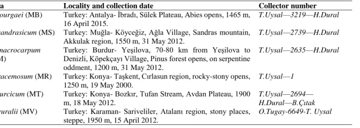
The cross section of scape is circular and a single layered epidermis which encloses the scapes. Towards to inner from epidermis, there is seen cortex parenchyma with oval-shaped cells that varies among 4-10 rows own to species. Sclerenchyma layer continuously follows cortex towards the centre and it has 3-5 layers varying as particular to species. The vascular bundles of outer circles are smaller than inner ones. Phloem and xylem elements of vascular tissue can be separated easily. There are thickness with spirals and annular rings in xylem and also phloem tissue contains sieve and companion cells. Vascular cylinder has a great deal of vascular bundles and they are collateral type (Figure 1). The number of bundles in vascular cylinder shows differences between species (Table 2). The pith consists of oval shaped parenchymatous cells. The epidermis has a few anomocytic stomata excluding M. bourgaei. The stomata cells are localized together with other epidermal cells (mesomorphic type). It never contains trichomes.
Figure 1. Cross section of scapes. A-M. bourgaei B-M. sandrasicum C-M. racemosum D- M. macrocarpum E-M. turcicum. e- epidermis, co- cortex, sc-sclerenchyma, vb-vascular bundle, p-pith (scale bar: 200 µm)
3.2. Leaf anatomy
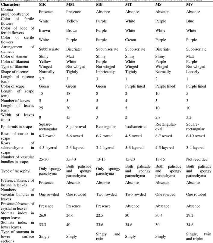
3.2.1. Mesophyll anatomy: The epidermis, single layered and made of cells thickened outer walls, consisting of quadrate, ovoid, and rectangular cells is place at both surfaces of the leaves. Both surfaces are covered by a thick cuticle layer in all species (Figure 2). The size of epidermal cells in the cross section is not significantly different among the species. As it is seen in M. turcicum and M. macrocarpum (Figure 2-e,d), adaxial parts of leaves are seen sharply undulated in M. sandrasicum and M. racemosum (Figure 2-b,c). Contrast to these, adaxial part of M. bourgaei is smooth. The leaves mesophyll is composed of different parenchymatic cells. M. bourgaei and M. racemosum have only spongy parenchyma in mesophyll (unifacial type) (Figure 2-a,c). The other species have palisade and spongy parenchyma (equifacial type) (Figure 2-b,d,e). Palisade parenchyma is only two layered in M. sandrasicum (Figure 2-b). Lacunae are present in M. racemosum. Also, the crystals such as raphide are determined in M. bourgaei, M. racemosum and M. macrocarpum. While vascular bundles are two rowed in M. bourgaei and M. turcicum, they are one-rowed in the remaining species.
Figure 2. The cross sections of examined taxa of Muscari. A-M. bourgaei B-M. sandrasicum C-M. racemosum D- M. macrocarpum E-M. turcicum. ue- upper epidermis, pp-palisade parenchyma, sp- spongy parenchyma, le-lower epidermis, vb- vascular bundle, cu-cuticle, cr- crystal, la- lacunae, st-stomata (bar: 100 µm)
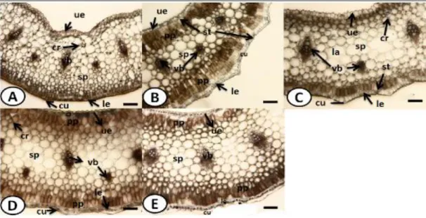
3.2.2. The characteristics of surface sections: Leaf surface of all studied species are amphistomatic. The stomata are anomocytic type. The stomata cell has usually a row with the epidermis on the both surface. In other words, they are described as mesomorphic type. The walls of upper and lower epidermal cells are smooth in all species (Figs. 3, 4). In M. bourgaei, the epidermal cells on upper surface are 190.9±39.1 µm long and 18.8±3.0 µm wide; the lower ones are 178.9±39.5 × 12.6±3.2 µm. The number of stomata on the lower surface in the same species is 7±1 per 30 µm2, and epidermal cells are 24±2 per 30 µm2. , The number of stomata on the upper surface is 6±1 per 30 µm2, and the epidermal cells are 12±2 per 30 µm2. For the upper and the lower surface, the stomatal index was calculated as 22.5; 33.6, respectively.
In M. sandrasicum, the upper epidermal cells are 186.6±66.8 µm long and 23.0±3.2 µm wide; the lower ones are 188.9±12.3 µm long and 20.6±2.6 µm wide. The number of stomata on the lower surface, is 7±1 per 30 µm2, and the number of epidermal cells are 16±2 per 30 µm2. The number of stomata on the upper surface is 6±1 per 30 µm2, and the number of epidermal cells are 14±2 per 30 µm2. For the upper and the lower surface, the stomatal index was calculated as 30.4; 30, respectively.
In M. racemosum, the upper epidermal cells are 206.3±37.9 µm long and 21.3±3.6 µm wide; the lower ones are 192.6±37.2 µm long and 16.3±3.2 µm wide. The number of stomata on the lower surface is 7±2 per 30 µm2, and the number of epidermal cells are 19±1 per 30 µm2. The number of stomata on the upper surface is 6±2 per 30 µm2, and the number of epidermal cells are 12±1 per 30 µm2. For the upper and the lower surface, the stomatal index was calculated as 26.9; 33.3, respectively.
In M. macrocarpum, the upper epidermal cells are 179.2±49.8 µm long and 28.4±2.8 µm wide; the lower ones are 186.4±48.3 µm long and 24.9±4.6 µm wide. The number of stomata on the lower surface is 4±2 per 30 µm2, and the number of epidermal cells are 11±1 per 30 µm2. The number of stomata on the upper surface is 6±2 per 30 µm2, and the number of epidermal cells are 9±1 per 30 µm2. For the upper and the lower surface, the stomatal index was calculated as 26.6; 40, respectively.
In M. turcicum, the upper epidermal cells are 228.8±50.3 µm long and 16.7±1.6 µm wide; the lower ones are 172.1±44.7 µm long and 21.4±3.2 µm wide. The number of stomata on the lower surface is 6±2 per 30 µm2, and the number of epidermal cells are 14±1 per 30 µm2. The number of stomata on the upper surface is 9±1 per 30 µm2, and the number of epidermal cells are 17±2 per 30 µm2. For the upper and the lower surface, the stomatal index was calculated as 30; 34.6, respectively.
Figure 3. The upper surface of transverse section of leaves of examined taxa. A-M. bourgaei B-M. sandrasicum C-M. racemosum D- M. macrocarpum E-M. turcicum. st-stomata, ec- epidermis cell (bar: 25 µm)
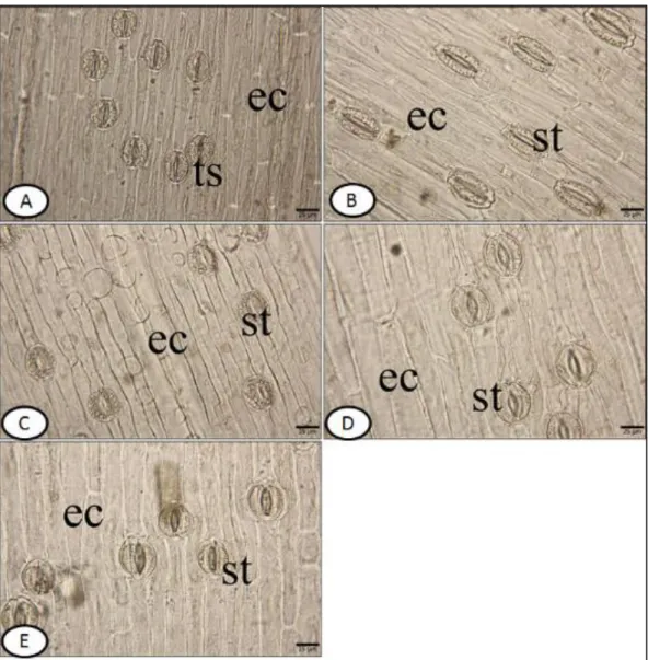
Figure 4. Lower surface of transverse section of leaves. A-M. bourgaei B-M. sandrasicum C-M. racemosum D- M. macrocarpum E-M. turcicum. ts- twin stomata, st-stomata, ec- epidermis cell (bar: 25 µm)
3.3. Numerical and Morphometric analyses
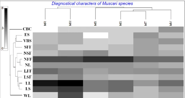
The numerical and morphometric analyses carried out that the studied Muscari species are clearly different in taxonomically and the relationships are exactly correlated with the previous taxonomical assignments [6,22,23,24]. According to information coming from dissimilarities of nonmetric, the most different species is M. racemosum and the species takes position in near of M. macrocarpum (Figure 6). Additionally, the most different two species are M. vuralii and M. racemosum, respectively (dissimilarity between them 31%). Besides, the less differentiated species are seen as M. turcicum and M. sandrasicum. The obtaining dendogram showed that the Muscari species are fairly similar in terms of morphological and anatomical characters (Figure 5). When we take a glance to dendogram, the species are mainly separated to 2 main clades. The first clade consists of M. macrocarpum and M. racemosum and so they are known taxonomically very close species. At the second clade, four species take position together and similarities among them are fairly high (the least 74%). In this clade, the closest two species are M. turcicum and M. sandrasicum with similarity 83%. As related to these species, M. vuralii is seen very close with similarity 80%. Finally, M. bourgaei was joined externally with lower similarity, 74%. We reduced the numerical characters to twelve in order to find the best diagnostical features via programme PRIMER7 and so, it was determined that four of them (number of fertile flower and leaf, length of scape and leaf) was more discriminative (Figure 5). Furthermore, it was seen that M. racemosum and M. macrocarpum were closer comparison to remaining to each other in terms of length of leaves. After than these characters, sterile and fertile flowers could be assessed as secondary important that all of these were used before classical taxonomy of Muscari [6]. On the other hand, the dendrogram showed that the anatomical characters, the number of vascular bundles and epidermis in scape, are less diagnosable to morphological ones discussed above.
Figure 5. A dendrogram showing taxonomic relationships of Muscari species based on morphological and anatomical data. (CBC: color of bulb coat, ES: epidermis of scape, VBS: Vascular bundles in scape, SFF: Shape of fertile flowers, NSF: Number of sterile flowers, NFF: Number of fertile flowers, NL: Number of leaves, LFF: Length of fertile flowers, LSF: Length of sterile flowers, LL: Length of leaves, LS: Length of scape, WL: Width of leaves)
Figure 6. The non-metric multi-plot analysis of Muscari species .
4. Conclusions and discussion
The obtained findings and our comparisons with literature revealed that there was a respectable correlation between anatomy and morphology of the studied species in terms of their taxonomy [25]. However, the results showed that morphologic characters are more distinctive comparing to anatomical features in diagnosis of the Muscari species (Figure 5). When considered the basic anatomical features of Muscari genus, we confirmed with previous reports that Muscari species had the general scape characteristics like a uniform layer occurring from epidermis, cortex, sclerenchyma [16,17,18,26). Besides this, we determined that the number of cortex cells, sclerenchyma layers and vascular bundles in the studied species could be different. As mentioned before in different reports the number of cortex layers is mostly variable and it could be display some differences as specifically own to species [16,17,18]. While cortex are absolutely identical in M. bourgaei and M. sandrasicum (6-7 rows)(Figure 1-a,b), sclerenchyma shows differences in rows (Table 2).
According to current literature anatomical differences comprising the mesophylls tissue within Asparagaceae family are reported in many times by different papers but it is not usually specified whether these are important aspects of systematic until soon [17,27,28,29,30,31,32,33,34]. A short time ago, we reported that mesophylls are very determinative to separate some Ornithogalum L. species taxonomically [35]. Again, we declared by this paper that mesophyll had got very high potential in point of classification of the Muscari species. From our findings, we can say that mesophylls consist of very different cell types and row of vascular bundles which have got a taxonomical importance. Although M. bourgaei and M. racemosum have unifacial mesophylls remaining species have equifacial one. Although this feature has been a taxonomical importance to separate taxa in level of species, it does not include any meaning to determine their taxonomical position in level section or subgenera. Because, the species positioned in different sections of Muscari could be display similar mesophyll features. Additionally, we determined that the rows of vascular bundles changes constantly ranged from 1 to 2 in the studied Muscari taxa. M. bourgaei and M. turcicum have two rows in mesophyll; the other three species have one row. Our findings is correlated with previous reports and the number of rows of vascular bundles shows a respectable variation in Muscari, they could be a taxonomical characters to distinguish species [17,27,28,32,35,36,37]. Generally, stomatas are ranged singly in members of Asparagaceae family [17,27,29,30,38]. It is determined that the studied species have generally single stomata type but M. bourgaei rarely displays different stomata feature which is characterized by the presence of twins at the low surface of the leaves. As correlated with our results, it is reported that M. vuralii has twin and triplet stomatas at the low surface of the leaves [17]. Unlike we couldn’t observe any twin and triplet stomata in the remaining Muscari species. As a general contribution about this subject, we think that stomata number and arrays might be changed as dependent on ecological and climatic factors. So, we predicted that these characters would not have any taxonomical values in limitation of Muscari species. Calcium oxalate crystals, either raphides or styloids, are present in the root cortex of most Asparagales. Raphides are common in Asparagales and many other monocotyledons, so their absence from some taxa is often of systematic significance [39]. We found that three of the investigated species (M. bourgaei, M. racemosum and M. macrocarpum) have raphides.
According to previous reports, some members of Asparagaceae family have variable lacunars having different diameters [15,16,18,29,33,34,35,37,40]. On the other hand, we observed lacunars only in M. racemosum. Therefore, we satisfied that lacunars would be used for separation of the some Muscari species as regards to its presence or absence. These intercellular spaces originate rhexigenously, as has been observed in some species of the Liliaceae s. l. by Fuchsig (1911) [41].
The dendrogram formed based on anatomical and morphological characters discriminated the six Muscari species from each other. Morphometrically analysis were used to solve taxonomical problems in very broad intervals and taxonomical levels, like Asparagaceae family which merged with Liliaceae based on PCA analysis or the other families such as Asteraceae, Rosaceae [42,43,44]. Non-metric analysis were performed and its results are congruent with similar researches [45].
As a general conclusion, the differences between the anatomical structure of vegetative organs of species such as the number of cortex parenchyma layer and of vascular bundles in vascular cylinder, mesophyll type in leaf, stomata type could be very useful distinguishing Muscari taxa. Besides these, the most discriminative for their taxonomy are morphologic characters which are such as fertile flowers, length of leaves and scape.
Acknowledgements
We would like to thank Dr. Osman Tugay and Dr. Hakkı Demirelma for their field work. References
[1] Garbari, F., Greuter, W. (1970). The taxonomy and typification of Muscari Miller (Liliaceae) and allied genera, and on the typification of generic names. Taxon 19, 329–335.
[2] Garbari, F. (1966). Contributo allo studio citologico dei Muscari Italiani. Caryologia 19, 419–428.
[3] Garbari, F. (1968). Il genere Muscari (Liliaceae): contributo alla revisone citotassonomica. Giornale Botanico Italiano 102, 87–105.
[4] Garbari, F. (1969). Nuove osservazioni citologiche sui generi Muscari Leopoldia. Giornale Botanico Italiano 103, 1–9.
[5] Davis, P.H., Stuart, D.C. Muscari Miller. (1980). In Tutin, T.G., Heywood, V.H., Burges, N.A., Moore, D.M., Valentine, D.H., Walters, S.M., & Webb, D.A. (Eds.), Flora Europaea 5. Cambridge University Press, London, pp. 46–49.
[6] Davis, P.H. (1984) Muscari Mill. In Davis, P.H. (Ed.) Flora of Turkey and the east Aegean Islands 8. Edinburgh University Press, Edinburgh, pp. 245–265.
[7] Speta, F. (1982). Uber die Abgrenzung und Gliederung der Gattung Muscari und uber ihre Beziehungen zu anderen Vertretern der Hyacinthaceae. Botanische Jahrbücher fur Systematik 103, 247–291.
[8] Speta, F. (1989). Muscari (subg. Leopoldia) mirum Speta, spec. nov., im Kreise seiner nachsten Verwandten. Phyton, 29, 105–117.
[9] Pfosser, M., Speta, F. (1999). Phylogenetics of Hyacinthaceae based on plastid DNA sequences. Annals of the Missouri Botanical Garden 86, 852–875.
[10] Tanker, N., Koyuncu, M., Coskun, M. (1998). Farmasötik Botanik, Ankara: Ankara Universitesi Eczacılık Fakültesi Yayınları, Ders Kitapları.
[11] Nakano, M., Tanaka, S., Kagami, S., Saito, H. (2005). Plantlet regeneration from protoplast of Muscari armeniacum Leichtl. ex Bak. Plant Biotechnology Journal 22, 249–251. https://doi 10.5511/plantbiotechnology.22.249.
[12] Miadokova, E., Masterova, I., Vlckova, V., Duhova, V., Toth, J. (2002). Antimutagenic potential of homoisoflavonoids from Muscari racemosum. Journal of Ethnopharmacology 81, 381–386.
[13] Yildirim, H. (2015). Muscari atillae (Asparagaceae): a new species from Eastern Anatolia, Turkey. Phytotaxa 213, 291–295. http://dx.doi.org/10.11646/phytotaxa.213.3.9
[14] Eker, I. Muscari, Güner, A., Aslan, S., Ekim, T., Vural, M., Babaç, M.T. (2012). Türkiye Bitkileri Listesi (Damarlı Bitkiler), İstanbul: Nezahat Gökyiğit Botanik Bahçesi ve Flora Araştırmaları Derneği yayını.
[15] Herrmann, N., Weiss, G., Durka, W. (2006). Biological flora of Central Europe: Muscari tenuiflorum Tausch. Flora 201, 81–101. doi:10.1016/j.flora.2005.03.002.
[16] Gursoy, M., Şık, L. (2010). Comparative anatomical studies on Muscari armeniacum Leichtlin ex Baker and Muscari neglectum Guss. in West Anatolia. Celal Bayar University Journal of Science 6, 61–72.
[17] Doğu, S., Dinc, M., Ayval, U. (2011). Anatomical characteristics of Bellevalia mathewii Özhatay & Koçak (Liliaceae). Biological Diversity and Conservation, 4, 14–18.
[18] Sezer, O., Ozgisi, K., Yaylaci, O.K., Koyuncu, O. (2013). Some morpho-anatomical studies on rare endemic Muscari sivrihisardaghlarensis. Biological Diversity and Conservation 6, 26–33.
[19] Vardar, Y. (1987). Botanikte Preparasyon Tekniği, Ege Üniversitesi Fen Fakültesi Basımevi.
[20] Clarke, K.R., Warwick, R.M. (1994). Change in marine communities: An approch to statical analysis and interpretation, Swindon, Natural Environment Research Council.
[21] Clarke, K.R., (1993). Non-parametric multivariate analyses of changes in community structure. Australian Journal of Ecology 18, 117–143. https://doi.org/10.1111/j.1442-9993.1993.tb00438.x.
[22] Karlén, T. (1987). Muscari sandrasicum (Liliaceae), a new species from Turkey. Willdenowia 16, 375–382. [23] Uysal, T., Ertuğrul, K., Dural, H., Kucukoduk, M. (2007). Muscari turcicum (Liliaceae/Hyacinthaceae), a new
species from south Anatolia, Turkey. Botanical Journal of Linnean Society 154, 233–236. https://doi.org/10.1111/j.1095-8339.2007.00646.x.
[24] Dogu, S., Bagci, Y. (2009). Muscari vuralii sp nov (Liliaceae/Hyacinthaceae) from South Anatolia, Turkey. Nordic Journal of Botany 27, 243–246. https://doi.org/10.1111/j.1756-1051.2009.00427.x.
[25] Dogu, S., Dinc, M. (2013). Anatomical characteristics of Muscari vuralii Y. Bagcı & Doğu (Hyacinthaceae). Modern Phytomorphology 3, 29–33.
[26] Cutter, E.G. (1971). Plant anatomy: Experiment and interpretation, Part 2, Organs. Addison-Wesley Publishing Company.
[27] Lynch, A.H., Rudall, P.J., Cutler, D.F. (2006). Leaf anatomy and systematics of Hyacinthaceae. Kew Bulletin 61, 145–159.
[28] Peruzzi, L., Caparelli, K.F., Cesca, G. (2007). Contribution to the systematic knowledge of the genus Ornithogalum L. (Hyacinthaceae): morpho-anatomical variability of the leaves among different taxa. Bocconea 21, 257–265.
[29] Kahraman, A., Celep, F., Dogan, M., Koyuncu, M. (2010). Morpho-anatomical studies on Bellevalia paradoxa Boiss. belonging to Liliaceae. Australian Journal of Crop Science 4, 150–154.
[30] Soykan, A., Meric, C. (2012). Morphological and anatomical studies of Tulipa orphanidea (Liliaceae). Phytologia Balcanica 18, 43–48.
[31] Nawaz, T., Hameed, M., Waqar-U-Nisa, Ahmad, M.S.A., Younis, A., Kanwal, H. (2012). Comparative Anatomy of Root and Stem of Some Native and Exotic Asparagus L. Species. Pakistan Journal of Botany 44, 153–158. [32] Raycheva, T., Stojanov, K. (2013). Comparative Anatomical Study of Five Species of Genus Asparagus in
[33] Tekin, M., Meric, C. (2013). Morphological and anatomical investigations on endemic Hyacinthella acutiloba in Turkey. Biological Diversity and Conservation 6, 161–168.
[34] Akyol, Y., Yetisen, K. and Ozdemir, C. (2014). The Morphological and Anatomical Studies on Fritillaria caucasica J.F. Adam (Liliaceae). Süleyman Demirel Üniversitesi Fen Edebiyat Fakültesi Fen Dergisi 9, 12–19, [35] Citak, B.Y., Dural, H., Buyukkartal, H.N. and Pinar, N.M. (2015). Morphological, anatomical, palynological, and
micromorphological characters of 2 endemic species of Ornithogalum (O. chetikianum and O. demirizianum) in Turkey. Turkish Journal of Botany 39(1), 48–59. doi:10.3906/bot-1401-35
[36] Selvi, S., Erdogan, E., Daskin, R. (2008). Morphological, anatomical and ecological studies on Hyacinthella lineata (Liliaceae). Ekoloji 17, 24–32.
[37] Yetisen, K., Ozdemir, C., Kucukoduk, M., Akyol, Y. (2012). A morphological and anatomical study of Hyacinthella glabrescens (Liliaceae). Phytologia Balcanica 18, 319–322.
[38] Guvenc, A., Coskun, M., Arıhan, O. (2011). Anatomical structure of cladodes of Ruscus L. taxa (Liliaceae) in Turkey. FABAD Journal of Pharmaceutical Science 36, 119–128.
[39] Kauff, F., Rudall, P.J., Conran, J.C. (2000). Systematic root anatomy of Asparagales and other monocotyledons. Plant Systematic and Evolution 223, 139–154.
[40] Jafari, A., Maassoumi, A.A., Bemani, M. (2008). A Biosystematical Investigation on Muscari species in Iran. Asian Journal of Plant Sciences 7, 228–232.
[41] Fuchsig, H. (1911). Vergleichende Anatomie der Vegetationsorgane der Lilioideen, Wien, Austria.
[42] Coşkunçelebi, K., Kandemir, A. and Beyazoğlu, O. (2002). Numerical taxonomic study on Ornithogalum (Liliaceae) in Black Sea region of Turkey. Biologia 57, 449–454.
[43] Makbul, S., Türkmen, Z., Coskuncelebi, K., Beyazoğlu, O. (2010). A Morfometric study on Scorzonera L. taxa (Asteraceae) from norteast Anatolia. Acta Botanica Croatia 69, 237–247.
[44] Cheikh-Affene, Z.B., Haouala, F. and Harzallah-Skhiri, F., (2015). Morphometric variation and taxonomic identification of thirteen wild rose populations from Tunisia. Acta Botanica Croatia 74, 1–17. https://doi.org/10.1515/botcro-2015-0008.
[45] Hilpold, A., López-Alvarado, J., Garcia-Jacas, N., Farris, E. (2015). On the identity of Centaurea population on Procida Island, Italy: Centaurea corensis rediscovered. Plant Biosystems 149, 1025–1035. doi: 10.1080/11263504.2014.983578