Contents lists available at
ScienceDirect
Food and Chemical Toxicology
journal homepage:
www.elsevier.com/locate/foodchemtox
Bioactive compounds in seaweeds: An overview of their biological
properties and safety
Kannan RR. Rengasamy
a,∗, Mohamad Fawzi Mahomoodally
b,∗∗,
Muhammad Zakariyyah Aumeeruddy
b, Gokhan Zengin
c, Jianbo Xiao
d, Doo Hwan Kim
aaDepartment of Bio-resources and Food Science, Konkuk University, Seoul, 05029, South Korea bDepartment of Health Sciences, Faculty of Science, University of Mauritius, Réduit, Mauritius cDepartment of Biology, Science Faculty, Selcuk University, Campus, Konya, Turkey
dInternational Research Center for Food Nutrition and Safety, Jiangsu University, Zhenjiang 212013, China
A R T I C L E I N F O Keywords: Marine foods Pharmacology Biopharmaceutical Biomedicine Metabolites Non-communicable A B S T R A C T
Seaweeds are among the significant currently exploited marine plant resources which are gaining full applica-tions in culinary, cosmetic, pharmaceutical, and biotechnological processes. Much attention has been devoted to seaweeds based on their proven health benefits and is considered as a rich source of structurally different bioactive metabolites for the discovery of novel functional food-based pharmacophores/drugs. Nonetheless, there is still a dearth of updated compilation and analysis of the in-depth pharmacological activities of these compounds. This review, therefore, aims to provide a piece of up-to-date detailed information on the major compounds isolated from various seaweed species together with their in-vitro and in-vivo biological properties. These compounds were found to possess broad pharmacological properties and inhibitory enzyme activities against critical enzymes involved in the aetiology of noncommunicable diseases. However, their toxicity, clinical efficacy, mechanisms of action, and interaction with conventional foods, are still less explored and require more attention in future studies.
1. Introduction
The importance of dietary food habits was already mentioned in the
earlier quote by Hippocrates in 460 BC that “Let food be thy medicine and
medicine be thy food’’. The improper dietary habits or unhealthy diet is a
critical concern for the currently alarming health disorders including
diabetes, obesity, cancer, and cardiovascular diseases. The intake of
unhealthy foods and the low intake of fruits and vegetables causes
about 2.7 million deaths including 14% of gastrointestinal cancer
deaths (14%), ischaemic heart disease deaths (11%) and nearly stroke
deaths (9%) (WHO Fact Sheet, accessed on June 19, 2018,
http://www.
who.int/dietphysicalactivity/fruit/en/index2.html). It is well known
that vegetables and fruits are important sources of phytocompounds
which perform a key role in the prevention of a panoply of diseases. The
global phytonutrients/nutraceutical market value, regarding value, is
projected to reach $4.63 Billion in 2020, at a CAGR of 7.2% from 2015
to 2020 (Markets and Markets, 2015). Likewise, the global market for
the botanical and plant-derived drug was estimated $29.4 billion in
2017 and is projected to escalate to 39.6 billion in 2022 with a
compound annual growth rate (CAGR) of 6.1% (Research, 2017). For
the past decades, researchers have shown great interest towards the
isolation of health-promoting substances from these fruits and
vege-tables. Both the Natural Health Products (NHP) and Nutraceuticals and
Functional Foods (NFF) focused much research on bioactive foods from
natural resources to develop healthy foods (Goldberg, 2012;
Nice,
1997).
Marine life offers 70% of earth's surface with the vast diversity of
life and biodiversity in the seas is only partially explored although
marine represents a rich source of novel metabolites with various
ap-plications includes cosmeceutical, nutraceuticals, agrochemicals,
pharmaceuticals and other industrially relevant chemicals (Faulkner,
2012). Recent research emphasises that drug discovery from marine
resources is increasing alarmingly and various biomolecules are in the
clinical pipeline.
2. Global trends in marine biodiscovery
In 2018, the world market for drugs derived from marine sources
https://doi.org/10.1016/j.fct.2019.111013
Received 27 September 2019; Received in revised form 20 November 2019; Accepted 29 November 2019
∗Corresponding author.
∗∗Corresponding author.
E-mail addresses:[email protected](K.R. Rengasamy),[email protected](M.F. Mahomoodally).
Food and Chemical Toxicology 135 (2020) 111013
Available online 30 November 2019
0278-6915/ © 2019 Elsevier Ltd. All rights reserved.
Table 1 Seaweed polysaccharides and their biological properties. Polysaccharide Source Biological properties Model used Findings Reference Fucoidan (1 ) Ascophyllum nodosum, Cladosiphon okamuranus, Fucus spiralis, F. distichus, F. evanescens, F. vesiculosus, F. serratus, Laminaria digitata, L. saccharina Anticoagulant, anti-inflammatory, antiadhesive and antiangiogenic In-vitro and in-vivo Inhibits the leucocyte recruitment in an inflammation model in rats. In In-vitro, P-selectin-mediated neutrophil adhesion to platelets showed that only fucoidans from A. nodosum, F. distichus, F. evanescens, F. serratus, F. spiralis, L. digitata and L. saccharina could serve as P-selectin inhibitors. Besides, all fucoidans, except that from C. okamuranus, displayed anticoagulant activity by APTT while fucoidans from F. distichus, F. evanescens, F. serratus, L. digitata and L. saccharina, showed strong antithrombin action in the platelet aggregation assay. These fucoidans also inhibited HUVEC tubulogenesis. Lastly, fucoidans from F. distichus, and F. vesiculosus, F. serratus, L. digitata and L. saccharina, blocked MDA-MB-231 breast carcinoma cell adhesion to platelets. Cumashi et al. (2007) Kelp Tyrosinase inhibition In-vitro Showed competitive inhibition of tyrosinase toward L-tyrosine (IC50 = 0.82 mg/mL), and the inhibitory constant Ki obtained from double-reciprocal plots was 0.99 mg/mL. Yu and Sun (2014) Fucus vesiculosus Anti-atopic dermatitis In vivo Ameliorated atopic dermatitis, accompanied by the decreased inflammatory cell infiltration, splenocytes proliferation, and CD4 +T cell response Tian et al. (2019) F. evanescens Antitumor In-vivo Administration of fucoidan at 10 mg/kg, displayed moderate antimetastatic and antitumor activities. It also potentiates the antimetastatic effects of cyclophosphamide in C57Bl/6 mice with transplanted Lewis lung adenocarcinoma. Alekseyenko et al. (2007) Sargassum fusiforme Anti-angiogenic In-vitro Inhibits the migration of HMEC-1 and tube formation dose-dependently. Cong et al. (2016) S. fusiforme Anti-cancer In-vitro and In-vivo Inhibited lung cancer cell growth through the disruption of angiogenesis via blocking VEGFR2/Erk/VEGF signalling and targeting VEGFR2/VEGF. Chen et al. (2016) Sargassum hornery, Eclonia cava, Costaria costata Anti-cancer In-vitro Blocks colony formation in colon cancer cells cell line and human melanoma. Ermakova et al. (2011) Cladosiphon okamuranus Anti-cancer In-vitro and In-vivo In ATL patients, fucoidan inhibits the growth of HTLV-1-infected T-cell lines and peripheral blood mononuclear cells. Haneji et al. (2005) Adenocystis utricularis Antiretroviral In-vitro Inhibitor of anti-HIV-1 activity against both drug-resistant and wild-type HIV-1 strains by blocking of viral entry and revealed no virucidal activity. Trinchero et al. (2009) A. utricularis Antiviral In-vitro Galactofucans potentially inhibited HSV 1 and 2, without any cytotoxic effect, while the uronofucoidans displayed no antiviral activity. Ponce et al. (2003) C. okamuranus Cardioprotective In-vivo Cardioprotective effect was noticed with Fucoidan against isoproterenol-induced myocardial infarction in rats. Fucoidan also improved lactate dehydrogenase, creatinine phosphokinase, aspartate transaminase and alanine transaminase. Also, fucoidan enhanced the antioxidant defence system in treated rats by reducing oxidative stress induced by isoproterenol. Moreover, fucoidan treatment reverses the effects of isoproterenol by decreasing total cholesterol, triglycerides, LDL cholesterol and increasing HDL cholesterol. Thomes et al. (2010) C. okamuranus Anti-proliferative In-vitro Oversulfated fucoidan dose-dependently reduced the U937 cell proliferation, induces the apoptosis by an activation-dependent pathway of caspase-3 and -7. On the other hand, the weak activity of native fucoidan suggests that the sulfate group substitution and sulfate content influence the anti-proliferative activity in U937 cells. Teruya et al. (2007) C. okamuranus Gastric protection In-vitro Protect the gastric mucus layer and stimulate ulcer healing power owing to its anti-peptic and basic fibroblast growth factor (bFGF) stabilising activity. Shibata et al. (2000) C. okamuranus Antiprion In-vivo Dietary fucoidan, administered orally for six days after infection, delays the disease onset thoroughly in infected mice with scrapie, but not when given before the infection. Doh-ura et al. (2007) F. evanescens Anticoagulant In-vitro & In-vivo Showed anticoagulant activity through plasma antithrombin III mediated. Thrombin inhibition. Kuznetsova et al. (2003) F. vesiculosus Anti-inflammatory In-vitro Fucoidan inhibits the excess PGE2 and NO production in LPS-stimulated BV2 microglia. Also diminished the iNOS, MCP-1, COX-2, MCP-1, TNF-α and L-1β Park et al. (2011a) (continued on next page )
Table 1 (continued ) Polysaccharide Source Biological properties Model used Findings Reference expression. Besides, fucoidan suppresses the NF-κB activation and down-regulation of an extracellular JNK, MAPK, ERK and AKT pathways. F. vesiculosus Anti-obesity In-vitro Fucoidan reduced lipid accumulation by stimulating lipolysis via increasing HSL and by expression of phosphorylated HSL and reduction of glucose uptake into adipocytes. Park et al. (2011b) Undaria piaantifida Immunostimulatory In-vitro Fucoidan enhanced the probiotic properties of lactic acid bacteria on immune functions by enhancing the production of IL-12 in response to a strain of LAB, Tetragenococcus halophilus KK221, disseminating production of IFN-γ. In in-vivo study with ovalbumin immunized mice, the enhanced immunobalance of T helper type 1/type 2 (Th1/Th2) was observed. Kawashima et al. (2012) L. japonica Antioxidant In-vitro Exhibited scavenging effects on hypochlorous acid and superoxide radical and inhibition of LDL oxidation induced by Cu 2+. ( Zhao et al., 2005 ) L. japonica Anti-inflammatory In-vitro & In-vivo In an In-vivo air pouch inflammation model, coadministration of fucoidan or Cistanche tubulosa extract synergistically suppressed nitric oxide production, carrageenan-induced vascular exudation, prostaglandin E2 concentrations. Kyung et al. (2012) Lessonia vadosa Anticoagulant and elicitor In-vitro Native fucoidan showed good anticoagulant activity and activation of defence enzyme activities of PAL, LOX and GST in tobacco plants. Chandía and Matsuhiro (2008) U. pinnatifida Antiplasmodial In-vitro & In-vivo Fucoidan fractions inhibited the P. falciparum merozoites mediated erythrocytes invasion and IC50 values againt chloroquine sensitive P. falciparum 3D7 stain for the three fucoidan fractions were 9.17, 7.28, and 1.95 μg/ml and 7.03, 4.74, and 2.21 μg/ml in chloroquine resistant P. falciparum K1 strain. About 37% suppressive effect against the control group and a delay in death associated with anemia was observed in P. berghei-infected mice with fucoidan. Chen et al. (2009) U. pinnatifida Anti-allergy In-vivo The suppressive effect of Th2 cytokines production in bronchoalveolar lavage fluid wa snoticed, and IFN-γ amount was not increased in fucoidan treated mice. Maruyama et al. (2005) U. pinnatifida Antitumor In-vitro & In-vivo Fucoidan mediated tumor destruction via the response of Th1 and NK cells. Maruyama et al. (2006) U. pinnatifida Antitumor In-vitro Displayed antitumor activity against PC-3, HeLa, A549, and HepG2 cells. Synytsya et al. (2010) Alginate (2 ) Commercial sodium alginate Inhibition of putrefactive compound In-vitro & In-vivo In human fecal culture and rat cecum, inhibited the putrefactive compound formation. Kuda et al. (2005) Eucheuma cottonii and Sargassum polycystum Antidiabetic In-vitro IC50 of (0.075–0.103) mg/ml, also a mixed-type inhibition. Zaharudin et al. (2018) Commercial sodium alginate Antibacterial In-vitro & In-vivo Approximately 70–90% inhibition againt L. monocytogenes V. parahaemolyticus and S. typhimurium to human enterocyte-like HT-29-Luc cells was observed with sodium alginate (0.1%). In addition, sodium alginate potentially inhibited 70% of S. typhimurium invasion. Incubation with sodium alginate for 18 h also Increased transepithelial electrical resistance of HT-29-Luc monolayer cells was also observed with 18 h incubation of sodium alginate. Moreover, decreased liver pathogen count was noticed in alginate fed mice. Kuda et al. (2015) Commercial sodium alginate Antibacterial In-vivo Alginate-based coating containing lactate and diacetate was effective in controling The controlled growth of L. monocytogenes and enhanced microbial safety of sliced and filleted smoked salmon was reported with alginate coated lactate and diacetate. Neetoo et al. (2010) Commercial sodium alginate Antibacterial In-vivo Alginate-based antimicrobial coatings enhanced the microbiological safety of poached and deli turkey products by controling L. monocytogenes growth. Juck et al. (2010) – Gastroesophageal reflux disease treatment Systematic review & meta- analysis Effective in the treatment of symptomatic gastroesophageal reflux disease and were superior to placebo and antacids. Compared to proton pump inhibitors or histamine-2 receptor antagonists, alginates appear less effective. Leiman et al. (2017) Commercial sodium alginate Anticancer In-vitro Markeb et al. (2016) (continued on next page )
Table 1 (continued ) Polysaccharide Source Biological properties Model used Findings Reference The novel paclitaxel-loaded alginate nanoparticle promoted decreased viability, cell-cycle arrest and induced apoptosis in patient's breast cancer cells superior to those of paclitaxel alone. Commercial sodium alginate Anti-inflammatory In-vivo Amelioration of mRNA expression in inflammation-related molecules and protected indomethacin-induced mucin depletion in the small intestine was reported in mice pretreated with sodium alginate prior to the administration of indomethacin. Horibe et al. (2016) Commercial sodium alginate Anti-inflammatory In-vivo Prevention of methotrexate-induced small intestinal mucositis and decreased hemoglobin, hematocrit levels and red blood cell counts in rats. Yamamoto et al. (2013) Commercial sodium alginate Anti-inflammatory In-vivo Sodium alginate prevented the increase in SOD, GPx, catalase activity and microvascular permeability and also prevented decreases in white and red blood cells in small intestinal damage induced by indomethacin. Yamamoto et al. (2014) Commercial Antioxidant In-vitro Low molecular weight alginates produced by thermal treatment of alginate polymer showed scavenging activity against ABTS and superoxide radicals. Kelishomi et al. (2016) L. hyperborean Wound healing In-vivo Calcium alginate enhanced skin collagen Iexpression from day 3 to day 14 with higher collagen I/III in alginate-group ratio than vaseline (control)-group at day 7 and 14. In addition, higher level of hydroxyproline in skin homogenate of alginate-group than the vaseline (control)-group from day 3 to day 14. Wang et al. (2015) Laminarin (3 ) Laminaria spp. Anti-inflammatory In-vivo Combined laminarin and fucoidan treatment enhanced diarrhoeal scores, body-weight loss, and clinical variables linked with a dextran sodium sulfate experiment in pigs, together with s decrease in colonic IL-6 mRNA abundance. O'Shea et al. (2016) Eisenia bicyclis Antibacterial In-vitro Laminarin (0.1%) inhibited the adhesion About 70–90% inhibition of L. monocytogenes, S. typhimurium and V. parahaemolyticus adhesion to human enterocyte-like HT-29-Luc cells with laminarin (0.1%). Kuda et al. (2015) Commercial Hepatoprotective In-vivo The increase in serum ALT, AST and LDH activities -reflecting hepatic alterations -was reduced after lipopolysaccharides injection in laminarin-treated rats than control groups. Laminarin also decreased serum monocytes number, TNF-α and nitrite. Neyrinck et al. (2007) NI Immunostimulatory In-vitro Showed immunostimulatory effect through the transcription factor pathway in macrophages by increasing the release of H2 O2 ,NO, calcium, MCP-1, LIF, VEGF, and G-CSF with enhancing expression of STAT1, STAT3, c-Fos, c-Jun, and COX-2 mRNA in RAW 264.7 cells. Lee et al. (2012a) Commercial Anticancer In-vitro Through mitochondrial pathway, induces the apoptosis of human colon cancer LOVO cells. Ji et al. (2012) Commercial Anticancer In-vitro Induce apoptosis of LoVo cells. The TRAIL, DR4, DR5, Bid, tBid, FADD and Bax expression levels were upregulated, while the Bcl-2, pro-caspase-3 and 8, expression levels were downregulated. Moreover, the casapse-8, -3, -6 and -7 activities were increased. Ji and Ji (2014) E. bicyclis Anticancer In-vitro The colony formation of human melanoma SK-MEL-28 and colon cancer DLD-1 cells were inhibited by laminarin and its enzymatic hydrolysed products. Menshova et al. (2014) Laminaria digitata Anticancer In-vitro In HT-29 colon cancer cells, laminaric induces apoptosis through ErbB signaling pathway. Park et al. (2013) Carrageenan (4 )‘ Commercial Anti-inflammatory In-vitro Carrageenan did not induce IL-6, IL-8, or MCP-1 (CCL2) in HT-29 and HCT-8 cell lines at 0.1, 1.0, and 10.0 mg/mL. McKim et al. (2016) Commercial Anticancer In-vitro & In-vivo In B16–F10 and 4T1 bearing mice, carrageenan inhibited tumor growth in and enhanced immune response by increasing the number of tumor-infiltrating dentritic cells, M1 macrophages, and additional stimulated CD4 +CD8 +T lymphocytes in spleen. Luo et al. (2015) Commercial Anti-allergic In-vivo λ-Carrageenan was identified as potentially new ligand for TLR4/MyD88 which triggers innate immunity, induced Th1-cytokines, PRRs recognised λ-carrageenan, and suppression of IgE production via reduced histamine release. Tsuji et al. (2003) (continued on next page )
was around $ 10,486.8 billion, which is forecasted to touch $21,955.6
billion by 2025 at a CAGR of 11.25% for the five-year period of
2019–2025 (Infinium Global Research, 2019). Marine-based US FDA
approved drugs mainly consist of marine metabolites or their synthetic
analogues. Also, most of the marine-derived medicines isolated from
various marine resources, but the contribution of marine algae is only
about 30% (Blunt et al., 2007).
3. Recent developments in algal drug research
Seaweeds or marine macroalgae reside in the littoral zone and are
now considered as primary resources of the oceans in terms of
eco-nomic and ecological significance (Dhargalkar and Pereira, 2005).
Taxonomically, seaweeds are grouped into three major phyla: (i)
Phaeophyceae (brown algae), which are primarily brown in color due
to its fucoxanthin content – xanthophyll pigment fucoxanthin (ii)
Chlorophyceae (green algae) - primarily dominated by chlorophyll ‘a’
and ‘b’, and other specific xanthophyll pigments; and (iii)
Rhodophy-ceae (red algae) primarily comprised of phycocyanin and phycoerythrin
(O’Sullivan et al., 2010). Approximately more than 1500 brown, 900
green and 4000 red seaweeds are available worldwide (Dawes, 1998).
The subtropical and tropical waters are entirely occupied by red and
green seaweeds, while cold temperate waters are predominantly
occu-pied by brown seaweeds (Khan and Satam, 2003).
The interest in the discovery of health-promoting substances of
marine origin is increasing especially from marine plants such as
sea-weeds, seagrass and mangroves for the past decades (El Gamal, 2010;
Rengasamy et al., 2014b). Among these, seaweeds or marine
macro-algae have much attracted functional food researchers. Recent shreds of
evidence suggest that seaweeds have been regularly consumed as food
in East Asian countries including Korea, China, Japan, and this dietary
habit as widespread throughout Europe, North America and, Southern
American countries (McHugh, 2003). Noticeably, long-life expectancy
and the lower rate of cardiovascular diseases among the Japanese
people are likely to be associated with their dietary habits including
their regular consumption of seaweeds (Shimazu et al., 2007).
The recent review by
Rengasamy et al. (2014a)
compiled various
bioactive compounds isolated from seaweeds and their role as enzyme
inhibitors to treat multiple diseases including cancer, diabetes,
in-flammation, dementia and others. Seaweeds are not only targeted for
drug development to treat various human health illness, but also play a
significant part as plant growth regulators, fungicides, pesticides, and in
part in plant growth such as auxin, cytokinin, gibberellins, betains
(Stirk et al., 2014), oligosaccharides, and phenolic compounds
(Rengasamy et al., 2014b,
2015). The biological properties of various
bioactive metabolites from marine algae have been recently extensively
reviewed by many researchers including enzyme inhibitors (Rengasamy
et al., 2014a), phlorotannins (Karadeniz and Kim, 2015;
Sanjeewa
et al., 2016) polysaccharides (Wang et al., 2014), protein hydrolysates
and bioactive peptides (Harnedy and FitzGerald, 2013;
Samarakoon
and Jeon, 2012), alkaloids (Güven et al., 2010), halogenated terpenoids
(Wang et al., 2013) and pigments (D’Orazio et al., 2012;
Dumay et al.,
2015;
Kim and Pangestuti, 2011). In this context, this review mainly
focuses on the bioactive compounds isolated from seaweeds and their
in-depth biological properties (see detailed activities in
Tables 1–6
and
Fig. 5).
4. The primary bioactive compound in seaweeds
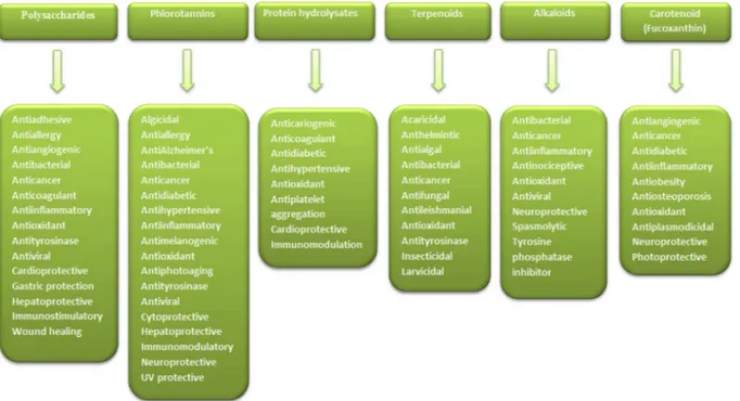
4.1. Polysaccharides
Polysaccharides are carbohydrate biopolymers consisting of simple
sugars linked by glycoside bonds and are classified into structural
polysaccharides, mucopolysaccharides and storage polysaccharides.
The study of the structure, biosynthesis and functions of sugar
mole-cules including polysaccharides are known as glycobiology and has
Table 1 (continued ) Polysaccharide Source Biological properties Model used Findings Reference Chondrus ocellatus Antitumor In-vitro & In-vivo From the histopathological in λ-carrageenan-treated mice indicated that λ-carrageenan was the causative agent for tumor cell pycnosis and necrosis in different degree. In H22 and S180 tumor cells, λ-carrageenan showed antitumor activity in-vitro. ( Zhou et al., 2004 ) C. ocellatus Antitumor In-vivo λ-carrageenan enhances antitumor activities of Fluorouracil (5-Fu) and progress the immunocompetence damaged by 5-Fu. Zhou et al. (2006) Gigartinaceae and Tichocarpaceae algae Antioxidant In-vitro & ex vivo Exhibited reducing power and inhibition of hydroxyl radicals and superoxide anion radicals. Sokolova et al. (2011) Gigartina skottsbergii Anti-viral In-vitro Reduced infectivity of the viruses BoHV-1 strain Cooper and SuHV-1 strain Bartha; this effect was more pronounced against BoHV-1. Diogo et al. (2015) Stenogramme interrupta Anti-viral In-vitro Showed anti-viral activity against herpes simplex virus Cáceres et al. (2000) A549, alveolar carcinoma; AKZ; ALT, alanine aminotransferase; APTT, activated partial thromboplastin time; AST, Aspartate transaminase; ATL, Adult T-cell leukemia; BoHV-1, bovine herpesvirus type 1; ERK, signal-regulated kinase; G-CSF, Granulocyte colony-stimulating factor; GPx, glutathione peroxidase; GST, glutathione-S-transferase; HeLa, cervical cancer; HepG2, hepatocellular carcinoma; HIV, human immune deficiency virus; HMEC-1, human microvascular endothelial cells; HSL, hormone sensitive lipase; HSV, herps simples virus; HTLV-1, human T-cell leukemia virus type 1; HUVEC, human umbilical vein endothelial cell; JNK, c-Jun N-terminal kinase; LDH, lactate dehydrogenase; LDL, low density lipoprotein; LIF, Leukemia Inhibitory Factor; LOX, lipoxygenase; MAPK, p38 mitogen-activated protein kinase; MCP-1, monocyte chemoattractant protein-1; NK cells, nature killer cells; PAL, phenylalanine-ammonia lyase; PC-3, prostate cancer 3; PRRs, pattern recognition receptors; STAT, Signal Transducer and Activator of Transcription; SuHV-1, suid herpesvirus type 1; VEGF, Vascular endothelial growth factor.
Table 2 Seaweed phlorotannins and their biological properties. Phlorotannins Source Biological activity Model used Findings Reference Phloroglucinol (5) E. cava Antioxidant In-vitro Showed scavenging effects against free radicals: DPPH, HO• and O2 •− and protect against H2 O2 -mediated DNA damage. Ahn et al. (2007) E. maxima Antioxidant In-vitro Potent DPPH radical scavenger at 0.13 μM Rengasamy et al. (2013) E. maxima Anti-Alzheimer's In-vitro Inhibits acetylcholinesterase (AChE) at 50% at a concentration of 579.32 μM. Kannan et al. (2013) E. maxima Antidiabetic In-vitro Inhibited 50% of α-glucosidase at a concentration of 1991 μM. Rengasamy et al. (2013) E. kurome Antibacterial In-vitro Displayed an MBC (Minimum bactericidal concentration) value of 0.79 of μmol/ mL against Campylobacter jejuni. Nagayama et al. (2002) -Anti-cancer In vitro Phloroglucinol engineered Ag nanoparticles displayed cytotoxic effect and morphological features of apoptotic cell death in MCF-7 cell lines Kumar et al. (2018) Dibenzo [1,4] dioxine-2,4,7,9-tetraol (6) E. maxima Antioxidant In-vitro Displayed an EC50 value of 0.01 μM against DPPH radical. Rengasamy et al. (2013) E. maxima Anti-Alzheimer's In-vitro Caused a 50% inhibition of AChE at a concentration of 84.48 μM. Kannan et al. (2013) E. maxima Antidiabetic In-vitro Exhibited α-glucosidase inhibition with an IC50 value of 33.69 μM. Rengasamy et al. (2013) Eckol (7) E. cava Anti-influenza In-vitro Showed inhibitory effects on Influenza Virus NA (rvH1N1) with IC50 value of 89.5 μM and A/Chicken/Korea/MS96/96 (H9N2, with IC50 value of 152.1 μM. Ryu et al. (2011) E. cava Skin protective In vitro Inhibition of PM 2.5 -induced cell apoptosis by eckol was through MAPK signaling pathway Zhen et al. (2019) E. stolonifera Anti-tyrosinase In vitro Reduced cellular melanin content and tyrosinase activity, Also downregulated melanogenesis enzymes expression such as tyrosinase, tyrosinase-related protein (TRP)-1, and TRP-2 in B16F10 melanoma cells Manandhar et al. (2019) E. bicyclis , E. cava and E. kurome Antioxidant In-vitro Exerted scavenging effect against DPPH and superoxide anion with EC50 value of 26 μM and 107 μM, respectively. Shibata et al. (2008) E. stolonifera Antioxidant In-vitro Exerted DPPH scavenging effect (EC 50 = 10.6 μM) and also inhibited ROS production in tacrine-treated HepG2 cells. Lee et al. (2012b) E. maxima Antioxidant In-vitro Potent DPPH radical scavenger at 0.01 μM EC50 value. Rengasamy et al. (2013) E. cava Antibacterial In-vitro Displayed antibacterial activity against Staphylococcus aureus at MIC values varying from 125 to 250 μg/mL and against Salmonella strains at MIC values of 125–250 μg/mL. The combinations of eckol and ampicillin exhibited a synergistic or additive effect. Choi et al. (2010) E. kurome Antibacterial In-vitro Showed bactericidal activity against S. aureus ,Bacillus cereus ,Escherichia coli ,C. jejuni ,S. typhimurium ,S. enteritidis ,and Vibrio parahaemolyticus with MBC values in the range 0.08–1.08 μmol/mL. Nagayama et al. (2002) E. stolonifera Anti-hypertension In-vitro Exhibited marked inhibitory activity against ACE with an IC50 value of 70.82 μM. Jung et al. (2006) E. stolonifera Hepatoprotective In-vitro In tacrine-treated HepG2 cells, eckol potentially inhibits the Fas-mediated cell-death protein expression and also inhibit the cytochrome c release from the mitochondria to cytosol. Lee et al. (2012b) E. maxima Anti-Alzheimer's In-vitro Caused a 50% inhibition of AChE at a concentration of 76.70 μM. Kannan et al. (2013) E. stolonifera Anti-photoaging In-vitro Caused a reduced expression of MMP-1 human dermal fibroblasts by inhibiting AP-1 dependent reporter gene activity and NF-κB. Joe et al. (2006) E. cava Anti-photoaging In-vitro Showed photoprotective effect against UV-B -induced cell damage. Heo and Jeon (2009b) E. bicyclis Anti-diabetic In-vitro Exhibited α-amylase inhibition (87.5% inhibition) and antiglycation activity (96.2% inhibition) at 1 mM. Okada et al. (2004) E. maxima Anti-diabetic In-vitro Displayed α-glucosidase inhibition at 11.163 μM IC50 value. Rengasamy et al. (2013) Dieckol (8) E. cava Immunomodulatory In-vivo The ionising radiation suppressed immune cell differentiation and proliferation was enhanced. Dieckol increased thymidine incorporation by splenocytes as much as 8.8-fold above that in irradiated mice without dieckol treatment. Also, the number of CD4 +helper T cells, CD8 +cytolytic T cells, CD45R/B220 +pan B cells, and CD11b +macrophages showed a marked increase in dieckol-treated irradiation group compared with irradiation-only control group at three days after irradiation. Park et al. (2010) E. stolonifera Anti-photoaging In-vitro Caused a reduced expression of MMP-1 human dermal fibroblasts by inhibiting AP-1 dependent reporter gene activity and NF-κB. Joe et al. (2006) E. cava Anti-photoaging In-vitro Displayed photoprotective effect against UV-B -induced cell damage. Heo et al. (2009) E. cava Anti-allergy In-vitro Le et al. (2009) (continued on next page )
Table 2 (continued ) Phlorotannins Source Biological activity Model used Findings Reference Histamine release was inhibited dose-dependently from both KU812 and RBL2H3 cell lines. E. stolonifera Antibacterial In-vitro Exhibited antibacterial activity against MSSA and MSRA S. aureus with MIC values ranged from 32 to 128 μg/mL. The combination of dieckol with ampicillin or penicillin displayed a synergistic activity against MRSA. Lee et al. (2008) E. bicyclis Anti-diabetic In-vitro Exhibited inhibitory activity on glycation (86.7% inhibition) and α-amylase (97.5% inhibition) at 1 mM. Okada et al. (2004) E. cava Anti-diabetic In-vitro Showed inhibitory activity against α-glucosidase (IC50 = 10.8 μmol/L) and α-amylase (IC 50 = 124.9 μmol/L). It also displayed a non-competitive type of inhibition against α-glucosidase. Lee et al. (2009) E. cava Matrix metalloproteinases inhibition In-vitro In human osteosarcoma cell, dieckol Inhibits the mRNA gene and protein levels of iNOS, COX-2, MMP-1, MMP-3, and MMP-13. Dieckol also inhibits the JNK and p38 MAPK phosphorylation. Ryu et al. (2009) E. cava Anti-inflammatory In-vivo The PGE 2 ,NO, and HMGB-1 production significantly inhibited in the serum of mice with LPS-induced septic shock. Yang et al. (2016) E. cava Cytoprotective Ex vivo In neonatal mouse cochlea, dieckol showed a dose dependent partial protective effect against gentamicin-induced hair cell. Chang et al. (2016) E. bicyclis ,E. cava and E. kurome Antioxidant In-vitro Exerted scavenging effect against DPPH and superoxide anion with EC50 value of 13 μM and 7.6 μM, respectively. Shibata et al. (2008) 2-Phloroeckol (9) E. stolonifera Hepatoprotective In-vitro In tacrine-treated HepG2 cells, eckol potentially inhibits the Fas-mediated cell-death protein expression and also inhibit the cytochrome c release from the mitochondria to cytosol. Lee et al. (2012b) E. stolonifera Antioxidant In-vitro Exerted DPPH scavenging effect (EC 50 = 35.2 μM) and also inhibited ROS production in tacrine-treated HepG2 cells. Lee et al. (2012b) E. stolonifera Anti-inflammatory In-vitro Showed inhibition of NO production (EC 50 = 85.3 μmol/L) in LPS-stimulated RAW 264.7 cells. Wei et al. (2016) Dioxinodehydroeckol (10) E. Cava Anti-cancer In-vitro In MCF-7 human breast cancer cells, Dioxinodehydroeckol significantly induced proliferative inhibition and apoptosis. Kong et al. (2009) E. stolonifera Antioxidant In-vitro Showed DPPH scavenging activity (EC 50 = 8.8 μM) which is more effective than L-ascorbic acid (EC 50 = 10.3 μM). Kim et al. (2009) E. cava UV protective In-vitro Protects the human keratinocyte cells from UVB-induced apoptosis. Ryu et al. (2015) 6,6′ Bieckol (11) E. cava Anti-HIV In-vitro Displayed inhibition against lytic effects, HIV-1 induced syncytia formation and viral p24 antigen production at EC50 values of 1.23, 1.72 and 1.26 μM respectively. Also, selective inhibition against HIV-1 RT enzyme (EC 50 = 1.07 μM). Artan et al. (2008) E. cava Anti-inflammatory In-vitro Through negative regulation of the NF-κB pathway, 6,6′ Bieckol down-regulated COX-2, iNOS, and pro inflammatory cytokines in LPS-stimulated macrophages. Yang et al. (2012) E. stolonifera Anti-inflammatory In-vitro Showed inhibition of NO production (EC 50 = 63.9 μmol/L) in LPS-stimulated RAW 264.7 cells. Wei et al. (2016) E. stolonifera Anti-inflammatory In-vitro Down-regulated NF-κB activation in LPS-stimulated microglial cells through JNK, p38 MAPK and Akt. Kim et al. (2016) E. cava Anti-allergy In-vitro Histamine release was inhibited dose-dependently from both KU812 and RBL2H3 cell lines. Le et al. (2009) 8,8′-Bieckol (12) E. cava Anti-HIV In-vitro Displayed HIV-1 RT and protease inhibition at IC50 = 0.51 and 81.5 μM respectively. Ahn et al. (2004) E. kurome Antibacterial In-vitro Showed bactericidal activity against S. aureus ,Bacillus cereus ,Escherichia coli ,C. jejuni ,S. typhimurium ,S. enteritidis ,and Vibrio parahaemolyticus with MBC values in the range 0.03–0.54 μmol/mL. Nagayama et al. (2002) E. bicyclis ,E. cava and E. kurome Antioxidant In-vitro Exerted scavenging effect against DPPH and superoxide anion with EC50 value of 15 μM and 6.5 μM, respectively. Shibata et al. (2008) 7-Phloroeckol (13) E. cava Anti-influenza In-vitro Exhibited inhibitory effects on Influenza Virus NA (rvH1N1), A/Hong Kong/8/68 (H3N2), A/Chicken/Korea/MS96/96 (H9N2) and, A/PR/8/34 (H1N1), with IC50 values of 44.2 μM, 37.4 μM, 32.2 μM and 41.2 μM, respectively. Ryu et al. (2011) Phlorofucofuroeckol-A (14) E. kurome Antibacterial In-vitro Nagayama et al. (2002) (continued on next page )
Table 2 (continued ) Phlorotannins Source Biological activity Model used Findings Reference Showed bactericidal activity against S. aureus ,Bacillus cereus ,Escherichia coli ,C. jejuni ,S. typhimurium ,S. enteritidis ,and Vibrio parahaemolyticus with MBC values in the range 0.08–0.66 μmol/mL. E. kurome Algicidal In-vitro Showed anti-algicidal activity against red tide dinoflagellates such as K. mikimotoi and C. polykrikoides. Nagayama et al. (2003) E. stolonifera Anti-hypertension In-vitro Exhibited ACE inhibition with an IC50 value of 12.74 μM. Jung et al. (2006) E. stolonifera Anti-diabetic In-vitro Exerted inhibition against AGE (IC 50 = 165.20 μM) and aldose reductase (IC50 = 125.45 μM). Jung et al. (2008) E. stolonifera Anti-inflammatory In-vitro Inhibited LPS-induced NO and PGE 2 production and by down-regulating inducible NO synthase and COX-2 protein expressions. Kim et al. (2009) E. stolonifera Anti-inflammatory In-vitro In LPS-stimulated RAW 264.7 cells, Phlorofucofuroeckol-A inhibits the production of NO (EC 50 = 6.95 μmol/L). Wei et al. (2016) E. bicyclis ,E. cava and E. kurome Antioxidant In-vitro Exerted scavenging effect against DPPH and superoxide anion with EC50 value of 15 μM and 6.5 μM, respectively Exerted scavenging effect against DPPH and superoxide anion with EC50 value of 12 μM and 8.4 μM, respectively Shibata et al. (2008) E. stolonifera Antioxidant In-vitro Showed radical scavenging activities against DPPH (EC 50 = 4.7 μM). Also suppressed the intracellular ROS concentration in LPS-induced RAW 264.7 cells Kim et al. (2009) Phlorofucofuroeckol B (15) E. stolonifera Antioxidant In-vitro Exerted DPPH scavenging effect (EC 50 = 4.9 μM) and also inhibited the intracellular ROS in tacrine-treated HepG2 cells. Lee et al. (2012b) E. stolonifera Anti-inflammatory In-vitro Displayed anti-inflammatory activity based on the inhibition of NO production (EC 50 = 12.1 μmol/L) in LPS-stimulated RAW 264.7 cells. Wei et al. (2016) E. arborea Anti-allergy In-vitro Exhibited dose-dependent inhibition of histamine release from rat basophile leukemia-2H3 cells (IC50= 7.8 μM). Sugiura et al. (2006) Triphlorethol-A (16) E. cava Antioxidant In-vitro Showed scavenging effect against intracellular ROS and DPPH radical and prevented lipid peroxidation. Kang et al. (2005) E. cava Antioxidant In-vitro Increased cellular antioxidant defense by inducing HO-1 via ERK–NF–E2 related factor 2(Nrf2)-ARE signaling pathway, thereby protecting cells from oxidative stress. Kang et al. (2007) E. cava Sedative In vivo 50 mg/kg dose decreased sleep latency) in C57BL/6N mice and increased the amount of non-rapid eye movement sleep (NREMS, without affecting rapid eye movement sleep Yoon and Cho (2018) Trifucodiphlorethol A (17) F. vesiculosus L. Chemopreventive In-vitro Exerted scavenging effect against DPPH (IC50 = 14.4 μg/ml) and peroxyl radicals (IC50 = 3.5 μg/ml). Also inhibited cytochrome P450 1A (IC 50 = 20.0 μg/ml) and aromatase (Cyp19) activity (IC50 = 3.3 μg/ml). Parys et al. (2010) Trifucotriphlorethol A (18) F. vesiculosus L. Chemopreventive In-vitro Exerted scavenging effect against DPPH (IC50 = 13.8 μg/ml) and peroxyl radicals (IC50 = 3.2 μg/ml). Also inhibited aromatase (Cyp19) activity (IC50 = 5.6 μg/ml) and cytochrome P450 1A (IC50 = 17.9 μg/ml). Parys et al. (2010) Fucotriphlorethol A (19) F. vesiculosus L. Chemopreventive In-vitro Exerted scavenging effect against DPPH (IC50 = 10.0 μg/ml) and peroxyl radicals (IC50 = 3.3 μg/ml). Also inhibited cytochrome P450 1A (IC 50 = 33.7 μg/ml) and aromatase (Cyp19) activity (IC50 = 1.2 μg/ml). Parys et al. (2010) Diphlorethohydroxycarmalol (20) Ishige okamurae Anti-diabetic In-vitro Diphlorethohydroxycarmalo inhibits the high glucose-induced glucotoxicity and apoptosis at 10 or 50 μg/mL. Diphlorethohydroxycarmalol also decreases NO level, intracellular ROS generation and thiobarbituric acid reactive substances increased by high glucose. Lee et al. (2012c) I. okamurae Anticancer In-vitro In human promyelocytic leukemia (HL60) cells, Diphlorethohydroxycarmalol induces the apoptosis in via a reduction in the Bcl-2 levels and simultaneous mitochondrial signaling through Bax, ultimately leading to mitochondrial dysfunction. Kang et al. (2012) I. okamurae Radioprotective In-vitro & In-vivo Protected cells from apoptosis through ROS scavenging effect and also protects bone marrows cells and intestinal progenitor cells. Ahn et al. (2011) I. okamurae Antioxidant In-vitro Displayed scavenging effect on ABTS radical and intracellular ROS, and also prevents H2 O2 -induced cell damage. Heo and Jeon (2009a) I. okamurae Photoprotective In-vitro Prevents UV-B radiation-induced cell damage in human fibroblast cell line. Heo et al. (2010a) I. okamurae Antityrosinase In-vitro Showed potent inhibitory effect against tyrosinase with an IC50 value of 142.20 μM compared to the positive control arbutin (IC50 = 384.82 μM). Heo et al. (2010a) I. okamurae Antimelanogenic In-vitro Heo et al. (2010a) (continued on next page )
offered enormous untapped potential in the discovery of new drug
targets. The high molecular weight polysaccharides and their
de-gradation products of low molecular weight oligosaccharides are
eco-nomically very important owing to the numerous biological properties
with minimal toxicity. Most polysaccharides are used as stabilisers,
thickeners, and emulsifiers in food industries (Tseng, 2001). Seaweeds
or marine macroalgae contain a wide range of polysaccharides which
are described to possess a plethora of pharmacological activites
in-cluding anticancer, antiinflammatory, and excellent antioxidant
activ-ities. The significant polysaccharides found in marine algae are
algi-nates, agarans, carrageenan, fucoidan, laminarin, and ulvans
(Rengasamy et al., 2014b). Although polysaccharides have potential
biological properties, their viscosity and poor solubility make them
inefficient for pharmaceutical applications. This problem has been
overcome with the discovery of oligosaccharides which are derived
from hydrolysis of polysaccharides either by using acid hydrolysis or
enzyme hydrolysis method. Recent research emphasises the importance
of oligosaccharides such as alginate oligosaccharides (derived from
al-ginate), fucoidan oligosaccharides (derived from fucoidan), laminarin
oligosaccharides (derived from Laminarin) and carrageenan
oligo-saccharides (derived from carrageenan). The polyoligo-saccharides and
oli-gosaccharides prepared from seaweeds or marine macroalgae and their
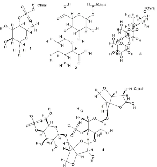
biological properties are summarised in
Table 1
and the chemical
structures of important polysaccharides are shown in
Fig. 1.
4.2. Phlorotannins
Phlorotannins, more commonly known as algal polyphenols, are
polymers of phloroglucinols which comprise up to 15% of dry weight of
brown algae. Laminariacea have been documented to be the most
abundant source of phlorotannins in marine algae. The molecular
weight of phlorotannins ranges from 126 kDa to 650 kDa. In the past
two decades, there have been a considerable literature on the isolation
and pharmacological properties of phlorotannins from brown algal
species such as Eisienia bycycles, Ecklonia cava, E. stolonifera and E.
maxima (
Kannan et al., 2013;
Rengasamy et al., 2013;
Rengasamy et al.,
2014a,
b). Phlorotannins have also been found to possess numerous
biological/pharmacological
properties
such
as
antimicrobial
(Nagayama et al., 2002), antioxidant (Kim et al., 2009), anti-HIV (Artan
et al., 2008), antiproliferative (Kong et al., 2009), anticancer (Parys
et al., 2010), anti-inflammatory (Kim et al., 2009), antidiabetes
(Kannan et al., 2013;
Rengasamy et al., 2013,
2014a), anti-Alzheimer
disease (Kannan et al., 2013), antihypersensitive (Jung et al., 2006),
anticoagulant (Li et al., 2007), and radioprotective (Moon et al., 2008).
Further comprehensive evidence on the isolation and pharmacological
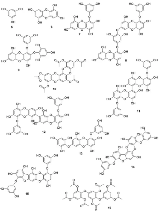
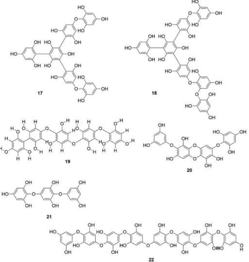
properties of brown algal phlorotannins are listed in
Table 2
and the
chemical structures of important phlorotannins are shown in
Fig. 2A
and B.
4.3. Protein hydrolysates
Protein hydrolysates are mixtures of amino acids generally
re-cognised as peptides or peptones, which are made from purified protein
by acid hydrolysis or using proteolytic enzymes and further subjected to
purification. To date, various protein hydrolysates have been reported
from seaweeds with potent pharmacological properties. Lately much
consideration has been diverted to the seaweed proteins and protein
hydrolysates. Seaweed protein hydrolysates have been presented to
possess many biological potential such as antibacterial activity
(Beaulieu et al., 2016), anti-hypertension (Pan et al., 2016;
Qu et al.,
2010;
Sai-kun et al., 2012;
Sheih et al., 2009;
Suetsuna et al., 2004),
anticoagulant (Indumathi and Mehta, 2016), antiplatelet aggregation
(Cian et al., 2012), antioxidant (Beaulieu et al., 2016;
Cian et al., 2012,
2013) and chelating properties (Cian et al., 2016). Wakeme jelly
pep-tide and Peppep-tide Nori S are the commercially available
anti-hy-persensitive peptides from the Japanese seaweed Undaria finnatifida
Table 2 (continued ) Phlorotannins Source Biological activity Model used Findings Reference Exerted a melanin inhibition with an IC50 value of 37.73 μM and the inhibition was potent than retinol, a positive control (IC 50 = 50.25 μM). I. okamurae Neuroprotective In-vitro Prevents H2 O2 -induced damage in neuronal cells and reduces the Bax expression. Heo et al. (2012) 2-(4-(3,5 dihydroxyphenoxy)-3,5-dihydroxyphenoxy) benzene-1,3,5-triol (DDBT) (21) S. patens Anti-diabetic In-vitro Suppressed the hydrolysis of amylopectin by human salivary and pancreatic α-amylases. Displayed inhibitory effects against α-amylase (IC 50 = 3.2 μg/mL), α-glucosidase from rat intestinal (IC50 = 25.4 μg/mL) and sucrase and maltase (IC50 = 114 μg/mL). Kawamura-Konishi etal. (2012) Octoplorethol A (22) I. foliacea Anti-diabetic In-vivo In type 2 diabetic db/db mice, Octoplorethol A significantly improves hyperinsulinemia and impaired glucose. Lee et al. (2016) ACE, angiotensin-converting enzyme; AGE, advanced glycation endproducts; ARE, antioxidant response element; DPP-IV, dipeptidyl peptidase IV; DPPH, 1,1-diphenyl-2-picrylhydrazyl; FRAP, ferric reducing antioxidant power; HO•, hydroxy; KU812, human basophilic leukemia; LPS, lipopolysaccharide; MIC, minimum inhibitory concentration; MSRA, methicillin resistant; MSSA, methicillin-susceptible; O2•−, superoxide anion radical; ORAC, oxygen radical absorbance capacity; RBL2H3, rat basophilic leukemia; ROS, reactive oxygen species.
Table 3 Biological properties of seaweed protein hydrolysates. Source Biological properties Model Findings Reference Palmaria palmate Antioxidant In-vitro Hydrolysed fractions post-treatment with either chymotrypsin or trypsin, showed DPPH scavenging, FRAP, and ORAC. Bondu et al. (2015) Antioxidant In-vitro Showed antioxidant effect with ORAC value of 45.17–467.54 and FRAP value of 1.06–21.59 μmol trolox equivalents/ g. Harnedy and FitzGerald (2013b) Antihypertensive In-vitro < 10-kDa fraction hydrolysed with chymotrypsin showed ACE inhibition with an IC50 of 460.05 mg/mL. Bondu et al. (2015) Antihypertensive In-vivo The tridecapeptide IRLIIVLMPILMA derived from papain hydrolysate of P. palmata exhibited renin inhibitory activity. In spontaneously hypertensive rats, seaweed protein hydrolysate showed a drop-in blood pressure. Fitzgerald et al. (2014) Cardioprotective and antidiabetic In-vitro Showed inhibition against ACE (IC50 = 0.19–0.78 mg/mL) and DPP-IV (IC 50 = 1.65–4.60 mg/mL). Harnedy and FitzGerald (2013a) Renin inhibitory In-vitro Fraction obtained from P. palmata protein hydrolysate exhibited 58.97% renin inhibition at 1 mg/mL. Fitzgerald et al. (2012) Undaria pinnatifida Antihypertensive In-vitro & In-vivo Showed inhibition against ACE In-vitro .Four tetrapeptides administrated orally into spontaneously hypertensive rats displayed antihypertensive activity. Suetsuna and Nakano (2000) Antihypertensive In-vitro & In-vivo Decreased blood pressure was observed with synthetic Phe-Tyr, and Ile-Tyr Tyr-His, Lys-Tyr, Ile-Tyr and Phe-Tyr in spontaneously hypertensive rats when administrated orally. Suetsuna et al. (2004) Porphyra yezoensis Antihypertensive In-vitro About 55% of ACE inhibition was noticed with an IC50 value of 1.6 g/L. Qu et al. (2010) Anticoagulant In-vitro The purified peptide derived from pepsin hydrolysate displayed prolonged APTT l35 s–320 swith an IC50 of 0.3 μM. Indumathi and Mehta (2016) Porphyra columbina Antioxidant In-vitro The residual cake hydrolysate exhibited ABTS and DPPH radical scavenging activity with IC50 value of 1.01 and 0.91 g/L, respectively. High copper chelating activity (≈97.5%) was also noticed. Cian et al. (2013) Antihypertensive In-vitro Residual cake hydrolysate showed 45.65% inhibition against. Cian et al. (2013) Antihypertensive In-vitro Hydrolysates showed ACE inhibition by uncompetitive mechanism, the highest activity being IC50 %, 1.2 g/L. Cian et al. (2015) Antioxidant In-vitro Displayed ABTS and DPPH scavenging, β-carotene bleaching, and copper-chelating activity Cian et al. (2015) Antiplatelet aggregation In-vitro The peptides showed antiplatelet aggregation activity, and the activity of peptides produced from alkaline protease was increased after simulated digestion process. Cian et al. (2015) Chelating agent and anticariogenic In-vitro P. columbina hydrolysate showed high iron-chelating activity (33%), copper-chelating activity (β-carotene oxidation rate: Ro ;0.7 min −1 ), and inhibition of phosphorus and Ca 2+ release (87 and 81%, respectively). Cian et al. (2016) Immunomodulation, Antihypertensive, Antioxidant In-vitro and ex vivo Both cold-water protein extract (PF) and PF hydrolysates (PFH) displayed immunosuppressive properties on rat splenocytes by enhancing IL-10 production and inhibiting the production of TNF-α and IFN-γ. PFH also showed > 35% of ACE inhibition and antioxidant effect (DPPH, TEAC, ORAC and copper-chelating activity). Cian et al. (2012) Enteromorpha clathrata Antihypertensive In-vitro Alcalase was found to be more suitable for the preparation of ACE inhibitory peptides from E. clathrata proteins than alkaline protease and trypsin. Under optimum condition, a hydrolysate with ACE inhibition (IC 50 = 0.66 mg/mL) was obtained. Sai-kun et al. (2012) Antihypertensive In-vitro Fractions less than −10 kDa exhibited higher ACE inhibition than > 10 kDa fraction. The identified active peptide namely Pro-Ala-Phe-Gly showed ACE inhibition (IC50 = 35.9 μM). Pan et al. (2016) Ulva rigida Antihypertensive In-vitro The hydrolysate produced under optimal proteolysis with pepsin plus bromelain, showed ACE inhibiton (IC50 = 0.483 mg/mL). Fraction < 1 kDa fraction exhibited the highest ACE inhibition (IC50 = 0.095 mg/mL). Purification by chromatographic techniques followed by Edman degradation yielded Ile-Pro (IP) and Ala-Phe-Leu (AFL) and these two peptides potentially inhibited ACE, with IC50 values of 0.020 and 0.023 mg/mL, respectively. Paiva et al. (2016)
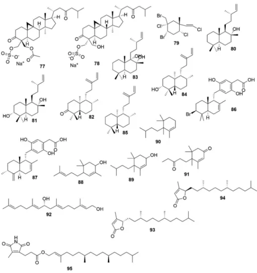
Table 4 Biological properties of seaweed terpenoids. Terpenoids Source Biological activity Model used Findings References Udoteafuran (23 ) Udotea flabellum Antibacterial In-vitro Growth inhibition of S. aureus . Fenical and Paul (1984) Udoteatrial (24 ) U. flabellum Antibacterial In-vitro Growth inhibition of S. aureus . Flexilin (25 ) U. conglutinata Antimicrobial In-vitro Inhibited S. aureus ,Vibrio splendida ,Dreschleria haloides ,and Candida albicans . U. conglutinata Cytotoxic In-vitro Inhibition of cell division in the sea urchin egg first cleavage (ED 100 = 16 μg/ml). Halimedatrial (26 ) Halimeda spp. Antimicrobial In-vitro Inhibited S. aureus ,Bacillus subtilis ,Serratitia marinorubra , Vibrio splendida ,V. harvevi ,V. leiognathi ,Lulworthia sp…, Alternaria sp…, D. haloides ,C. albicans . Halimeda spp… Cytotoxic In-vitro Inhibition of cell division in the sea urchin egg first cleavage (ED 100 = 1 μg/ml). Elatol (27 ) Laurencia dendroidea Acaricidal and repellent activity Exhibited strong repellent activity against T. urtica with moderate toxicity. Born and Bianco (2012) L. dendroidea Larvicidal Potent larvicidal effect with an LC50 value of 10.7 ppm. Bianco et al. (2013) L. dendroidea Antileishmanial activity In-vitro In Leishmania amazonensis, elatol inhibits promastigote and intracellular amastigote forms with an IC50 of 4.0 μM and 0.45 μM, respectively. Santos et al. (2010) L. microcladia Anti-tumor In-vitro and In-vivo Prompted cell cycle arrest in the G1 and the sub-G1 phases, leading cells to apoptosis. Elatol also decreased the expression of cyclin-D1, cyclin-E, cyclindependent kinase (cdk)2 and cdk4. A decrease in bcl-xl and an increase in bak, caspase-9 and p53 expression was also observed. Treatment in-vivo with elatol also decreased tumor growth in C57Bl6 mice. Campos et al. (2012) Diterpenoid seco-dolastane (4R,9S,14S)-4alpha-acetoxy-9beta, 14alpha-dihydroxydolast-1 (15),7-diene (28 ) C. cervicornis Acaricidal and repellent activity Exhibited moderate toxicity, but a high degree of repellent activity against T. urticae . Mertensene (29 )and violacene (30 )and two derivatives (dibromomertensene (31 )and dihydromertensene (32 ) Plocamium cartilagineum Insecticidal activity Toxicity of violacene to Schizaphis graminum aphids was higher than other compounds, causing 92% mortality of aphid after 48 h. It was similar to insecticides used as aphicides. Violacene, dibromomertensene, and dihydromertensene decreased the reproduction index of aphids. Also, dibromomertensene and violacene protected tomato plants against the tomato moth Tuta absolute. Argandona et al. (2000) Pterocladiella capillacea Anti-cancer In vitro Mertensene induced G2/M cell cycle arrest and caspase dependent apoptosis of Human Colon Adenocarcinoma HT29 cell line via the modulation of ERK-1/-2, AKT and NF-kB signaling. Tarhouni-Jabberi etal. (2017) Telfairine (33 ) P. telfairiae Insecticidal activity Exhibited 100% larvicidal activity against Culex pipiens pallens at 10 ppm in solution. Watanabe et al. (1989) Isoparguerol (34 ), isoparguerol-16-acetate (35 ), isoparguerol-7,16-diacetate (36 ), parguerol-16-acetate (37 ), deoxyparguerol (38 ), parguerol-7,16-diacetate (39 ), deoxyparguerol-7-acetate (40 ) Jania rubens Anti-tumor In-vitro The isolated compounds inhibited viability of Ehrlich carcinoma tumor cells (90, 90, 100, 80, 90, 70, 80% inhibition, respectively). Isoparguerol derivatives displayed slightly greater efficacy than parguerol derivatives. Awad (2004) J. rubens Anthelmintic In-vitro Anthelmintic activities (against earthworms -Allolobophora caliginosa ). Isoparguerol and parguerol derivatives were more effective than deoxyparguerol series. These compounds displayed high anthelmintic activity when compared with the same concentration (10%) of the reference anthelmintic drug mebendazole. Awad (2004) (continued on next page )
Table 4 (continued ) Terpenoids Source Biological activity Model used Findings References Neoirietetraol (41 ) Laurencia yonaguniensis Toxicity In-vivo Displayed toxicity to the Artemia salina (brine shrimp) with an LC50 of 40.1 μM. Takahashi et al. (1998) L. yonaguniensis Antibacterial In-vitro Displayed least antibacterial activities against Alcaligenes aquamarinus and Escherichia coli . Takahashi et al. (2002) Stypoquinonic acid (42 ) Stypopodium zonale Tyrosine Kinase Inhibitor In-vitro Showed inhibitory effect on tyrosine kinase (IC50 = 79.7 μg/mL). Wessels et al. (1999) S. zonale Antibacterial In-vitro Inhibited the growth of Bacillus megaterium and E. coli. Wessels et al. (1999) Dehydrothyrsiferol (43 ) Laurencia viridis Antitumor In-vitro Inhibited human breast cancer cell lines, viz. ZR-75-1, Hs578T and T47D with IC50 of 16.0, 18.9 and 13.5 μM, respectively. Pec et al. (1999) Stypolactone (44 ) S. zonale Antitumor In-vitro Showed weak cytotoxic property against A-549 (human lung carcinoma) and HT-29 and H-116 (human colon carcinoma) cell lines, with IC50 values of > 25.0 μg/ml, in each case. Dorta et al. (2002) Aplysiaterpenoid A (45 ) Plocamium telfairiae Insecticidal Displayed strong insecticidal activities against the mosquito larvae (Anopheles gambiae )and German cockroach (Blatella germanica ). Watanabe et al. (1990) Telfairine (46 ) P. telfairiae Insecticidal Displayed strong insecticidal activities against the mosquito larvae (Anopheles gambiae )and German cockroach (Blatella germanica ). Watanabe et al. (1990) Dictyterpenoid A (47 )and B (48 ) Dilophus okamurae Feeding-deterrent Displayed feeding-deterrent activity against abalone Haliotis discus hannai. Suzuki et al. (2002) Scopariol (49 ), isorigidol (50 ), (+)-3-(Z)-bromomethylidene-10β-bromo-β-chamigrene (51 ) Laurencia scoparia Anthelmintic In-vitro Showed anthelmintic effect against Nippostrongylus brasiliensis Davyt et al. (2001) 2β,3α-epitaondiol (52 ), flabellinol (53 ), flabellinone (54 ), stypotriolaldehyde (55 ), stypohydroperoxide (56 ) Stypopodium flabelliforme Neurotoxic In-vitro Moderate toxicity to murine neuro-2a cells (LC 50 = 2–25 μM) was observed for alln the compounds. 2β,3α-epitaondiol, flabellinol, and flabellinone possessed potent sodium channel blocking activity. In addition, stypotriolaldehyde displayed a biphasic effect on the concentration of intracellular Ca 2+ in rat cerebellar granule neurons. Sabry et al. (2005) 2β,3α-epitaondiol (52 ), flabellinol (53 ), flabellinone (54 ) S. flabelliforme Antitumor In-vitro Exhibited moderate cytotoxic effect with NCI–H460 human lung cancer cell line. Sabry et al. (2005) (6R)-6-hydroxydichotoma-3,14-diene-1,17-dial (57 ) Dictyota menstrualis Feeding-deterrence Displayed feeding-deterrent properties against the amphipod Parhyale hawaiensis Dana. Pereira et al. (2000) 3,7-dihydroxy-dihydrolaurene (58 ) Laurencia obtusa Cytotoxicity In-vitro Displayed cytotoxic effect on K562, MCF7, PC3, HeLa, A431, and CHO cell lines, with IC50 values of > 300, 201.7, 182.3, 121.3, 176.4, 234.7 μM, respectively. Kladi et al. (2006) Perforenol B (59 ) L. obtusa Cytotoxicity In-vitro Displayed cytotoxic effect on K562, MCF7, PC3, HeLa, A431, and CHO cell lines, with IC50 values of 67.4, 28.2, 54.8, 50.9, 73.2, 80.2 μM, respectively. Kladi et al. (2006) (1S*, 2R*, 6R*, 8S*, 9R*)-8-bromo2,5,6,9-tetramethyltricyclo-[7.2.0.0]undec-4-en-3-one (60 ) L. obtusa Cytotoxicity In-vitro Displayed cytotoxic effect on K562, MCF7, PC3, HeLa, A431, and CHO cell lines, with IC50 values of > 300, > 300, 126.2, 111.3, 137.1, > 300 μM, respectively. Kladi et al. (2006) 7-hydroxylaurene (61 ) L. obtusa Cytotoxicity In-vitro Displayed cytotoxic effect on K562, MCF7, PC3, HeLa, A431, and CHO cell lines, with IC50 values of 64.2, 15.8, 18.1, 40.5, 23.9, 78.2 μM, respectively. Kladi et al. (2006) (continued on next page )
Table 4 (continued ) Terpenoids Source Biological activity Model used Findings References Isolaurenisol (62 ) L. obtusa Cytotoxicity In-vitro Displayed cytotoxic effect on K562, MCF7, PC3, HeLa, A431, and CHO cell lines, with IC50 values of 127.4, 95.5, 103.2, 88.6, 122.0, 165.5 μM, respectively. Kladi et al. (2006) (E)-2-tridecyl-2-heptadecenal (63 ) L. obtusa and L. microcladia Cytotoxicity In-vitro Displayed cytotoxic effect on K562, MCF7, PC3, HeLa, A431, and CHO cell lines, with IC50 values of 82.7, 51.4, 71.6, 51.8, 45.8, 107.6 μM, respectively. Kladi et al. (2006) Perforenone A (64 ) L. obtusa Cytotoxicity In-vitro Displayed cytotoxic effect on K562, MCF7, PC3, HeLa, A431, and CHO cell lines, with IC50 values of > 300, > 300, 138.3, 117.7, 105.1, > 300 μM, respectively. Kladi et al. (2006) 3-epi-perforenone A (65 ) L. obtusa Cytotoxicity In-vitro Displayed cytotoxic effect on K562, MCF7, PC3, HeLa, A431, and CHO cell lines, with IC50 values of > 300, > 300, 144.4, 154.2, 151.9, > 300 μM, respectively. Kladi et al. (2006) Laurinterol (66 ) L. microcladia Cytotoxicity In-vitro Displayed cytotoxic effect on K562, MCF7, PC3, HeLa, A431, and CHO cell lines, with IC50 values of 128.3, 67.2, 76.6, 83.9, 74.6, 165.8 μM, respectively. Kladi et al. (2006) Bromolaurenisol (67 ) L. microcladia Cytotoxicity In-vitro Displayed cytotoxic effect on K562, MCF7, PC3, HeLa, A431, and CHO cell lines, with IC50 values of 112.7, 78.3, 92.4, 105.8, 81.6, > 200 μM, respectively. Kladi et al. (2006) Amijiol (68 )Amijiol acetate (69 )Dolabellatrienol (70 ), Dolastane mijiol-7-10-diacetate (71 ), Pachydictyol A (72 ), Isopachydictyol A (73 ), 8b-hydroxypachydictyol A (74 ),Isodictyohemiacetal (75 ), and Dictyol C (76 ) Dictoyota dichotoma DNA protective, cytotoxicity, antioxidant In-vitro Showed DNA protective effect, and cytotoxicity against HepG2, WI-38 and MCF-7 cell lines. Also displayed antioxidant effect by means of ABTS and erythrocytes hemolysis. Ayyad et al. (2011) Capisterone A (77 )and B (78 ) Penicillus capitatus Antifungal In-vitro Showed effective antifungal activity against the marine fungi (Lindra thallasiae) . Puglisi et al. (2004) (1S,2S,4R,5R,1′E)- 2-bromo-1-bromomethyl-1,4-dichloro-5-(2′-chloroethenyl)-5-methylcyclohexane (79 ) Plocamium hamatum Antifungal, antibacterial and antialgal In-vitro Showed potent antialgal activity towards Chlorella fusca , antinfungal effect on Ustilago violacea and Mycotypha microspora ,and antibacterial effect against Bacillus megaterium. König et al. (1999) Labda-14-ene-8-ol (80 ), Labda-14-ene3α,8α-diol (81 ), ent-Labda-13 (16),14-diene-3-one (82 ),Labda-14-ene-8α,9α-diol (83 ), ent-Labda-13 (16),14-diene-3α-ol (84 ), ent-Labda-13 (16),14-diene (85 ) Ulva fasciata Antibacterial In-vitro Inhibited the growth of Vibrio alginolyticus, V. parahaemolyticus and V. vulnificus and with MIC values ranging from 30 to 250 μg/ml. Chakraborty et al. (2010) Peyssonoic acid A (86 ) Peyssonnelia sp. Antimicrobial and antineoplastic In-vitro Inhibited the growth of the bacterial pathogen of marine algae, Pseudoalteromonas bacteriolytica (IC50 = 799 μM), and Lindra thalassiae (IC 50 = 506 μM), and also showed least antineoplastic activity against ovarian cancer cells (IC50 = 34.5 μM). Lane et al. (2010) Peyssonoic acid B (87 ) Peyssonnelia sp. Antimicrobial and antineoplastic In-vitro Inhibited the growth of P. bacteriolytica (IC50 = 377 μM), and L. thalassiae (IC50 = 331 μM), and also showed least antineoplastic activity against ovarian cancer cells (IC50 = 13.5 μM). Lane et al. (2010) 2,5,5-Trimethyl-4-(4-methylpent-3-enyl)-2-cyclohexen-1-ol (88 ) Ulva fasciata Antioxidant In-vitro Displayed DPPH and ABTS scavenging effect (IC50 = 13.74 mM and 66.8% inhibition at 50 μM, respectively). Chakraborty and Paulraj (2010) 4-Isopentyl-3,4,5,5-tetramethyl-2-cyclohexen-1-ol (89 ) U. fasciata Antioxidant In-vitro Displayed DPPH scavenging effect (IC50 = 23.60–20.83 mM). Chakraborty and Paulraj (2010) 6-Isopentyl-1,5,5,6-tetramethyl-1-cyclohexene (90 ) U. fasciata Antioxidant In-vitro Displayed DPPH and ABTS scavenging effect (IC50 = 80.56 mM and 12.8% inhibition at 50 μM, respectively). Chakraborty and Paulraj (2010) 3,4,5,5-Tetramethyl-4-(3-oxopentyl)-2-cyclohexene-1-one (91 ) U. fasciata Antioxidant In-vitro Displayed DPPH and ABTS scavenging effect (IC50 = 10.24 mM and 78% inhibition at 50 μM, respectively). Chakraborty and Paulraj (2010) (continued on next page )
and Porphyra yasoensis, respectively (Harnedy and FitzGerald, 2013).
Although protein hydrolysates possess various biological properties,
isolation of anti-hypertension inhibitory peptides from seaweed is now
getting much momentum among researchers worldwide. The detailed
information on the proteins, peptides, and other protein hydrolysates
isolated from seaweeds are given in
Table 3.
4.4. Terpenoids
Terpenoids, sometimes referred to as terpenes, are a huge group of
natural products commonly found in plants. They are a unique class of
hydrocarbon moiety comprising of terpenes attached to an
oxygen-containing group. In the current market, more than 60–75% drugs are
employed for the treatment/management of infectious diseases and
cancer, among these more than 23000 molecules belong to the class of
terpenoid (Wang et al., 2005). For instance, the currently available
commercial antimalarial drug Artemisinin and the anticancer drug
paclitaxel (Taxol®) are terpenoid biomolecules, and many terpenoid
molecules are in the clinical pipeline. Terpenoids are commonly
clas-sified as monoterpenoids, sesquiterpenes, diterpenes, sesterterpenes
based on their chemical structure. Marine organisms are well-known to
be a reservoir of these four categories of terpenes which have been
documented to have numerous pharmacological properties. Seaweeds
or marine macroalgae are a vast source of structurally diverse
terpe-noids especially red seaweeds, which are reported to have high amount
of terpenoids. Among the seaweeds, the family Rhodomelaceae is
considered as a terpenoid pool due to its vast chemical diversity and
structurally different terpenoids. More than 1058 molecules harnessed
naturally have been identified and characterised from this family which
accounts to 20% of the total halogenated compounds characterised
from all marine organisms (Wang et al., 2013). The comprehensive
information on the pharmacological properties of terpenoids from
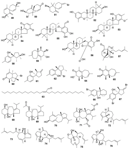
seaweeds are shown in
Table 4
and the chemical structures of important
terpenoids are shown in
Fig. 3A–C.
4.5. Alkaloids
In 1819 Meissner proposed the term “alkaloid” which originated
from the Arabic words “al kaly” and the Greek “eidos”, meaning
alkali-like. Alkaloids are heterocyclic nitrogenous compounds having Br-, I-,
Cl-, and S- in their structure (Güven et al., 2010,
2013). Alkaloids are
well known diverse group of natural biomolecules reported to have
enormous health benefits and biological potential. The well-known
example is Galanthamine; a commercially available anticholinesterase
inhibitor used to treat dementia over the years. A more recent book
written by
Aniszewski (2015)
described the applications of alkaloids in
pharmaceutical and agricultural industries. Although alkaloids are
ex-tensively studied, research on marine algal-based alkaloids is still
under-explored. The first alkaloid molecule was isolated from marine
red algae Phyllophora nervosa is hordenine in 1969 (Güven et al., 1969,
1970). The alkaloids isolated from various marine algae and its
phar-macological properties are listed in
Table 5
and the chemical structures
of important alkalaoids are shown in
Fig. 4.
4.6. Photosynthetic pigments
Plants possess various photosynthetic pigments such chlorophylls,
carotenoids, anthocyanins, betains, and research on the biological
properties of these colourful pigments is gaining much attention due to
its health-promoting effects. Like other terrestrial plants seaweeds,
marine macroalgae are listed as one of the primary resources of these
beneficial health pigments including carotenoids, fucoxanthin
(xan-thophyll pigments found in brown seaweeds), phycocyanin and
phy-coerythrin (found in red seaweeds). The importance of carotenoids
especially β-carotene, α-carotene, β-cryptoxanthin, lycopene, lutein
and zeaxanthin has already been well documented (Rodriguez-Amaya,
Table 4 (continued ) Terpenoids Source Biological activity Model used Findings References Crinitol (92 ) Sargassum tortile Antibacterial In-vitro Showed inhibition against Bacillus subtilis (MIC = 50 μg/ml), Brevibacterium ananoniagenes (MIC = 100 μg/ml), Streptococcus mutans (MIC = 50 μg/ml), Staphylococcus aureus (MIC = 400 μg/ml), Propionibacterium acnes (MIC = 25 μg/ml), Trichophyton mentagrophytes (MIC = 25 μg/ml). Kubo and Smith (1998) Racemobutenolid A (93 )and B (94 ) Caulerpa racemosa Cytotoxicity In-vitro Exhibited moderate cytotoxic effect on HL-60 (IC50 = 49.3 μM). Yang et al. (2015) 4′,5′-dehydrodiodictyonema A (95 ) C. racemosa Enzyme inhibitory activities In-vitro Showed inhibitory effects against protein tyrosine phosphatase 1B (PTP1B), T-cell PTPase (TC-PTP) and cell division cycle 25 homolog B (CDC25B) with IC50 values 2.30, 12.56, and 42.92 μM, respectively. Yang et al. (2015)
Table 5 Biological properties of seaweed alkaloids. Alkaloid Source Biological activity Model used Findings References Caulerpin (96 ) Caulerpa racemosa Antinociceptive In-vivo In the abdominal constriction test, the alkaloid exerted a reduction in the acetic acid-induced nociception at 0.0945 μmol (0.0103–1.0984). Caulerpin also caused a favourable nociception inhibition in the hot plate test at 100 μmol/kg, p.o. De Souza et al. (2009) C. racemosa Anti-inflammatory In-vivo 100 μmol/kg, p.o og caulerpin showed a high anti-inflammatory activity. This effect was further established on (i) capsaicin-induced ear edema model (% inhibition of 55.8) and (ii) the carrageenan-induced peritonitis (number of recruit cells decreased by 48.3%) C. racemosa Anti-viral In-vitro Showed antiviral activity against bovine viral diarrhea virus (EC 50 = 2.0 μM). Pinto et al. (2012) Caulerpa sp. Spasmolytic Ex vivo using guinea pig ileum Repressed phasic contractions induced by carbachol, histamine, and serotonin in a non-selective manner. A dose-dependent inhibition against serotonin-induced cumulative contractions. It also relaxed KCl-pre-contracted ileum and carbachol in a dose-dependent manner. Ayyad and Badria (1994) Caulerpa lentilifera, C. racemosa, Caulerpa microphysa and Caulerpa sertularoides Antibacterial In-vitro Presented moderate antibacterial action against 8 bacterial species isolated from algal surface. Vairappan (2004) Almazole C (97 ) Haraldiophylum sp. Antibacterial In-vitro Showed antibacterial activity against Gram-negative pathogens. Fresneda et al. (2007) Lophocladine A (98 )and B (99 ) Lophocladia sp. Anticancer In-vitro Exhibited cytotoxicity to MDAMB-435 breast cancer and NCI–H460 human lung tumor cell lines. The activity was correlated with microtubule inhibition. Gross et al. (2006) Lophocladia sp. Neuroprotective In-vitro Showed affinity for NMDA receptors and found to be a δ-opioid receptor antagonist. 4. Martefragin A (100 ) Martensia fragilis Antioxidant Ex vivo In in rat liver microsomes, Martefragin A inhibited NADPH-dependent lipid peroxidation (IC50 = 2.8 μM). Takahashi et al. (1998) 5. Racemosin A (101 ) Caulerpa racemosa Neuroprotective In-vitro Reduced the Aβ25–35-induced SH-SY5Y cell damage with a 14.6% increase in cell viability at 10 μM compared to the positive control EGCG (16.57% increaseat 10 μM). Liu et al. (2013) 6. Racemosin C (102 ) Caulerpa racemosa Tyrosine phosphatase-1B inhibitory activity In-vitro Exhibited significant PTP1B (human protein tyrosine phosphatase-1B) inhibitory activity with IC50 values of 5.86 μM compared to the positive control oleanolic acid (IC50 = 3.03 μM). Yang et al. (2014) 7. Azocinyl morpholinone (103 )[3-(2-ethyl-6-((3Z,7Z)-1,2,5,6-tetrahydroazocin-5-yl)hexyl) morpholin6-one] (104 ) Gracilaria opuntia Antioxidant In-vitro Showed scavenging effect against DPPH radicals (IC50 = 0.086 mg/mL). Makkar and Chakraborty (2018) G. opuntia Anti-inflammatory In-vitro and In-vivo Inhibited COX-2 (IC50 = 0.84 mg/mL) and 5-lipoxidase (IC 50 = 0.85 mg/mL). Makkar and Chakraborty (2018)