http://journals.tubitak.gov.tr/veterinary/ © TÜBİTAK
doi:10.3906/vet-1401-48
A detailed evaluation of the skeletal elements of the skull
in the grey heron (Ardea cinerea)
Şükrü Hakan ATALGIN1,*, Emine Ümran BOZKURT BÜYÜKÇOPUR2, İbrahim KÜRTÜL3 1Department of Anatomy, Faculty of Veterinary Medicine, Balıkesir University, Balıkesir, Turkey 2Department of Anatomy, Faculty of Veterinary Medicine, Kırıkkale University, Kırıkkale, Turkey 3Department of Anatomy, Faculty of Veterinary Medicine, Mustafa Kemal University, Hatay, Turkey
1. Introduction
In terms of structural and functional aspects, comparative morphologists describe the skull of avian species as a component possessing the rostrum, orbits, and braincase (1). Another view is that skull bones in birds are classically divided into neurocranium and splanchnocranium (2,3).
The skull bones of avian species articulate each other with a certain degree of movement permitted. This motion, cranial kinesis, is a relative movement between specific components of the skull. The movement of the upper beak is generated through the nasofrontal (cranio-frontal) joint. The upper beak rotates either rostro-dorsally (protraction) or caudo-ventrally (retraction), relative to the braincase. Bock (3) and Bühler (4) have indicated that this movement passively stabilizes the mandible position while the jaw is closed, works as an enhancement mechanism of the jaw opening strength, provides endurance of the primary vision axis during the tending, leads to very fast closure of the jaw due to the consequent movement of the upper and lower bills, lowers the effects of shocks, and extends the attachment area for jaw muscles. Zusi (5) has added that the basic advantage of this movement is to transform the 3-dimensional kinematics potential of the joint into a skillfully used complex instrument.
Moreover, Hoese and Westneat (6) and Meekangvan et al. (7) have indicated that this movement has a role in voice production. Finally, Gussekloo et al. (8)have reported that there is no relation between the cranial kinesis and pterygoid–palatinal complex.
The grey heron, a species of the family Ardeidae commonly found in boggy and shallow water areas in Europe, Asia, Africa, Madagascar, and Indonesia, is a long-legged and long-necked bird. In general, species that belong to this family have smaller heads and long, sharped-edged, and lance-like beaks. Skeletal features of the birds in this family show essential differences when compared to those of other Ciconiform birds. These differences indicate a phylogenetic relation between the closely related bird groups. Bones are also related functionally to each other to provide attachment areas for muscles. These functional relations can be used in the systemic taxonomy of the birds on the species and subspecies level (9).
Payne and Risley (10) have examined 13 skull bone characteristics out of the skull samples from 53 different species of the family Ardeidae, which they consider to be essential to understanding the evolutionary relationship among the Ardeidae. These characteristics comprise beak shape, plate shape, caudal edge of the palatine, lateral Abstract: This study was designed to reveal, in detail, the features of the skull bones in the grey heron, a species of the family Ardeidae,
and to compare the findings with previous related literature reports and with Nomina Anatomica Avium. The articulation between the nasal and frontal bones was observed to be through the movable frontonasal joint. The upper beak fused with the neurocranium via this joint and with the palatine and vomer. It was also bound indirectly through the zygomatic bone articulating with the quadrate bone. The frontal bone possessed the frontal depression dorsally. Wideness of the frontal bone and supranasal pila seemed to be unique for the grey heron. The quadrate bone was observed to play a primary role in the beak action. No medial process was present in the mandible. Overall, the results hereby indicate that the wider jaw opening and sharp and pointed nature of the beaks in the grey heron enable it to feed on such creatures as fish, frogs, and crustaceans.
Key words: Grey heron, skull bones
Received: 14.01.2014 Accepted: 18.04.2014 Published Online: 17.06.2014 Printed: 16.07.2014
process of the palatine, interorbital foramen, subraorbital foramen, largeness of the lacrimal, ventral extension of the lacrimal, lateral groove of the lacrimal, lateral process of the ectethmoid, ventral tubercle of the ectethmoid, ventral edge of the ectethmoid, and basitemporal edge. They only found small differences in the shapes of the ectethmoid and lacrimal and the position of the temporal fossa among
Ardea cinerea, Ardea herodias, and Ardea cocoi. They also
mentioned that Ardea species differ from Egretta species by not forming an edge in the midline of the skull roof through the temporal fossa. Another constant variation between the 2 species is the shape of the caudal edge of the palatine.
Pascotto and Donatelli (11), Gussekloo et al. (8), Çakır (12), and Zusi (5) have studied the cranial osteology of different avian species. Meekangvan et al. (7)have revealed the mechanism of the cranial kinesis in the skull of the grey heron. Payne and Risley(10) have also documented the skull characteristics of different Ardea species phylogenetically. Lobon and Buscalioni (9) have examined, with functional and mathematical models, the cranial osteology of the different avian species some of which belong to the family Ardeidae. To our knowledge, however, there has been no gross morphological study defining in detail the skeletal elements of the skull in the grey heron, whose feeding and drinking kinematics relate greatly to the cranial skeletal morphology. Detailed gross anatomical data need to be acquired to further analyze cranial kinesis in avian species and to contribute to phylogenetic studies. This study, therefore, aimed to document the features of the skull bones in the grey heron, which might be an interesting and valuable contribution to the functional analysis of the cranial kinesis, as well as to the avian skull morphological evolution.
2. Materials and methods
The heads of 3 adult grey herons of both sexes (there was no significant weight difference between the 2 sexes) weighing 2.5 and 2.6 kg were used in the study. The heads were taken from already dead animals obtained in Turkey. The skulls were dissected and cleaned. They were then put in a 5% KOH solution for maceration and remnants of the soft tissue were cleaned. Finally, the skulls were examined in detail and the characteristics were measured. Endocranial components were disregarded. Photographs were taken with a digital camera (Canon PowerShot SD700 IS). Nomina Anatomica Avium (2) was employed for anatomical nomenclature.
3. Results 3.1. Ossa cranii
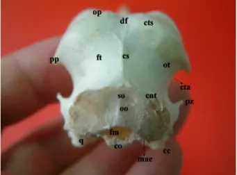
As a part of the occipital bone (Figure 1, oo), the foramen magnum was oval and 0.5 cm in diameter (Figure 1, fm)
and the occipital condyle was prominent ventrally (Figure 1, co). The exoccipital bone fused with the lateral process of the parasphenoid bone forming the ventral surface of the external acoustic process (Figures 1; 4, mae) through the paroccipital process. The area muscularis aspera (Figure 2, ama) was widely present on the orbital surface of the laterosphenoid bone (Figure 2, ls). The temporal bone (Figures 1 and 2, ot) was eminent between the parietal bone (Figures 1–3, op) and the squamooccipital region of the occipital bone (Figure 1, so). It was separated from this region by the sharp and high transverse nuchal crest (Figures 1–3, cnt). A prominent sagital crest (Figures 1 and 2, cs) was determined in the midline between the transverse nuchal and superior temporal crests (Figures 1–3, cts). The inferior temporal crest, (Figure 2, cti) originating from the lateral edge of the transverse nuchal crest and lying cranially, formed the cranial border of the subtemporal fossa (Figure 2, fst) which was a triangle-like area with its apex pointing upward. There was a short zygomatic process (Figures 1 and 2, pz) on the cranial edge of the inferior temporal crest. The caudal border of the subtemporal fossa was constructed by the transverse nuchal crest. Thus, the ventral border was formed by a ventral crest (Figure 2, vc) possessing a notch suitable for the otic capitulum of the quadrate bone and uniting the ventral edges of the transverse nuchal and inferior temporal crests.
The temporal fossa (Figures 1–3, ft) was very deep and wide. The inferior temporal crest (Figures 1 and 2, cta) formed the concave ventral surface of the temporal fossa. The distance between the jugal arch (Figures 2 and 4, aj)
Figure 1. Caudal view of the skull in the grey heron.
oo, os occipitale; fm, foramen magnum; co, condylus occipitalis; cnt, crista nuchalis transversa; ot, os temporale; ft, fossa temporalis; df, depressio frontalis; cs, crista sagittalis; pp, processus postorbitalis; pz, processus zygomaticus; cta, crista temporalis anterior; mae, meatus acusticus externus; q, os quadratum; cc, condylus caudalis; so, squama occipitalis.
and the edge of the postorbital process (Figures 1 and 2, pp) was 1.5 cm, and that between the jugal arch and base of the postorbital process was 2 cm. Therefore, the postorbital process reached only one-fourth of the distance between the skull roof and jugal arch.
The squamoparietal part of the parietal region (Figures 1–3, op) showed 2 parietal tubercles in compliance with the parietal lobes of the brain. The superior temporal crest formed the border of os parietale with os temporale.
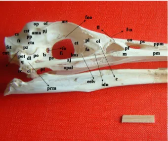
The frontal bone (Figures 2 and 3, of) fused rostrally with the nasal bone through the frontonasal joint (Figures 2 and 3, f-n), rostrolaterally with the lacrimal bone via the frontolacrimal suture (Figure 2, f-l), and caudally with the parietal bone without showing an eminent line. The squamofrontalis possessed a frontal depression (Figures
1 and 3, df) throughout the length of the frontal bone, which deepened medially. It ended at the frontonasal joint while extending caudally between the 2 parietal regions as a slight hollow. The width of the frontal region between the 2 supraorbital borders (Figures 2 and 3, ms) was 2 cm. The olfactory nerve sulcus (Figure 2, sno) was present on the orbital surface of the frontal bone and dorsal to the interorbital septum. Foramen nervi olfactorii (Figure 2, fno) was seen as a narrow fenestra in the caudal portion of this sulcus.
A process (Figures 2 and 3, pj) resembling the zygomatic process of the frontal bone present in mammals was determined at the caudal edge of the supraorbital border of the frontal bone. It was a very short, flat, and wide one not reaching the ventral end of the postorbital process. The measures taken between the cranial edges of the right and left process indicated that the skull roof at the parietal region was wider (2.8 cm) than at the frontal region. Observed dorsally, the skull of the grey heron was rectangular. The transversal length of the nasofrontal joint region was 1.2 cm. A slight impression for the nasal gland was present on the orbital surface of the frontal region, near the supraorbital border.
The mesethmoid bone was located rostral to the interorbital septum, joining the formation of the nasal septum. The dorsal lamina of the mesethmoid bone did not reach the nasofrontal joint area.
The ectethmoid bone (Figure 2, et) contributed to the construction of the antorbital fenestra (Figure 2, fa) through its lateroventrally directed process (Figure 2, eelv). Figure 2. Lateral (right side) view of the skull in the grey heron
(Scale bar 2 cm).
cnt, crista nuchalis transversa; cti, crista temporalis inferior; vc, ventral crista; fst, fossa suptemporalis; ft, fossa temporalis; ot, os temporale; pz, processus zygomaticus; cs, crista sagittalis; cts, crista temporalis superior; cta, crista temporalis anterior; pp, processus postorbitalis; ama, area muscularis aspera; op, os parietale; of, os frontale; pj, processus jugalis; ms, margo supraorbitalis; fno, foramen nervi olfactorii; sno, sulcus nervi olfactorii; et, os ethmoidale; eelv, lateroventral process of the ethmoid bone; ol, os lacrimale; pl, processus lacrimalis; fl, frontolacrimal sutura; c, crista lacrimalis; f-n, frontonasal joint; on, os nasale; ps, pila supranasalis; m, maxilla; ppm, processus premaxillaris; pm, processus maxillaris; aj, arcus jugalis; opal, os palatinum; pt, os pterygoideum; fo, foramen opticum; fi, fonticuli interorbitales; ls, latero sphenoid; ps, para sphenoid; lms, lamina parasphenoidale; q, os quadratum; po, processus orbitalis; pot, processus oticum; prm, processus mandibularis; dl, depressio lateralis; cc, condylus caudalis; cl, condylus lateralis; fa, fenestra antorbitalis; idn, incisura ductus nasolacrimalis; pf, processus frontalis.
Figure 3. Dorsal view of the skull in the grey heron (Scale bar
2 cm).
pm, os premaxillare; on, os nasale; ps, pila supranasalis; f-n, frontonasal joint; ol, os lacrimale; df, depressio frontalis; ms, margo supraorbitalis; of, os frontale; op, os parietale; pj, processus jugalis; cts, crista temporalis superior; ft, fossa temporalis; cnt, crista nuchalis transversa.
The basisphenoid bone (Figure 4, bs) articulated caudally with the basioccipital bone (Figure 4, bo), and, via its lateral edge, joined the ventral surface of the external acoustic process (Figures 1 and 4, mae).
The parasphenoid bone (Figure 2, ps) was present in front of the basisphenoid bone, had parasphenoid rostrum in a crest shape, and parasphenoid lamina (Figure 2, lms), which narrowed cranially. The laminas from both sides united dorsally in the midline and fused with the ethmoid cranially (Figure 2, et). They also joined the formation of the interorbital septum and constructed the ventral border of the interorbital fonticulus (Figure 2, fi).
The lacrimal bone (Figures 2 and 3, ol) was a highly developed bone extending through the nasofrontal joint thereby articulating with the nasal bone. The unification formed among the lacrimal, frontal, and nasal bones via the articular surface of the frontonasal joint was a sutura type articulation. The lacrimal bone made up the lateral border of the antorbital fenestra. Connection of the lacrimal bone with the jugal arch was provided by its ventral process. The ventrally located larger process of the lacrimal bone formed the antorbital fenestra by combining with the laterally directed process of the ectethmoid bone.
The lacrimal bone possessed the lacrimal process (Figure 2, pl) and a large and prominent crest on the external surface. The highly deep incisura for the nasolacrimal duct (Figure 2, idn) was determined right under this crest.
A well-developed fonticulus interorbitalis (Figure 2, fi) was observed on the interorbital septum, with a dorsoventral diameter of 1.1 cm and anterioposterior diameter of 1.3 cm. The interorbital septum was made up of the ectethmoid and mesethmoid bones cranially, the laterosphenoid bone caudolaterally, and the frontal bone dorsally. The diameter of the oval optic foramen (Figure 2, fo), located on the caudal portion of the interorbital septum, was 0.2 cm.
3.2. Ossa faciei
The nasal bone (Figures 2 and 3, on) united caudally with the frontal bone via the nasofrontal joint (Figures 2 and 3, f-n), and ventrally with the premaxillare through the long premaxillar process (Figure 2, ppm), which constructed the bony nostrils dorsally as well. There was a narrow groove determined between the premaxillar process of the nasal bone and maxilla, beginning in front of the nostrils and ending without reaching the apex of the upper beak. The convex supranasal pilum (Figure 3, ps), limiting both nostrils dorsally, was 0.5 cm in length.
In reliance with the shape of the premaxillare (Figures 2 and 3, pm), the upper beak was highly narrow and long, ending in a sharp edge. The dorsal border of the lateral surface of the premaxillare articulated with the nasal bone, and the ventral border, making a line between the palatine process and lateral surface, was very sharp. The premaxillare united caudally with the maxilla without displaying an eminent border, while the frontal process (Figure 2, pf) bounded the caudal borders of the nostrils. At the same time, it also formed the floor of the nostrils through a horizontal component. The palatine process of the premaxillare joined with a suture caudally to the palatine bone via the palatine process yet both of the palatine processes united longitudinally in the midline.
The nasal process of the maxilla (Figure 2, m) fused with the maxillary process of the nasal bone. Through the jugal process, the maxilla also joined the formation of the jugal arch. The ventral border of the maxilla showed a consistency to the ventral border of the lateral surface of the premaxillare, making a sharp border separating the maxillopalatine process from the lateral surface of the maxilla.
The horizontal premaxillar process (Figure 4, oppm) of the palatine bone (Figures 2 and 4, opal) made a bony construction of the palate by combining with the palatine process of the premaxillare. The palatine was in contact dorsally at the level of the orbit with the presphenoid and mesethmoid. It also formed the choana (Figure 4, ch) with the contribution of the vomer medially.
The palatine extended caudally forming the medial (Figure 4, crm) and lateral (Figure 4, crl) crests by curving like a cone. There were deep, long, and narrow ventral Figure 4. Ventral view of the skull in the grey heron (Scale bar
2 cm).
oppm, os palatinum processus maxillare; ch, choana; opal, os palatinum; v, vomer; crm, crista medialis; crl, crista lateralis; fv, fossa ventralis; spp, sutura pterygopalatina; pt, os pterygoideum; bs, Basisphenoidale; aj, arcus jugalis; q, os quadratum; bo, basioccipitale; cm, condylus medialis; cl, condylus lateralis; cc, condylus caudalis; si1, sulcus intercondylaris 1; si2, sulcus intercondylaris 2; f, a foramen in os pterygoideum; mae, meatus acusticus externus.
fossae between the crests (Figure 4, fv). The choanal fossa was observed between the medial crests. The caudal edge of the medial crest was longer than that of the lateral crest. The pterygoid process of the palatine articulated caudally with the pterygoid via a suture (Figure 4, spp). The vomer (Figure 4, v) was a sharp-edged bone possessing a deep groove ventrally and uniting caudally with the pterygoid via a suture. The bilateral pterygoids (Figures 2 and 4, pt) were short rod-like bones articulating caudally with the quadrats and cranially with the palatine bones. Each bone had a foramen (Figure 4, f) on their edges fusing with the quadrate bones. The jugal constructed the jugal arch (Figures 2 and 4, aj) by articulating cranially with the jugal process of the maxilla and caudally with the quadratojugal cotyla of the quadrate bone, and its length was 4.8 cm. There was a pneumatic foramen (Figure 4, fpq) at the medial aspect of the caudal border of the quadrate bone (Figures 1, 2, and 4, q) and right under the squamose capitulum, which indicated the pneumatic nature of the quadrate bone in the grey heron. There was a deep medial depression on the medial surface of the body of the quadrate bone while there was a shallow lateral depression (Figure 2, dl) on the lateral surface.
The medial (Figure 4, cm), lateral (Figures 2 and 4, cl) and caudal (Figures 1, 2, and 4, cc) condyles of the mandibular process (Figure 2, prm) of the quadrate bone were separate form each other. The intercondyler groove (Figure 4, si1) separating the lateral condyle from the others was deep while the intercondyler groove (Figure 4, si2) between the medial and caudal condyles was shallow. The quadratojugal cotyla was present as a small part joining to the jugal arch on the lateral surface of the lateral condyle.
The orbital process (Figure 2, po) was long, flat, and wide, and its front edge lying towards the optic foramen. Its ventral border showed no particular peculiarity while the dorsal border was deeply concave.
The otic process (Figure 2, pot) was short. It articulated with the ventral articular surface of the suptemporal fossa of the temporal bone through the otic capitulum laterally and squamosal capitulum medially. The otic capitulum was larger than the squamosal capitulum and both were separated by the intercapitular crest.
On the mandible (Figure 5, m), the dental bone (Figure 5, d) was sharply pointed, the dorsal part (Figure 5, pd) was hollow, and the ventral part was bumpy. The length of the mandibular symphysis (Figure 5, ps) was 3.5 cm.
Nearly 5/17 of the ventral border of the mandible was occupied by the angular bone (Figure 6, oa). The mandibular angle was almost 180°. There was the wide fossa aditus canalis neurovascularis (Figures 5 and 6,
facn) on the medial surface of the angular bone. The retroarticular process (Figures 5 and 6, pr) was large, containing a wide surface pointing ventrocaudally and facing caudodorsally. The medial process of the mandible (Figure 6, pmm) was oval, high, and bumpy, displaying a pneumatic articular foramen (Figure 6, fpa). The coronoid bone had an exclusive and prominent coronoid process (Figure 6, pc). The fenestra caudalis mandibulae was present on the medial surface of the splenial bone.
Figure 5. Dorsal view of the mandible in the grey heron (Scale
bar 2 cm).
Pr, processus retroarticularis; facn, fossa aditus canalis neurovascularis; m, mandibula; pd, pars dorsalis; ps, pars symphysialis; d, os dentale.
Figure 6. Dorsal view of the caudal part of the mandible in the
grey heron (Scale bar 2 cm).
Pr; processus retroarticularis; fpa, foramen pneumaticum articulare; ppm, processus medialis mandibulae; pc, processus coronoideus; oa, os angulare; facn, fossa aditus canalis neurovascularis.
4. Discussion
As reported in the duck and goose (1), this study has revealed that the occipital region in the grey heron is in vertical position and contains no cerebellar prominence (6). Yet, the supcondylar fossa and basilar tubercle are lacking, and the ventrally transverse basal crest and cerebellar surface of the supraoccipital region are inconspicuous in the grey heron. A process observed on the caudal edge of the supraorbital region of the grey heron examined in this study has not been mentioned in Nomina Anatomica
Avium (2) or elsewhere. In the study, no fossa for the
nasal gland has been observed on the external surface of the frontal region. Instead, there is an impression present on the orbital surface of the frontal region and near the supraorbital border.
Pascotto and Donatelli (11) have indicated that the postorbital process is an extension of the parietal bone in the species of the family Coraciiformes while Baumel (2) has reported it to be a process of the temporal bone. On the other hand, our study determined that in the grey heron it was formed by the contribution of the parietal and temporal bones, and was present lateral to the superior temporal crest between the 2 bones. Pascotto and Donatelli (11) and Baumel (2) have reported the presence of the orbital depression on the proximal surface of the postorbital process. In this study, however, there was no orbital depression and the area was formed horizontally in the grey heron.
In the grey heron examined in our study, the cranial kinesis has been determined to be the prokinetic type because the semimovable nasofrontal joint is formed in front of the orbit, as indicated by the literature (7). Thus, the fact that the dorsal lamina of the mesethmoid does not reach the nasofrontal joint and the lacrimal bone articulates with the frontal and nasal bones through the suture, contributes to the idea that the grey heron examined in our study has indeed the prokinetic type cranial kinesis. This gives the grey heron a prokinetic skull and an extremely wide gap to display its feeding behavior.
The antorbital fenestra was determined in the grey heron to be formed medially by the lateral process of the ectethmoid, ventrally by the caudal process of the lacrimal bone, dorsomedially by the internal surface of the frontal bone, and dorsolaterally by the medial surface of the lacrimal bone. Pascotto and Donatelli (11) have reported, however, that the lacrimal bone does not contribute to the formation of the antorbital fenestra in motmots.
In this study, the supraorbital process of the lacrimal bone has been shown to form the cranial 1/7 of the supraorbital border. Baumel (2) has not reported the presence of the supraorbital process in the lacrimal bone even though Bahadır (1) has documented such process and a crest on the external surface of the lacrimal bone in the
duck and goose. Although Baumel (2)has indicated that the temporal fossa is in the area between the transverse nuchal and temporal crests, our study has found that it is bounded by the superior, inferior, and anterior temporal crests as reported by Pascotto and Donatelli (11). There are 3 depressions: the laterocaudal, lateroventral, and laterodorsal impressions, reported on the lateral surface of the quadrate bone in species of the family Momotidae (11). In our study of the grey heron, there is only a lateral depression, which may be an equivalent to the laterodorsal depression. The upper beak of the grey heron is parallel with the orbital fenestra, orbit, and skull, and the skull is the extended type. This has resulted in the basioccipital and sphenoid bones being located horizontally and the foramen magnum directing caudally. The nasofrontal joint of the grey heron is formed in front of the orbit as indicated by Meekangvan et al. (7),which allows the prokinesis type jaw movement. However, there are some factors documented limiting the free action of the upper beak: nonflexible nature of the nasofrontal joint area, suture type articulation of the lacrimal bone with the frontal and nasal bones, and shortage of the orbital fenestra. Gussekloo et al. (8) have suggested that the structure formed by the pterygoid and palatine bones has no relationship with the movement mechanism of the head. By contrast, the articular surface of the flat and wide palatine bone allowing for very limited action for the nasofrontal joint has been determined to be an essential movement limiting factor. These results are similar to those reported by Bock (3). In spite of the indicated factors limiting the free action of the upper beak, the strong nature of the orbital process of the quadrate bone and its extension to the optic foramen as well as the concave nature of its dorsal edge indicate a lever-like action. Osseous articulation of the quadrate bone in the skull of the grey heron leads us to think that the beaks act concomitantly, as reported by Zusi (5). In contrast to the reports by Hoese and Westneat (6) in sparrows, the upper beak of the grey heron moves along with the skull because of the mandible moving ventrally and the quadrate bone protruding forward. Consequently, this leads to an increase in the opening of both jaws even though the free movement of the nasofrontal joint is limited. Thus, the joint between the mandible and the 3 condyles of the quadrate bone causes an increase in the distance between the mandibular rami during the ventral movement of the mandible. Zusi (5) has reported this mechanism leading to lateromedial movement. In this study, the temporal fossa has been located on the dorsocaudal aspect of the cranial cavity, lying between the parietal and squamaoccipital regions, and 2 temporal fossa are separated by a crest between the transverse nuchal and superior temporal crests. These findings are also in parallel with the reports by Payne and Risley (10). The highly wide and deep nature of the
temporal fossa and wide area muscularis aspera indicate that the musculature of the lower beak is highly developed.
Consequently, wider jaw opening and sharp and pointed nature of the beaks in the grey heron enable it to
feed on fish, frogs, and crustaceans. Further morphological, functional and geometrical studies on the muscles and ligaments in the grey heron will surely be very helpful in understanding the kinetic mechanisms of the skull joints.
References
1. Dursun N. Evcil Kuşların Anatomisi. Ankara, Turkey: Medisan Yayınevi; 2002 (in Turkish).
2. Baumel JJ. Handbook of Avian Anatomy. Nomina Anatomica Avium. 2nd ed. Cambridge, MA, USA: Nuttall Ornithological Club; 1993.
3. Bock WJ. Kinetics of the avian skull. J Morphol 1964; 114: 1–42.
4. Bühler P. Functional Anatomy of the Avian Jaw Apparatus
Form and Function in Birds, Vol. 2. New York, NY, USA: Academic Press; 1981.
5. Zusi RL. Patterns of Diversity in the Avian Skull. In: Hanken J, Hall BK, editors. The Skull: Patterns of Structural and Systematic Diversity. Vol. 2. Chicago, IL, USA: University of Chicago Press; 1993. pp. 391–437.
6. Hoese WJ, Westneat MW. Biomechanics of cranial kinesis in birds: testing linkage models in the white-throated sparrow (Zonotrichia albicollis). J Morphol 1996; 227: 305–320. 7. Meekangvan P, Barhorsta AA, Burtona TD, Chatterjee S,
Schovanec L. Nonlinear dynamical model and response of avian cranial kinesis. J Theor Biol 2006; 240: 32–47.
8. Gussekloo SWS, Vosselman MG, Bout RG. Three-dimensional kinematics of skeletal elements in avian prokinetic and rhynchokinetic skulls determined by roentgen stereophotogrammetry. J Exp Biol 2001; 204: 1735–1744. 9. Lobon JM, Buscalioni AD. Avian skull morphological
evolution: exploring exo- and endocranial covariation with two-block partial least squares. Zoology 2006; 109: 217–230. 10. Payne RB, Risley CJ. Systematics and evolutionary relationships
among the herons (Ardeidae). No. 150. University Of Michigan, MI, USA: Miscellaneous Publicatıons Museum of Zoology; 1976.
11. Pascotto MC, Donatelli RJ. Cranial osteology in Momotidae (Aves: Coraciiformes) J Morphol 2003; 258: 32–48.
12. Çakır A. Kelaynak kuşunda (Geronticus eremita) neurocranium kemikleri. J Turkish Vet Med Assoc 2001; 72: 35–38.