ORIGINAL ARTICLE
Spleen Salvaging Treatment Approaches in Non-parasitic Splenic
Cysts in Childhood
Hasan Özkan Gezer1&Pelin Oğuzkurt1
&Abdulkerim Temiz1&Emineİnce1&
Semire Serin Ezer1&Nazım Emrah Koçer2&Şenay Demir3
&Akgün Hiçsönmez1
Received: 20 July 2015 / Accepted: 9 October 2015 / Published online: 17 October 2015 # Association of Surgeons of India 2015
Abstract The aim of this study was to evaluate our experi-ence with primary non-parasitic splenic cysts (NPSC) which are relatively rare in children and consist almost exclusively of single case reports or small case series in the literature. The medical records of all patients who presented to our clinic with NPSC between 2005 and 2015 were evaluated retrospectively. There were 22 children whose ages ranged from 2 months to 14 years (mean 9.2±4.7 years). The size of the cysts was in the range of 5 to 200 mm (mean 55.4±48.2 mm). Ten patients underwent surgery for splenic cysts. Partial splenectomy (n = 2), total cyst excision (either open n=4 or laparoscopically n=1), and total splenectomy (n = 3) were performed. The non-operated patients were asymptomatic and followed with ultrasound (US). The follow-up period in non-operated patients ranged from 6 months to 5 years (mean 2.27 ± 1.29 years). Complete regression was observed in four (33 %) non-operated patients. The regressed cyst mea-surements were 10, 16, 30, and 40 mm, respectively. Approximately half of the NPSC is diagnosed incidentally. Small (<5 cm) asymptomatic cysts should be under regular follow-up with US/physical examination for regression. If
surgery is required, we prefer open cyst excision as it gives excellent results and preserves splenic immune function.
Keywords Splenic cysts . Children . Splenectomy . Cystectomy
Introduction
Non-parasitic splenic cysts (NPSC) are rare in childhood, making it difficult to classify, define pathogenesis, and draw evidence-based conclusions for the ideal management of this disease [1]. The non-operative management of splenic trauma and the growing use of imaging modalities in the evaluation of abdominal pain have contributed to the diagnosis of increased number of splenic cysts in children [2]. They are usually asymptomatic and are often discovered incidentally when in-vestigating other symptoms or examining a patient after trau-ma [3]. Therefore, the rising incidence of diagnosed splenic cysts may be due to the increased use of abdominal US and other types of imaging [3,4].
* Hasan Özkan Gezer hozkangezer@yahoo.com.tr Pelin Oğuzkurt poguzkurt@yahoo.com Abdulkerim Temiz aktemiz@yahoo.com Emineİnce inceemine@mynet.com Semire Serin Ezer semireserin@yahoo.com Nazım Emrah Koçer nemrahkocer@yahoo.com
Şenay Demir
drsenaydemir@hotmail.com Akgün Hiçsönmez
akgunhicsonmez@gmail.com
1 Department of Pediatric Surgery, Başkent University Faculty of
Medicine, Dadaloğlu Mah 39 sokak, No: 6, Yüreğir, Adana 01210, Turkey
2 Department of Pathology, Başkent University Faculty of Medicine,
Ankara, Turkey
3 Department of Radiology, Başkent University Faculty of Medicine,
The original classification of NPSC was based on the pres-ence (primary or true cysts) or abspres-ence (secondary or pseudocysts) of cellular lining, according to Fowler’s defini-tion as well as Martin’s later simplificadefini-tion [5]. Several studies focusing particularly on NPSC have been published to better understand the pathogenesis, classification, and optimum treatment [6]. This report presents 22 children, with 10 having received an operation and had a histopathological diagnosis of NPSC. The aim of this study was to evaluate our experience with NPSC which are relatively scant, consisting almost ex-clusively of single case reports or small case series. We also aim to define a rational approach that focuses on surgical treatment during childhood in the light of literature.
Materials and Methods
This study was conducted at Başkent University Faculty of Medicine after the approval of the ethical committee and was in conformance with the Declaration of Helsinki. All parents of patients provided informed consent for this study. During a 10-year period (between 2005 and 2015), 22 consecutive pa-tients with non-parasitic splenic cyst were enrolled at our sur-gical center. Inclusion criterion was defined as patients with NPSC who had been treated operatively or non-operatively. Demographic data was collected and analyzed retrospectively from the database of the hospital information system. Supplementary information was obtained from standardized telephone interviews with the patients during the follow-up.
Parameters of patients including age, sex, history of trau-ma, presenting symptoms, physical examination, laboratory and radiological findings, therapeutic approaches, and out-comes were evaluated.
The complete blood count, serum biochemistry, and the echinnococcus-antibody titre test were conducted in all patients. Ultrasonography (US) was the initial imaging method. Computed tomography (CT) and magnetic resonance imaging (MRI) were also performed.
Cyst aspiration was performed preoperatively in patients who had severe symptoms, such as abdominal pain and/or vomiting due to the mass effect of the cyst, in order to relieve these symptoms for palliative purposes.
Small (<5 cm) and asymptomatic splenic cysts were followed non-operatively without any specific treatment. They underwent US, initially at 6-month intervals and, 1 year later, annually. Surgery is performed in symptomatic patients such as abdominal pain and vomiting due to mass effect or in patients that have a cyst greater than 5 cm in diameter. All surgical procedures were done 3 weeks after immunization against capsular bacteria (Streptococcus pneumoniae, Haemophilus influenzae b, and Neisseria meningitidis C), in case total splenectomy might be required during surgery. Partial splenectomy, total cyst excision either open or
laparoscopically, and total splenectomy were performed. The patients followed with US, initially at 6-month intervals and, later on, annually postoperatively. The pathologic diagnosis during the microscopic study was performed by the pathology department.
Statistical analysis was performed using Microsoft Office Excel (Version 2007, Microsoft Corporation, Redmond, Washington, USA). All numerical data are expressed as mean values±SD or as proportions.
Results
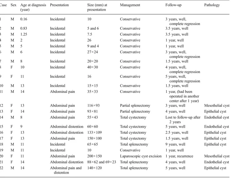
There were 22 children (13 males and 9 females) whose ages ranged from 2 months to 14 years (mean 9.2±4.7 years). None of the children had a history of antecedent trauma. Twelve patients (55 %) were symptomatic. Among the symptomatic patients, the main complaints were abdominal distension in three patients, abdominal pain in seven patients, and abdom-inal distension and pain in two patients. The physical exami-nation revealed splenomegaly in nine patients with cysts mea-suring more than 6 cm in diameter. The complete blood count and serum biochemistry were normal in all patients. The echinnococcus-antibody titre test was conducted, and the re-sults were negative in all cases. Details of all patients are shown in Table1.
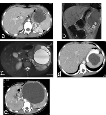
Ten of the 22 patients underwent surgery, and the age of those patients ranged from 8 to 14 years (mean 12±2.1 years). There were three male and seven female patients. All except one were symptomatic, having complaints such as abdominal pain and/or distension. US was chosen as the initial imaging method and revealed a round homogeneous anechoic area with thin septations and a smooth thin wall in all patients. CT (n=8) and magnetic resonance imaging (MRI) (n=1) were also performed before surgery. Contrast-enhanced CT scans demonstrated a spherical, well-defined lesion with attenuation similar to water. In two patients, tiny calcifications were de-tected in CT scans (Fig. 1a). The cyst size was measured between 55 and 200 mm (mean 100±36.2 mm). Four cysts were located in a single pole of the spleen. In the remaining six patients, there were multiple cysts (n=1; Fig.1b, c), extending to the hilus (n=1; Fig.1d) and invading both poles with a very limited remaining parenchyma (n=4; Fig.1e). Cyst aspiration was performed preoperatively in seven of the ten patients that had severe symptoms, such as abdominal pain and/or vomiting due to the mass effect of the cyst, in order to relieve these symptoms for palliative purposes.
All surgical procedures were done 3 weeks after immuni-zation against capsular bacteria (Streptococcus pneumoniae, Haemophilus influenzae b, and Neisseria meningitidis C), in case total splenectomy might be required during surgery. Partial splenectomy (n=2), total cyst excision (either open n=4 or laparoscopically n=1), and total splenectomy (n=3)
were performed. Splenectomies previously performed in chro-nological process of the disease were due to multiple cysts extending to the hilus and invading both poles with very limited remaining parenchyma. However, in all subsequent patients, total cyst excision was the initial plan. In single-pole cysts, we preferred either total cystectomy or partial splenec-tomy. In our series, conversion to a total splenectomy was not required. One patient who underwent partial splenectomy de-veloped a perisplenic abscess 15 days after the operation. He was treated with percutaneous drainage and antibiotics. Additionally, one patient who underwent laparoscopic cyst excision for a 130-mm cyst had recurrence at 1 month postoperatively and still following with US. The pathologic diagnosis during the microscopic study was an epithelial cyst in five patients, an endothelial cyst in three patients, and a mesothelial cyst in two patients (Fig.2).
The age of the 12 patients that were followed non-operatively ranged from 2 months to 14 years (mean 6,8±5 years). There were ten males and two females. Abdominal pain not related to splenic cyst was a complaint in three and in nine
patients splenic cysts were diagnosed incidentally. In all pa-tients, after the initial US, no further investigation was done. The cyst size was in the range of 5 to 40 mm (mean 20.4±11.7 mm). The cyst was localized in a single pole of the spleen in nine patients. Multiple cysts were seen in three pa-tients. The patients underwent a follow-up using US, initially at 6-month intervals and, 1 year later, annually. The follow-up period ranged from 6 months to 5 years (mean 2.27 ± 1.29 years). During the follow-up, no further imaging studies and intervention were required for the patients. One of those patients (whose cyst measured 35×33 mm in diameter and planned to be under follow-up) underwent an operation at an-other center 1 year later. The remaining patients are still being followed and are asymptomatic. Interestingly, in four patients in whom the cyst sizes were 30, 16, 40, and 10 mm, respectively, complete regression was observed, Also, in our series, we de-tected progression in the cyst size during 1-year follow-up pe-riod, from 5×5 mm to 7.5×5 mm and from 15×10 mm to 27× 28 mm, respectively. However the remaining six were followed up with no progress in size.
Table 1 Details of our cases Case Sex Age at diagnosis
(year)
Presentation Size (mm) at presentation
Management Follow-up Pathology
1 M 0.16 Incidental 10 Conservative 3 years, well,
complete regression 2 M 0.83 Incidental 5 and 6 Conservative 3.5 years, well
3 M 1.25 Incidental 7.5 Conservative 3.5 years, well
4 M 2 Incidental 26 Conservative 1 year, well
5 M 5 Incidental 9 and 4 Conservative 1 year, well
6 M 6 Incidental 27×24 Conservative 3 years, well,
complete regression
7 M 8 Incidental 20×20 Conservative 1.5 years, well
8 F 10 Incidental 40×30 Conservative 4 years, well,
complete regression
9 F 11 Incidental 16 Conservative 5 years, well,
complete regression
10 M 13 Incidental 15×15 Conservative 1.5 years, well
11 M 14 Abdominal pain 35×33 Conservative 1 year, (had been operated in another center after 1 year)
12 F 13 Abdominal pain 116×93 Partial splenectomy 3 years, well Mesothelial cyst 13 F 14 Abdominal pain 93×81 Partial splenectomy 4 years, well Epithelial cyst 14 M 8 Abdominal pain 55×43 Total cystectomy Lost to follow-up after
2 years
Endothelial cyst 15 F 9 Abdominal distention 60×60 Total cystectomy 5 years, well Endothelial cyst 16 F 13 Abdominal distention 133×109 Total cystectomy 2.5 years, well Epithelial cyst 17 F 13 Abdominal pain 150×100 Total cystectomy 1.5 years, well Epithelial cyst 18 M 11 Incidental 65×65 Total splenectomy 9 years, well Epithelial cyst
19 M 11 Incidental 10 Conservative 1 year, well
20 F 11 Abdominal pain 200×150 Laparoscopic cyst excision 1 year, recurrence Mesothelial cyst 21 F 14 Abdominal distention 88×62 and 69×23 Total splenectomy 4 years, well Endothelial cyst 22 M 14 Abdominal pain and
distention
Discussion
Although the incidence may be rising, the overall prevalence of splenic cyst remains low, making it difficult to draw evidence-based conclusion on the ideal management of this disease [1].
In our study, 22 patients were diagnosed with NPSC, 10 of whom underwent surgery. We divided our patients into two main groups as treated operatively and non-operatively As per literature consisting of many adult series and some pediatric series [7], we considered that for small (<5 cm) and asymp-tomatic splenic cysts, a non-operative follow-up without any specific treatment is safe and surgery is required in symptom-atic patients or in patients that have a cyst greater than 5 cm or complicated cysts. In our series, 12 patients with small and/or asymptomatic cysts were subjected to non-operative treat-ment. For conservative treatment, we suggest a follow-up with US at 6-month intervals initially, and if the cyst size did not increase, the follow-up period changed to yearly after 1 year. NPSC do not produce any specific symptoms until they reach a significant size [8]. Large cysts may cause abdominal discomfort or pain due to distension of the capsule or they may present as a palpable mass [8]. In the present study, 55 % of all patients and 90 % of the operated patients were
symptomatic with main complaints of sensation of fullness and pain or discomfort in the left upper abdominal quadrant, as well as mild abdominal distension. In some patients, the symptoms due to the mass effect of the cyst were so severe that they required cyst aspiration in order to relieve these symptoms. The aspiration procedure had not been previously used for palliative purposes in the literature. Percutaneous aspiration and sclerosis, formerly used for treatment, fail to provide long-term control as a primary procedure because of high recurrence rates. Additionally, after aspiration, a dense perisplenic inflammatory reaction was reported to occur that compromised splenic mobilization, and consequently, the pos-sibility of future splenic surgery [9]. However we did not encounter any difficulty in subsequent surgery following as-piration. Seven of the nine patients that underwent surgery had previous cyst puncture without sclerosis. During the proce-dure, aspiration of the cyst fluid not only helped to reduce the size of the cyst but also helped diagnosis by analyzing the fluid obtained [10]. We consider that percutaneous drain-age without chemical sclerotherapy can be used safely for palliative purposes, especially for patients that have severe symptoms.
For the etiology of a cyst detected in the spleen, parasitic causes should also be considered. Serological tests for
Fig. 1 a Abdominal CT scan after intravenous contrast showing a lobulated, multiseptate, thin-walled, low-density-content cystic mass 6× 6.5 cm in diameter; millimetric calcifications were seen in the anterior part of the cyst. b, c T1- and T2-weighted abdominal MRI scan of confirmed multiseptate multiple cysts with a dense fluid content which might be connected to each other. d Abdominal CT scan of case 6 showing low-density-content cystic mass at the hilum. e Abdominal CT scan revealed a cystic mass, axial dimension 133 × 109 mm, with peripheral millimetric calcification
Fig. 2 Microphotograph reveals the spectrum of splenic cysts. a, b Cystic formation lined by flattened cells. Lining cells are immunohistochemically positive for F8 antibody which represents the endothelial origin (a HE ×40, b F8 ×100). c, d Epidermoid cysts lined by squamous epithelium (c HE ×40, d HE ×100). e, f Mesothelial cyst lined by cuboidal cells that are immunohistochemically positive for calretinin antibody (e HE ×40, f calretinin ×40)
Echinococcus granulosus should be done [8]. In our patients, the hematological, biochemical, and serological investigations were normal.
We consider that US is usually the first radiological study for investigation of splenic diseases. It generally revealed a homogeneous, anechoic mass with thin walls typically consis-tent with the literature [11]. Different findings, such as septations, a heterogeneous pattern of internal echogenicity or hemorrhage, and posterior acoustic shadowing due to cal-cifications in the wall, may suggest a complex cyst [11]. Further imaging with a CT or MRI is indicated if a solitary symptomatic lesion is detected [12]. Calcification of the wall, which shows a long-lasting cyst, is not an indication by itself for cyst removal [13]. We used CT and MRI in order to view the location of the cyst in the spleen and the relation of the cyst with the parenchyma, vascular structures, and hilus, before planning subsequent surgery.
With regard to the understanding the importance of splenic immune function, conservative splenic surgery gained popu-larity in the treatment of NPSC. Patient age and the size, location, and nature of the cyst influence the selection of the applied surgical procedure [14]. The literature suggests oper-ative cyst removal if the patient is symptomatic or if the cyst is greater than 4 to 5 cm due to the higher possibility of rare complications [9], such as cyst rupture, either spontaneous or secondary to trauma, infection or hemorrhage, degenera-tion, hypersplenism, or hypertension due to renal compression [14]. However, none of these complications were seen in our patients even with a cyst as large as 200 mm in diameter. But, in five patients, as a result of mass effect, there was severe pain with vomiting which required intervention by percutaneous drainage for palliative purposes.
There is no standardized protocol for assessing and classi-fying the severity of the disease and type of surgery for NPSC [2]. We performed a total splenectomy in previous years for three of the ten patients that had multiple cysts, cysts covering all of the splenic parenchyma, and cysts settled in the mid-upper pole extending to the hilum, as described in the litera-ture [15]. Thereafter, with conservative splenic surgery gaining popularity, we performed partial splenectomy (n=2) preserving more than 25 % of the splenic parenchyma, which is the minimal splenic tissue required to preserve immunolog-ic protection [2] and total cyst excision (either open n=4 or laparoscopically n=1) regardless of the cysts’ location, size, and proximity to the hilum based on our subsequent experi-ences. Additionally, we considered that in all situations, the size of the splenic remnant and the relation of the cyst to the hilar vessels are the aspects that can be only definitively de-termined at the time of operation [3]. Because of this, in three patients, with huge splenic cysts with a limited parenchyma located close to hilar vessels, we performed total cyst excision with delicate bleeding control, preserving the hilar vessels and remaining splenic parenchyma. Now, we believe that the plan
of the surgery should be cyst removal initially in all cases until encountering a complicated situation during surgery.
In recent years, with widespread use of laparoscopy, many surgeons also trended towards minimally invasive approaches in the treatment of NPSC. Although unroofing technique is used in a small number of cases, and rarely in adults, it has been the preferred laparoscopic approach in childhood. However, our experience in laparoscopic spleen surgery is limited, and there are high recurrence rates (50–88 % was reported in children) in the literature [15,16]; we performed laparoscopic cyst excision (unroofing) in the last patient. Unfortunately, we observed a 130-mm cyst recurrence after 1 month and still following up the case at 6 months. Therefore, we preferred open surgery for all patients with resultant excel-lent results protecting splenic immune function without recurrence.
Several publications regarding the classification of NPSC are described in the literature. The pathogenesis of primary cysts is characterized by a preformed cystic cavity that is not secondary to trauma. A primary cystic cavity can result from the compaction of mesothelial cells found during development (congenital cysts), aberrant germ cells (dermoid cysts), or ves-sel malformation (angiomas). In secondary splenic cysts, a primary event, such as trauma, infarct, or inflammation, leads to secondary formation of the cystic cavity [6]. The splenic cysts reported in our series, which have undergone surgery, were congenital cysts of endothelial, epithelial, and mesothe-lial nature, according to the histopathological examination. Because of the lack of traumatic history in patients presented in our series, we also support the hypothesis that the majority of NPSC are congenital.
Twelve patients treated conservatively (non-operatively) with small and/or asymptomatic cysts were followed up to maximum of 5 years with US and physical examination. The follow-up period was at 6-month intervals initially, and if the cyst size did not increase, the follow-up time changed to year-ly after 1 year. In three patients, abdominal pain not related to the splenic cyst was the main complaint, and in nine patients, the cyst was found incidentally. Interestingly, in four patients (33 % of followed), we observed complete regression of the cyst that has not yet been reported in the literature. Also, in our series, we detected cyst progression in two cases during a 1-year follow-up period. However, the remaining six were followed up with no progress in size.
The main limitations of this study are its retrospective na-ture and that it was conducted in a single center.
Conclusion
NPSC are rare in the pediatric age group. However, the in-creasing use of various imaging techniques for non-specific abdominal pain has contributed to a higher rate of diagnoses
of these cases. We suggest that non-operative management may be applied for asymptomatic and smaller cysts. However, if the cyst becomes symptomatic or progresses in size more than 5 cm in diameter, a surgical intervention should be performed. Spleen preserving techniques are the most reli-able management option currently availreli-able in selected cases. Splenectomy must be performed only if spleen-saving proce-dures are not possible or if those proceproce-dures increase the risk of preoperative and postoperative complications for the patient.
Compliance with Ethical Standards This study was conducted at Başkent University Medical Faculty after the approval of the ethical com-mittee and was in conformance with the Declaration of Helsinki. Conflict of Interest The authors declare that they have no competing interests.
References
1. Heidenreich A, Canero A, di Pasquo A (1996) Laparoscopic ap-proach for treatment of a primary splenic cyst. Surg Laparosc Endosc 6:243–246
2. Hansen MB, Moller AC (2004) Splenic cysts. Surg Laparosc Endosc Percutan Tech 14:316–322
3. Dachman AH, Ros PR, Murari PJ, Olmsted WW, Lichtenstein JE (1986) Nonparasitic splenic cysts: a report of 52 cases with radiologic-pathologic correlation. AJR Am J Roentgenol 147:537–542
4. Boybeyi O, Karnak I, Tanyel FC, Ciftçi AO, Senocak ME (2010) The management of primary nonparasitic splenic cysts. Turk J Pediatr 52:500–504
5. Hodge MG, Ricketts RR, Simoneaux SF, Abramowsky CR, Elawabdeh N, Shehata BM (2012) Splenic cysts in the pediatric population: a report of 21 cases with review of the literature. Fetal Pediatr Pathol 31:54–62
6. Mirilas P, Mentessidou A, Skandalakis JE (2007) Splenic cysts: are there so many types? J Am Coll Surg 204:459–465
7. Sinha CK, Agrawal M (2011) Nonparasitic splenic cysts in children: current status. Surgeon 9:49–53. doi:10.1016/j. surge.2010.08.005
8. Trompetas V, Panagopoulos E, Priovolou-Papaevangelou M et al (2002) Giant benign true cyst of the spleen with high serum level of CA 19-9. Eur J Gastroenterol Hepatol 14:85–88
9. De Caluwé D, Phelan E, Puri P (2003) Pure alcohol injection of a congenital splenic cyst: a valid alternative? J Pediatr Surg 38:629–632 10. Smith ST, Scott DJ, Burdick JS et al (2001) Laparoscopic marsupialization and hemisplenectomy for splenic cysts. J Laparoendosc Adv Surg Tech A 11:243–249
11. Schlittler LA, Dallagasperina VW (2010) Non-parasitic splenic cysts. Rev Col Bras Cir 37:442–446
12. Morgenstern L (2002) Nonparasitic splenic cysts: pathogenesis, classification, and treatment. J Am Coll Surg 194:306–314 13. Till H, Schaarschmidt K (2004) Partial laparoscopic decapsulation
of congenital splenic cysts. A medium-term evaluation proves the efficiency in children. Surg Endosc 18:626–628
14. Morandi E, Castoldi M, Merlini DA et al (2012) Is there a role of percutaneous drainage in non-parasitic splenic cysts? Case report. G Chir 33:343–345
15. Schier F, Waag KL, Ure B (2007) Laparoscopic unroofing of splenic cysts results in a high rate of recurrences. J Pediatr Surg 42:1860–1863
16. Fisher JC, Gurung B, Cowles RA (2008) Recurrence after laparo-scopic excision of nonparasitic splenic cysts. J Pediatr Surg 43: 1644–1648. doi:10.1016/j.jpedsurg.2007.12.052

