The role of inducible nitric oxide synthase in urinary bladders of
cattle with enzootic hematuria and its struggle with uroplakin III
Hikmet KELEŞ
1, Mehmet Fatih BOZKURT
1, Aykut ULUCAN
2, Hasan Hüseyin DEMİREL
3,
Hayati YÜKSEL
4, Erkan KARADAŞ
1, Ayhan ÖZKUL
51Afyon Kocatepe University, Faculty of Veterinary Medicine, Department of Pathology, Afyonkarahisar; 2Bingöl University,
Vocational School of Health Services, Department of Medical Services and Techniques, Bingöl; 3Afyon Kocatepe University, Bayat
Junior Technical College, Department of Laboratory and Veterinary Health, Afyonkarahisar; 4Bingöl University, Faculty of
Veterinary Medicine, Department of Pathology, Bingöl; 5Ankara University, Faculty of Veterinary Medicine, Department of
Pathology, Ankara, Turkey.
Summary: Changes in uroplakin secretion in the urothelium leads a gradual ability lost in neoplastic conditions. Urothelium is one of the target regions where nitric oxide can damage. Therefore, in this study, the presence of iNOS and UPIII were investigated in bovine enzootic hematuria cases. The results were supported by comparing them with those achieved by critical markers for malignancy of tumor such as caspase 3, Bax, p53, PCNA, and VEGF. The presence of 8-OHDG, a good marker for oxidative DNA damage, was also investigated. For this purpose, urinary bladders with enzootic hematuria were collected from the slaughterhouses. In histopathological examinations, neoplastic, pre-neoplastic, and inflammatory lesions were observed in these urinary bladders. In immunohistochemical examinations, iNOS reacts as negative in the UPIII positive healthy urothelium which preserves the structure of AUM. On the other hand positive reaction has been observed in AUM modified and UPIII negative urothelium. In these cases, as an indicator of oxidative damage, 8-OHdG positivity was observed in the UPIII negative and iNOS positive areas. Additionally, increased cell proliferation, decreased apoptosis as well as p53 disorganization were clearly observed in most of the cases. Thus, the active role of nitric oxide, as a free radical derivative, has been shown in the enzootic hematuria cases of cattle.
Keywords: Bovine enzootic hematuria, iNOS, nitric oxide, urinary bladder, uroplakin III.
Enzootik hematürili sığırların idrar kesesinde indüklenebilir nitrik oksit sentaz’ın rolü ve uroplakin
III ile mücadelesi
Özet: Uroplakin sekresyonundaki değişiklikler, neoplazi varlığında ürotelyumda aşamalı bir fonksiyon kaybına neden olur. Ürotelyum nitrik oksit hasarına maruz kalabilen hedef bölgelerden biridir. Bu nedenle, bu çalışmada, enzootik hematüri vakalarında iNOS ve UPIII varlığı araştırıldı. Sonuçlar, apoptozis ve tümör gelişimi için kritik göstergeler olan caspase 3, Bax, p53, PCNA ve VEGF ile karşılaştırılarak desteklendi. Oksidatif DNA hasarı için iyi bir belirteç olan 8-OHdG varlığı da irdelendi. Bu amaçla, mezbahadan enzootik hematüri şüpheli idrar keseleri toplandı. Bu idrar keselerinin histopatolojik incelemelerinde, neoplastik, pre-neoplastik ve yangısal lezyonlar gözlendi. İmmunohistokimyasal incelemelerde, UPIII pozitif yani AUM yapısı korunan sağlıklı ürotelyumda iNOS negatif reaksiyon izlenirken, AUM yapısı modifiye UPIII negatif ürotelyumda iNOS pozitif reaksiyon görüldü. Bu vakalarda, UPIII negatif ve iNOS pozitif alanlarda oksidatif stresin bir göstergesi olarak, 8-OHdG pozitifliği de gözlendi. Ek olarak, hücre proliferasyonunda artma, apoptoziste azalma ve düzensiz p53 varlığı çoğu vakada gözlendi. Böylece, sığırların enzootik hematüri vakalarında bir serbest radikal türevi olan nitrik oksitin aktif rolü ortaya kondu.
Anahtar sözcükler: İdrar kesesi, iNOS, nitrik oksit, sığır enzootik hematürisi, uroplakin III.
Introduction
Enzootic hematuria (EH) is a serious carcinogenetic syndrome and forms due to chronic consumption of bracken fern (Pteridium aquilinum) in cattle and buffaloes (7). Bovine papillomavirus type 2 is associated with some urinary bladder tumors. However, it is claimed that the latent virus is activated by the effects of toxic substances in the bracken fern (3). Except neoplastic formations,
pre-neoplastic, inflammatory, and vascular changes were also seen in the urinary bladder (7, 24). Similar to several bracken-fern infested regions around the world, this syndrome is seen in the Black Sea coastal provinces in Turkey (24).
Nitric oxide (NO), a reactive nitrogen derivate, has an unpaired electron, and therefore is a free radical (FR) (2, 38). Excessive NO and its reactive products may
interfere with cell cycle, and change the nature of cellular apoptosis (11). Nitric oxide is produce by NO synthase (NOS) and mediates many physiological functions (19). Two isoforms of NOS are neuronal and endothelial NOS. The third and potentially dangerous type is inducible NOS (iNOS). Production, exposure time, and releasing of iNOS are also higher than those of the other two forms (10).
There is a close relationship between NO and carcinogenesis (17, 35). Also, enhancer effects of NO on tumor growth and metastasis have been shown (14). Lower tumor formation and metastasis rate were seen in the iNOS secretion inhibited tumors (8). After iNOS transduction of tumor cells, increased tumor growth rate was seen in human colon adenocarcinoma cell implanted mouse (36).
Bovine enzootic hematuria is a chronic problem in animal husbandry. Carcinogenetic and mutagenic effects of biologically active substances in bracken-fern are well known in the issue, but, the effects of FR on neoplastic transformation haven’t been observed in any study. In this study, unlike the previous studies, it was aimed to reveal iNOS struggles with Uroplakin III (UPIII) in different pathological lesions which include mainly neoplastic, preneoplastic and vascular changes.
Materials and Methods
Material: Urinary bladders were collected from the slaughterhouses in Turkey, Akçaabat county of Trabzon province. It was reported that all the animals had on bracken-infested pasture. Bladders were inspected for any neoplastic and/or non-neoplastic lesions and after macroscopical examinations, 70 of the bladders with pathological alterations were fixed in the 10% buffered formalin saline.
Histopathological method: After fixation, the specimens were processed and blocked with paraffin. Immediately afterwards, they were sectioned into 5 µm and stained with hematoxylin & eosin (HE). The histological patterns of these lesions were classified under light microscope (Olympus CX41 attached Kameram® Digital image analyze system) in accordance with the current and accepted criteria’s (31).
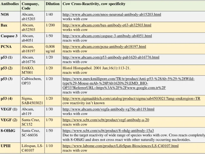
Immunohistochemical methods: Streptavidin-biotin-peroxidase complex method was used with SPlink HRP Broad Bulk Kit (D01-110, GBI®). Antibodies against iNOS, UPIII, Bax, caspase 3, PCNA, 8-OHdG, VEGF, and p53 were applied. Positive and negative controls were made as recommended by the manufacturers. Dilutions and cow cross-reactivity are given in Table 1. Four
Table 1. Antibodies, dilutions, and cow cross-reactivity. Tablo 1. Antikorlar, sulandırmaları ve sığır uyumluluğu.
Antibodies Company, Code
Dilution Cow Cross-Reactivity, cow specificity
NOS Abcam,
ab15203
1/40 http://www.abcam.com/nnos-neuronal-antibody-ab15203.html works with cow
Bax Abcam,
ab32503
1/200 http://www.abcam.com/bax-antibody-e63-ab32503.html works with cow
Caspase 3 Abcam, ab4051
1/50 http://www.abcam.com/caspase-3-antibody-ab4051.html reacts with cow
PCNA Abcam, ab18197
0,008 ug/ml
http://www.abcam.com/pcna-antibody-ab18197.html reacts with cow
p53 (1) Abcam, ab16776
1/20 http://www.abcam.com/p53-antibody-pab1620-ab16776.html reacts with cow
p53 (2) DAKO, M7001
1/20 Histol Histopathol. 2001 Jan;16(1):113-21. reacts with cow
p53 (3) Calbiochem, OP33
1/20 https://www.merckmillipore.com/TR/tr/product/Anti-p53-%28Ab-5%29-%28Wild-
type%29-Mouse-mAb-%28PAb1620%29,EMD_BIO-OP33?ReferrerURL=https%3A%2F%2Fwww.google.com.tr%2F reacts with cow
p53 (4) Sigma, SAB4503021
1/20 http://www.sigmaaldrich.com/catalog/product/sigma/sab4503021?lang=en®ion=TR cow reactivity isn’t known
VEGF (1) Abcam, ab119
1/30 http://www.abcam.com/vegfa-antibody-vg76e-ab119.html works with cow
VEGF (2) Santa Cruz, SC152
1/70 https://www.scbt.com/scbt/product/vegf-antibody-a-20 reacts with cow
8-OHdG Santa Cruz, SC-66036
1/50 https://www.scbt.com/scbt/product/8-ohdg-antibody-15a3
Due to the target reactivity of wide range of species works with cow. Cross reacts completely with 8-OHdG and does not cross react with other naturally occurring nucleotides. UPIII Lifespan,
LS-C40107
1/10 https://www.labome.com/product/LifeSpan-Biosciences/LS-C40107.html reacts with cow
different commercial antibodies for p53, and two for VEGF were tested. All immunohistochemical procedures, including control, were performed according to our previous study (14). Sections were visualized with 3-amino-9-ethylcarbazole (AEC), (GBI-C0112), 3,3'-diaminobenzidine (DAB), (Sigma-D4293), Vector VIP (Vectorlab- SK4600), and DAB black chromogens. Background colorized with Mayer’s Hematoxylin.
Results
Pathomorphological results: Macroscopically,
tumors were localized mostly in corpus and then, in trigonum, lateral wall, and vertex of urinary bladder, respectively. The histological patterns of lesions were
classified under light microscope and neoplastic, pre-neoplastic, inflammatory, and other lesions, were classified. Details of cases were given in Table 2 and findings were also visualized with macroscopical figures (Figures 1A-1F).
Immunohistochemical results: In sections stained by iNOS; urothelial cells, especially intact umbrella cells did not give positivity. Superficially settled cells, in erythrocyte exocytose-occurred hyperplastic epithelium, showed pretty much dense and strong positivity. Intensive positivity was also observed in severely degenerated and dead cells. All three layers of urothelium stained with iNOS in cases of papillary hyperplasia, and papilloma. In contrast to these stainings in urothelial layer and luminal
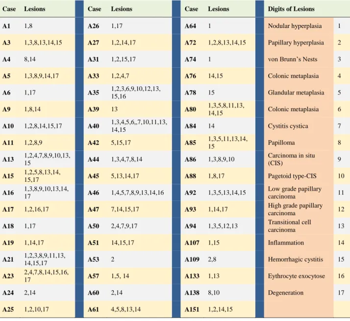
Table 2. Separate classification of neoplastic, pre-neoplastic, inflammatory, and the other lesions observed in every case. (A total 54 case). For the best visualization, lesions were shown by digits in this table and meaning of these digits were showed in the last two column.
Tablo 2. Her vakada görülen neoplastik, pre-neoplastik, yangısal ve diğer lezyonların ayrı ayrı sınıflandırılması. (Toplam 54 vaka). En iyi görsellik için, lezyonlar tabloda rakamlarla ifade edildi ve bu rakamların anlamları son iki sütunda gösterildi.
Case Lesions Case Lesions Case Lesions Digits of Lesions
A1 1,8 A26 1,17 A64 1 Nodular hyperplasia 1
A3 1,3,8,13,14,15 A27 1,2,14,17 A72 1,2,8,13,14,15 Papillary hyperplasia 2
A4 8,14 A31 1,2,15,17 A74 1 von Brunn’s Nests 3
A5 1,3,8,9,14,17 A33 1,2,4,7 A76 14,15 Colonic metaplasia 4
A6 1,17 A35 1,2,3,6,9,10,12,13,
15,16 A78 15 Glandular metaplasia 5
A9 1,8,14 A39 13 A80 1,3,5,8,11,13,
14,15 Colonic metaplasia 6
A10 1,2,8,14,15,17 A40 1,3,4,5,6,,7,10,11,13,
14,15 A84 14 Cystitis cystica 7
A11 1,2,8,9 A42 5,15,17 A85 1,3,5,11,13,14,
15 Papilloma 8 A13 1,2,4,7,8,9,10,13, 15 A44 1,3,4,7,8,14 A86 1,3,8,9,10 Carcinoma in situ (CIS) 9 A15 1,2,5,8,13,14,
15,17 A45 5,13,14,17 A88 1,8,17 Pagetoid type-CIS 10
A16 1,3,8,9,10,13,14,
17 A46 1,4,5,7,8,9,13,14,16 A92 1,3,5,13,14,15
Low grade papillary
carcinoma 11
A17 1,2,16,17 A47 7,14,15,17 A93 1,14,17 High grade papillary
carcinoma 12
A18 1,17 A50 2,4,7,9,17 A94 1,3,5,12,13 Transitional cell
carcinoma 13
A19 1,14,17 A51 14,15,17 A107 1,15 Inflammation 14
A21 1,2,3,8,9,11,13,
14,15,17 A53 2 A109 2,8 Hemorrhagic cystitis 15
A23 2,4,7,8,14,15,16,
17 A57 1,5, 14 A133 1,13 Eythrocyte exocytose 16
A24 2,14 A60 2,14 A138 8,10 Degeneration 17
surfaces of glandular metaplasia (GM), any positivity was seen in von Brunn’s nests (VBNs) and nodular hyperplasia. Although any positivity in the VBNs, luminally settled cells with pyknotic nuclei, in newly shaped glandular metaplasia from VBNs, showed cytoplasmic positivity. Dense cytoplasmic staining was seen in the dying cells during metaplastic transformation of VBN to GM. In invasive transitional cell carcinoma (TCC) cases prominent staining was observed in invasive tumor cells (Figures 2A-2C).
In sections stained by UPIII; normal superficial urothelial cells (umbrella cells) gave the best surface and cytoplasmic staining. Membrane-like immunopositive staining was predominantly observed on the luminal surface of epithelial cells both in normal and some pathological conditions. While there was weak positivity in the luminal surface of GM, any staining was obtained in the colonic metaplasia (CM) cases. Superficial epithelial cells, which were covering the area of neoplastic structures mainly in TCC showed membrane-like immunopositivity. A membrane like staining was also seen on the cell surfaces of both umbrella and the other cells, but cells in the CM with mucinous metaplasia displayed UPIII negativity.
When compared the positivity, completely opposite results were seen between UPIII and iNOS staining. The UPIII positive regions (asymmetric unit membrane- AUM structure protected normal urothelium) showed iNOS negative results and UPIII negative regions (AUM structure deformed urothelium) given iNOS positive result (Figures 2D-2E).
Similar to iNOS stain results, any 8-OHdG staining was detected in the areas where the firm's umbrella. Cytoplasmic positivity was observed in umbrella as well as in the closest cells to disintegrated areas. Absolutely negative 8-OHdG results were observed in the UPIII positive areas. It was determined that similar localizations of 8-OHdG positivity and the iNOS had significance (Figures 2F-2G).
In the presence of any epithelial lesion, individual VEGF positivity in just below lamina propria was interpreted to the opening of new vessels. In relation of tumor vascularization, individual cellular staining was detected among the neoplastic cells of invasive TCC, but negative results were obtained from mature capillaries in these cases. Evident cytoplasmic staining of p53, especially in pre-neoplastic pathology and epithelial tumors, showed parallelism with iNOS results. There were
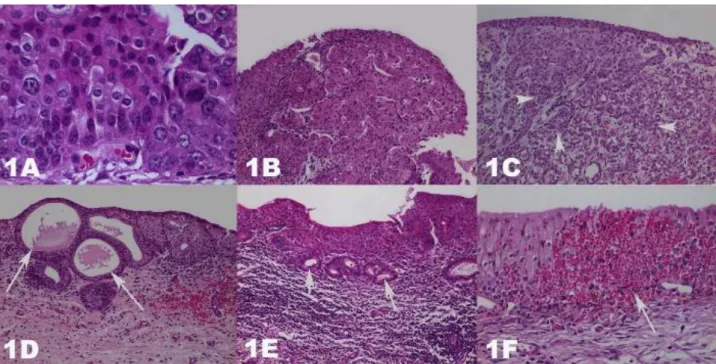
Figure 1A-F. Hematoxylin & Eosin stained urinary bladders. Magnifications are 40, 10, 4, 10, 10, and 20, respectively. A. Carcinoma in situ, anaplastic cells. B. High grade papillary carcinoma, cells showing anisocytosis and anisonucleosis. C. Transitional cell carcinoma, invasive neoplastic cells (arrow heads). D. Glandular metaplasia, gland-like metaplastic structures (arrows). E. Colonic metaplasia, gland like formations, which formed by mucin containing cells (arrows). F. Erythrocyte extravasation, free erythrocytes in the epithelial layers (arrow).
Şekil 1A-F. Hematoksilen & Eozin boyalı idrar keseleri. Resim büyütmeleri sırasıyla 40, 10, 4, 10, 10 ve 20’dır. A. Karsinoma in situ, anaplastik hücreler. B. Yüksek dereceli papiller karsinom, anizositozis ve anizonükleozis gösteren hücreler. C. Transisyonel hücreli karsinom, invaziv neoplastik hücreler (ok başları). D. Glanduler metaplazi, bez benzeri metaplastik yapılar (oklar). E. Kolonik metaplazi, müsin içeren hücrelerce oluşturulan bez benzeri oluşumlar (oklar). F. Eritrosit ekstravazasyonu, epitel katları arasında serbest eritrositler (ok).
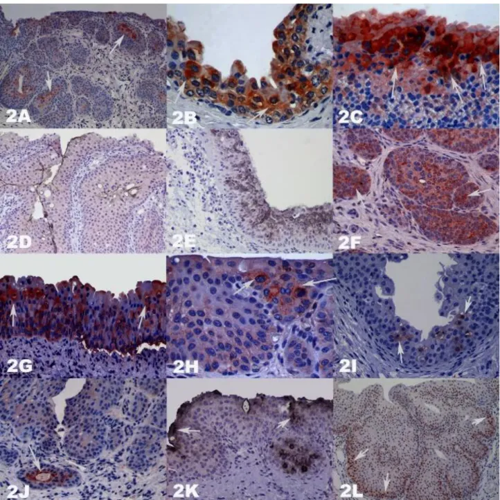
Figure 2A-L. Bladders stained with Streptavidin biotin peroxidase complex method with Mayer’s Hematoxylin counterstain. In general AEC chromogen was used for visualization DAB (UPIII), and Vector’s VIP (iNOS) chromogens were applied in double IHC stained D-E. DAB Black was used only K. Magnifications are 10, 40, 40, 20, 20, 20, 20, 40, 20, 20, 20, and 10, respectively. A. Luminal iNOS positivity in the GM formation started VBNs (arrows). B. Epithelial iNOS positivity in the damaged areas (arrows). C. Intense iNOS positivity in the erythrocyte exocytose occurred areas (arrows). D-E. UPIII and iNOS positivity. F. 8-OHdG positivity in the VBN (arrows). G. 8-OHdG positivity in the hyperplastic epithelium (arrows). H. Bax positivity in the hyperplastic epithelium (arrows). I. Cellular caspase 3 positivity in the hyperplastic epithelium (arrows). J. Luminal caspase 3 positivity in the GM formation started VBN (arrow). K. Cytoplasmic p53 positivity in hyperplastic epithelium, and GM formation started VBN (arrows). L. Basally intensive PCNA positivity in the hyperplastic epithelium (arrows).
Şekil 2A-L. Streptavidin biotin peroksidaz kompleks yöntemi ile boyanmış idrar keselerinde zemin boyaması Mayer's Hematoksilen ile yapılmıştır. Görünürlük için genellikle AEC kromojeni kullanıldı. Duble IHC ile boyanan D ve E’de DAB (UPIII) ve Vector's VIP (iNOS) kullanılırken, DAB black sadece K'da görülmektedir (oklar). Resim büyütmeleri sırasıyla 10, 40, 40, 20, 20, 20, 20, 40, 20, 20, 20 ve 10’dir. A. VBN’den GM’ye dönüşen yapıda lüminal iNOS pozitifliği (oklar). B. Hasarlı bölgelerde epitelyal iNOS pozitifliği (oklar). C. Eritrosit ekstravazasyonu şekillenmiş bölgelerde yoğun iNOS pozitifliği (oklar). D-E. UPIII ve iNOS pozitifliği. F. VBN'de 8-OHdG pozitifliği (oklar). G. Hiperplastik epitelde 8-OHdG pozitifliği (oklar). H. Hiperplastik epitelde Bax pozitifliği (oklar). I. Hiperplastik epitelde hücresel caspase 3 pozitifliği (oklar). J. VBN'den GM’ye dönüşen oluşumda luminal caspase 3 pozitifliği (ok). K. Hiperplastik epitelde ve VBN'den GM’ye dönüşen oluşumda sitoplazmik p53 pozitifliği (oklar). L. Hiperplastik epitelde bazal yerleşimli yoğun PCNA pozitifliği (oklar).
no staining in superficially settled cells and morphologically intact umbrella cells. Nuclear positivity was only detected in the cells which will remain on luminal surface in newly forming GM from VBNs by the metaplastic way. When the results of PCNA, Bax, and caspase 3 were examined, it was observed that apoptosis decreased in EH cases whereas cell proliferation increased (Figures 2H-2L).
Discussion and Conclusion
The mammalian urothelium consists of superficial (umbrella cells), intermediate, and basal cell layers (27). A major component of the luminal surface of superficial cells is the AUM (34). Main components of AUM are uroplakins (UPs) and four of these are named as uroplakin Ia, Ib, II, and III (34, 41). UPs shapes a barrier within the urine and tissue fluids and this is called as blood-urine barrier (5). UPs are specific for transitional epithelial tumors, as in normal transitional epithelium (22, 34). Changes in UP secretion leads a gradual lost in ability in the urothelium under neoplastic conditions and thus UPs may be a useful morphologic biomarker for urinary bladder pathologies (18, 43).
Immunohistochemical detection of UPs in domestic animals’ urothelial tumors has been reported only in cattle (1, 4) and dogs (29). Atypical staining was described in normal appeared areas which was near the tumor, and also near the urothelium which was above the endothelial-originated tumors in cow bladders (4). Similar to the previous studies, the positivity was predominantly observed on the luminal surface of epithelial cells under both normal and pathological conditions. According to our observations UP positivity in the surface cells on non-invasive lesions, such as hyperplastic areas, may be associated with well differentiation.
Absence in urothelial permeability was also determined for water and urine in UPs knock-out studies (13). In our cases, hyperplasia and its advanced forms were mostly found in areas where the permeability barrier is broken (UPIII negative). Accordingly, one may suggest that the urothelium tries to respond by cellular hyperplasia and other answers the absence of permeability barrier.
Expression, localization, and function of NO is mainly associated with iNOS in urothelium (18, 39). Nitric oxide can cause deterioration in superficial cells, thereby breaks permeability barrier (30).In our study, besides morphological differences in superficial urothelial cells, increase in iNOS secretion, and decrease in UPIII secretion were observed especially in pathological conditions. Consequently, we found iNOS positivity in UP negative and/or decreased areas, and iNOS negativity in UP positive areas.
A strong iNOS immunoreactivity were particularly found in AUM impaired and severe bleeding and
erythrocyte exocytosis shaped areas in our cases. This is the most likely originated since iNOS, hypoxia-inducible factor-1, and many other factors are produced during hemorrhage-caused tissue hypoxia (16).
8-hydroxy-2'-deoxyguanosine, a modified base, occurs in DNA after FR attacks. Immunohistochemical visualization of 8-OHdG will serve as a biomarker of oxidative damage in cellular DNA (2, 40). In our examination, prominent 8-OHdG positivity were seen in iNOS positive/ UPIII negative areas and this situation has given a strong basis for our theory about how free radicals may affect urinary bladder tumors in EH cases.
iNOS positivity is always defined in human TCC tissue and adjacent dysplastic area (12). However, no association between iNOS and p53 immunoreactivity were reported. A relationship between iNOS and p53 in human esophageal tumors has been mentioned (20). Increased NO levels may promote tumor growth via p53, and DNA repair mechanisms also degrade due to p53 deficiency (42). According to the researchers (28) intracytoplasmic p53 localization increases in endoplasmic reticulum-related stress conditions, and at the end of this, p53-associated apoptosis is blocked. Furthermore, cytoplasmic localization of p53 has been associated with metastasis and poor prognosis (23, 32). In our study, mostly intracytoplasmic p53 staining was observed in both neoplastic and pre-neoplastic tissues. Intracytoplasmic p53 accumulation have been interpreted as a finding of possible iNOS responsibility for cytoplasmic accumulation of p53, and failure of apoptotic effect by us.
A previous study (4) has mostly noticed p53 staining in vascular bladder cancers of cattle. A similar finding expressed by the researchers in epithelial invasive carcinomas and a cyclin D1 positivity also seen in p53 positive cells. Positive results for cyclin D1, specifically effective in G1/S phase of cell proliferation, have been described by these researchers. In our study, the course of cell proliferation was analyzed with PCNA and positivity was observed in neoplastic and pre-neoplastic pathologies from basally settled regions.
As it is well known, p53, Bcl-2, and Bax genes encode pro-apoptotic or anti-apoptotic affected proteins. Apoptotic process is interrupted in a p53 mutated cell, associated with DNA damage which cause genomic instability, on the other hand, Bcl-2 and Bax have adverse effects in process of cell death, over-release of Bcl-2 results cell proliferation, and over-secretion of Bax encourages cell death (37).
The balance between pro-apoptotic and anti-apoptotic regulator proteins is important for the future of the cell. Excessive apoptotic proteins (Bcl-2, etc.) are associated with cell survival but cell death results in an excess of Bax (45). One of the factors that affect apoptosis
is caspases and caspases have important roles in programmed cell death (6). Caspase 3, which is one of the most important executioner of apoptosis, is partially or fully served in many structural and regulatory protein cleavages (25). In our study, compared to the excessive PCNA positivity, a significant deprivation of apoptotic activity in these regions was present.
Some pro-apoptotic proteins like Bax can cause cell death by the caspase independent way (21). Therefore, the course of apoptosis was examined through both caspase 3 and Bax in this study. Both caspase 3 and Bax positive cells separately showed less positivity. This minority can be interpreted as the increased iNOS activity-caused impaired p53 localization (insufficient tumor suppression), and consequently decreased caspase, and Bax activation. iNOS and Bax negative results were obtained in the umbrella cells of undamaged AUM areas also support this theory.
The possibility of tumor growth, and metastasis are dependent to adequate blood flow which will provide with angiogenesis. Excessive release of iNOS, and VEGF induces angiogenesis in tumors. p53 even if suppress angiogenesis by suppressing the release of iNOS, and VEGF (9, 44). In our study; iNOS positivity in the vasculature components of benign or malignant tumors showed that NO activity continued in neoplastic cells.
Bleeding is common in the bladder tumors (7). In extracellular space, the regulation function of hemoglobin is lost, and Hb acts essentially as a NO scavenger (26). This may be as a result of S-nitrosilation of thiol groups or interaction with the iron in the heme (33). In our study, intense iNOS staining observed in the areas of bleeding can be interpreted in this context. Hemoglobin may clean the NO and thus hematuria reduces the level of NO.
Compared with UPIII and iNOS staining, completely opposite results were seen in our study. The UPIII positive regions (AUM structure protected normal urothelium) showed iNOS negative results and also UPIII negative regions (AUM structure deformed urothelium) showed iNOS positive result. Intracytoplasmic p53 accumulation in iNOS positive regions were noted as an important finding. We have interpreted that those findings may have been related with an endoplasmic reticulum-associated stress due to cytoplasmic accumulation of p53 and loss of its suppressor effect. Thus, apoptosis might have been disappeared on both caspase 3 and Bax in fern-associated carcinogenesis of cattle. In addition, 8-OHdG, as an example of oxidative DNA damage, positivity was seen in iNOS positive/UPIII negative areas. This finding is a strong evidence for active role of iNOS in fern-related urinary bladder carcinogenesis of cattle.
In conclusion; effect of FRs on the etiology were demonstrated in many different tumor types. To the best of knowledge of authors, this is the first study about the
effects of NO, which is a FR derivate, in the etiopathogenesis of fern associated-bladder tumors in cattle. The results showed that NO plays important roles in EH-associated tumors. And also, UPIII has an adverse effect to this NOS activity.
Acknowledgements
The authors declare that they have no conflict of interest. All experimental protocols were reviewed and approved by the Laboratory Animal Care Committee of Afyon Kocatepe University (AKUHEK-44-07). This research was supported by Afyon Kocatepe University, Scientific Research Coordination Unit (Project No: 08.VF.15).
References
1. Ambrosio V, Borzacchiello G, Bruno F, et al. (2001): Uroplakin expression in the urothelial tumors of cows. Vet Pathol, 38, 657-660.
2. Aruoma OI, Halliwell B (1995): 13-DNA Damage by Free Radicals: Carcinogenic Implications. 199-214. In: D Blake, G Paul (Eds), Immunopharmacology of Free Radical Species. Academic Press, London.
3. Campo MS, Jarrett WF, Barron R, et al. (1992): Association of bovine papillomavirus type 2 and bracken fern with bladder cancer in cattle. Cancer Res, 52, 6898-6904.
4. Carvalho T, Naydan D, Nunes T, et al. (2009): Immunohistochemical evaluation of vascular urinary bladder tumors from cows with enzootic hematuria. Vet Pathol, 46, 211-221.
5. Chang A, Hammond TG, Sun TT, et al. (1994): Permeability properties of the mammalian bladder apical membrane. Am J Physiol, 267, 1483-1492.
6. Cohen GM (1997): Caspases: the executioners of apoptosis. Biochem J, 326, 1-16.
7. Dawra RK, Sharma OP (2001): Enzootic bovine haematuria- Past, Present and Future. Vet Bull, 71, 1R-27R.
8. Dong Z, Staroselsky AH, Qi X, et al. (1994): Inverse correlation between expression of inducible nitric oxide synthase activity and production of metastasis in K-1735 murine melanoma cells. Cancer Res, 54, 789-793. 9. Forrester K, Ambs S, Lupold SE, et al. (1996): Nitric
oxide-induced p53 accumulation and regulation of inducible nitric oxide synthase expression by wild-type p53. Proc Natl Acad Sci USA, 93, 2442-2447.
10. Geller DA, Billiar TR (1998): Molecular biology of nitric oxide synthases. Cancer Metastasis Rev, 17, 7-23. 11. Hahm KB, Lee KJ, Choi SY, et al. (1997): Possibility of
chemoprevention by the eradication of Helicobacter pylori: oxidative DNA damage and apoptosis in H. pylori infection. Am J Gastroenterol, 92, 1853-1857.
12. Hayashi H, Kuwahara M, Fujisaki N, et al. (2001): Immunohistochemical findings of nitric oxide synthase expression in urothelial transitional cell carcinoma including dysplasia. Oncol Rep, 8, 1275-1279.
13. Hu P, Meyers S, Liang F-X, et al. (2002): Role of membrane proteins in permeability barrier function:
uroplakin ablation elevates urothelial permeability. Am J Physiol Renal Physiol, 283, F1200-1207.
14. Jadeski LC, Chakraborty C, Lala PK (2002): Role of nitric oxide in tumour progression with special reference to a murine breast cancer model. Can J Physiol Pharmacol, 80, 125-135.
15. Keles I, Bozkurt MF, Cemek M, et al. (2014): Prevention of cyclophosphamide-induced hemorrhagic cystitis by resveratrol: a comparative experimental study with mesna. Int Urol Nephrol, 46, 2301-2310.
16. Kiang JG, Bowman PD, Wu BW, et al. (2004): Geldanamycin treatment inhibits hemorrhage-induced increases in KLF6 and iNOS expression in unresuscitated mouse organs: role of inducible HSP70. J Appl Physiol, 97, 564-569.
17. Klotz T, Bloch W, Volberg C, et al. (1998): Selective expression of inducible nitric oxide synthase in human prostate carcinoma. Cancer, 82, 1897-1903.
18. Lobban ED, Smith BA, Hall GD, et al. (1998): Uroplakin gene expression by normal and neoplastic human urothelium. Am J Pathol, 153, 1957-1967.
19. Marletta MA, Yoon PS, Iyengar R, et al. (1988): Macrophage oxidation of L-arginine to nitrite and nitrate: nitric oxide is an intermediate. Biochemistry (Mosc), 27, 8706-8711.
20. Matsumoto M, Furihata M, Kurabayashi A, et al. (2003): Association between inducible nitric oxide synthase expression and p53 status in human esophageal squamous cell carcinoma. Oncology, 64, 90-96.
21. McCarthy NJ, Whyte MK, Gilbert CS, et al. (1997): Inhibition of Ced-3/ICE-related proteases does not prevent cell death induced by oncogenes, DNA damage, or the Bcl-2 homologue Bak. J Cell Biol, 136, Bcl-215-Bcl-2Bcl-27.
22. Moll R, Wu XR, Lin JH, et al. (1995): Uroplakins, specific membrane proteins of urothelial umbrella cells, as histological markers of metastatic transitional cell carcinomas. Am J Pathol, 147, 1383-1397.
23. Moll UM, Ostermeyer AG, Haladay R, et al. (1996): Cytoplasmic sequestration of wild-type p53 protein impairs the G1 checkpoint after DNA damage. Mol Cell Biol, 16, 1126-1137.
24. Özkul IA, Aydin Y (1996): Tumours of the urinary bladder in cattle and water buffalo in the Black Sea region of Turkey. Br Vet J, 152, 473-475.
25. Peña-Llopis S, Ferrando MD, Peña JB (2003): Fish tolerance to organophosphate-induced oxidative stress is dependent on the glutathione metabolism and enhanced by N-acetylcysteine. Aquat Toxicol Amst Neth, 65, 337-360. 26. Perutz MF (1996): Blood. Taking the pressure off. Nature,
380, 205-206.
27. Peter S (1993): Ultrastructure of the Urothelium. 23-33. In: P Rathert, S Roth, M Soloway (Eds), Urinary Cytology. 2nd Ed. Springer-Verlag, Berlin-Heidelberg.
28. Qu L, Huang S, Baltzis D, et al. (2004): Endoplasmic reticulum stress induces p53 cytoplasmic localization and prevents p53-dependent apoptosis by a pathway involving glycogen synthase kinase-3beta. Genes Dev, 18, 261-277. 29. Ramos-Vara JA, Miller MA, Boucher M, et al. (2003):
Immunohistochemical detection of uroplakin III, cytokeratin 7, and cytokeratin 20 in canine urothelial tumors. Vet Pathol, 40, 55-62.
30. Romih R, Korosec P, Sedmak B, et al. (2008): Mitochondrial localization of nitric oxide synthase in partially differentiated urothelial cells of urinary bladder lesions. Appl Immunohistochem Mol Morphol AIMM, 16, 239-245.
31. Roperto S, Borzacchiello G, Brun R, et al. (2010): A review of bovine urothelial tumours and tumour-like lesions of the urinary bladder. J Comp Pathol, 142, 95-108. 32. Schlamp CL, Poulsen GL, Nork TM, et al. (1997):
Nuclear exclusion of wild-type p53 in immortalized human retinoblastoma cells. J Natl Cancer Inst, 89, 1530-1536. 33. Stamler JS (1994): Redox signaling: Nitrosylation and
related target interactions of nitric oxide. Cell, 78, 931-936. 34. Sun TT, Liang FX, Wu XR (1999): Uroplakins as markers
of urothelial differentiation. 7-18. In: LS Baskin, SW Hayward (Eds), Advances in Bladder Research. Plenum Publishers Corporation, New York.
35. Thomsen LL, Miles DW (1998): Role of nitric oxide in tumour progression: Lessons from human tumours. Cancer Metastasis Rev, 17, 107-118.
36. Thomsen LL, Scott JM, Topley P, et al. (1997): Selective inhibition of inducible nitric oxide synthase inhibits tumor growth in vivo: studies with 1400W, a novel inhibitor. Cancer Res, 57, 3300-3304.
37. Weinberg RA (2007): The Biology of Cancer. 307-356. Garlan Science, New York.
38. Wiseman H, Halliwell B (1996): Damage to DNA by reactive oxygen and nitrogen species: role in inflammatory disease and progression to cancer. Biochem J, 313, 17-29. 39. Wolf H, Haeckel C, Roessner A (2000): Inducible nitric oxide synthase expression in human urinary bladder cancer. Virchows Arch Int J Pathol, 437, 662-666.
40. Wu LL, Chiou CC, Chang PY, et al. (2004): Urinary 8-OHdG: A marker of oxidative stress to DNA and a risk factor for cancer, atherosclerosis and diabetics. Clin Chim Acta, 339, 1-9.
41. Wu XR, Lin JH, Walz T, et al. (1994): Mammalian uroplakins. A group of highly conserved urothelial differentiation-related membrane proteins. J Biol Chem, 269, 13716-13724.
42. Xu W, Liu LZ, Loizidou M, et al. (2002): The role of nitric oxide in cancer. Cell Res, 12, 311-320.
43. Yu J, Manabe M, Wu XR, et al. (1990): Uroplakin I: a 27-kD protein associated with the asymmetric unit membrane of mammalian urothelium. J Cell Biol, 111, 1207-1216. 44. Zhang L, Yu D, Hu M, et al. (2000): Wild-type p53
suppresses angiogenesis in human leiomyosarcoma and synovial sarcoma by transcriptional suppression of vascular endothelial growth factor expression. Cancer Res, 60, 3655-3661.
45. Zheng JH, Viacava Follis A, Kriwacki RW, et al. (2016): Discoveries and controversies in BCL-2 protein-mediated apoptosis. FEBS J, 283, 2690-2700.
Geliş tarihi: 03.02.2017 / Kabul tarihi: 16.04.2017 Correspondence Author:
Assoc. Prof. Dr. Hikmet KELES Afyon Kocatepe University, Faculty of Veterinary Medicine, Department of Pathology,
03030, ANS Campus, Afyonkarahisar, TURKEY e-mail: [email protected]