Short Communication / Kısa Bilimsel Çalışma
A case of vaginal xanthoma in a cow; First report
Özlem ÖZMEN1, Tekinalp GELEN2, Ahmet AYDOĞAN1, Mehmet HALIGUR1,Yunus ÇETIN3
Mehmet Akif ErsoyUniversity, Faculty of Veterinary Medicine, 1Department of Pathology, 3Department of Obstetrics and
Gynecology, Burdur; 2AkdenizUniversity, Medical Faculty, Department of Pathology, Antalya, Turkey.
Summary: A case of vaginal xanthoma in a 3-year-old Holstein cow is described on the basis of macroscopic, microscopic and immunohistochemical findings. A pink-colored mass surgically excised from the vagina was hard, nodular and 328g in weight. Histopathologically, the mass was consisted of lobular areas surrounded by a stroma and composed of numerous foamy macrophages, abundant lipid material, rarely giant cells and cholesterol clefts. Immunohistochemically, the mass was positive for vimentin, proliferating cell nuclear antigen (PCNA) and CD 68, but negative for smooth muscle actin (SMA), glial fibriler acidic protein (GFAP) and S100 protein antibodies. To the authors’ knowledge, this is the first report of a vaginal xanthoma in a cow.
Keywords: Cow, immunohistochemistry, xanthoma, vagina.
Bir inekte vaginal ksantom olgusu; İlk rapor
Özet: Bu olguda, 3 yaşlı, Holştayn ırkı bir inekte saptanan vaginal ksantom makroskobik, mikroskobik ve immunohisto-kimyasal olarak tanımlandı. Vaginadan pembe renkli, sert kıvamlı, 328 gr ağırlığındaki bir kitle cerrahi olarak uzaklaştırıldı. Histopatolojik olarak, kitle stromayla sarılı lobuler alanlardan oluşuyordu ve çok sayıda köpüklü makrofajlar, yoğun lipid materyali, az sayıda dev hücre ile kolesterol yarıkları içeriyordu. Immunohistokimyasal olarak vimentin, prolifere hücre nükleer antijen (PCNA) ve CD 68 ile pozitif; düz kas aktin (SMA), glial fibriler asidik protein (GFAP) ve S100 protein ile negatif reaksiyon saptandı. Yapılan taramalara göre bu, bir inekte saptanan ilk vaginal ksantom olgusudur.
Anahtar sözcükler: İmmunohistokimya, inek, ksantom, vagina.
Xanthomas refer non-neoplastic but reactive lesions that typically form yellowish macules, papules, nodules or plaques in the skin associated with lipid deposits (Banajee et al., 2011; Balme et al., 2009). They are frequently localized in the subcutaneous or submucosal tissues like as oral and genital mucosa or tendons, tendon sheaths, brain, bone and esophagus in human (Muthusamy et al., 2008; Hirokawa et al., 2003; Bonhomme et al., 2000; Burgdorf, 1997; Jones et al., 1997). Histopathologically, they consist of collection of large, pale “foam cells” doubtless of reticuloendothelial origin with extensive granular cytoplasm by many minute droplets of cholesterol and other lipids (Jones et al., 1997). Varying degrees of fibrosis and clefts can be seen. Most xanthoma cells are mononuclear, but giant cells, especially of the Touton type with a wreath of nuclei, may be found (Dhanuthai and Torrungruang, 2007; Garner et al., 1999; Chastain and Graham, 1978). Particularly in humans but also commonly in animals, they are associated with hyperlipidemia (Balme et al., 2009; Dhanuthai and Torrungruang, 2007; Garner et al., 1999; Chastain and Graham, 1978).
According to veterinary literature, xanthomas are seen rarely in dogs, cats and goats (Ozmen and Haligur, 2012; Banajee et al., 2011; Balme et al., 2009; Romanucci et al., 2008; Vogelnest, 2001; Chastain and Graham, 1978). There is only one report of ocular xanthelasma (a kind of xanthoma of the eyelids) in a cow (Derakhshanfar and Molaei, 2007). The aim of this report is to describe clinicopathological features of vaginal xanthoma in a cow.
A 3-year-old, female, Holstein cow was presented to Veterinary Faculty clinics with complain of difficult and painful urination that started 2 weeks ago. The body condition score of the cow was good. She was calving 2 months ago and there was no difficult birth stated at anamnesis. The cow was clinically examined by rectal palpation and introducing a vaginal speculum. A round and firm mass was observed at the right vaginal wall. The mass caused mechanical pressure to urethra and orificium urethra externa. No lesions were found in the other genital tracts of the cow. Normal blood biochemical and hematological results were observed.
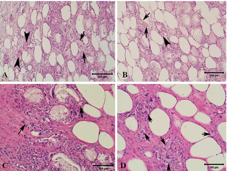
Figure 2. A-D: Microscopical features of xanthoma. Numerous foamy cells (arrows) in which excess fat material are stored inside histiocytes, with clear, foamy and vacuolated abundant eosinophilic cytoplasm, and giant cells (arrow heads), HE.
Şekil 2. A-D: Ksantomun mikroskobik görünümleri. Çok sayıda yoğun yağlı material içeren, açık renk, köpüklü ve vakuollü bol eozinofilik sitoplazmalı histiyositlerden oluşan köpük hücreleri (oklar) ile dev hücreler (ok başları), HE.
Figure 1. A: Macroscopical feature of the vaginal mass measuring 17x15x8.5cm in size. B: Cut surface of the vaginal mass, is subdivided into whitish lobular masses by thin septa.
Şekil 1. A: 17x15x8.5cm çapındaki vaginal kitlenin makroskobik görünümü. B: Vaginal kitlenin kesit yüzü, ince septumlarla bölünmüş beyazımsı lobüler yapılar görülüyor.
and its appetite was normal. The cow feed adlibitum by commercial cattle food and high milk performance was stated by the owner. Caudal epidural anesthesia was induced for surgical treatment by administration of 5mL of 2% lidocaine (Adokain; Sanovel, Turkey). Also, local anesthesia was performed with the infiltration of the same drug within the vaginal mucosa around the base of the mass. Following ligation of the stalk, the mass was surgically removed through the vaginal route. Normal saline (3 liters) were injected intravenously during the operation. Large spectrum antibiotic (Clemipen-Strep; Topkim, Turkey) was given twice daily for a period of 4 days intramuscularly as postoperative care.
After surgical removing the mass was presented to the Department of Pathology. For histopathological examination, tissue samples were fixed in 10% neutral buffered formalin and processed routinely for light microscopy. Five micron thick sections were taken from paraffin embedded tissues and stained with Hematoxylin- Eosin (HE), Periodic acid-Schiff (PAS), Ziehl-Neelsen and Gram’s stains to detect any microorganisms. Oil red O staining was used on frozen sections for to demonstrate fat.
Selected tissue sections were stained immunohisto-chemically in order to demonstrate proliferating cell nuclear antigen (PCNA; DAKO- Glostrup, Denmark, monoclonal mouse anti-PCNA, Clone:PC10, 1/100 dilution), S100 protein (Neomarker; S100 protein Ab-2, rabbit polyclonal antibody, 1/100 dilution), smooth muscle actin (SMA; DAKO, monoclonal mouse anti-human, Clone:1A4, 1/100 dilution), vimentin (DAKO, monoclonal mouse anti-vimentin Clone:V9, 1/100 dilution), CD68 (DAKO, monoclonal mouse anti-human CD68, Clone PG-M1, ready to use) and glial fibrillary acidic protein (GFAP; DAKO, polyclonal rabbit anti-glial fibrillary acidic protein, Clone Z 0334, 1/500 dilution) using a routine streptavidine-biotin peroxidase technique.
Briefly, after deparaffinization, endogenous peroxidase blocking (methanol with 0.3% H2O2; 10 min) and rinsing with phosphate-buffered saline (PBS; 0.01 M, pH 7.3), microwave heat retrieval (citrate buffer solution 1:100; 10 min) were performed for all antibodies (Abs), followed by rinsing in PBS. After preincubation for 30 min with 10% normal rabbit serum, the slides were incubated with primer antibodies. After incubation of primary antibody, slides were incubated with streptoavidine peroxidase for 20 min and after washing PBS biotinilated Abs for 30 min. Then all slides were rinsed with PBS, incubated with peroxidase substrate solution containing DAB, rinsed with distilled water, counter stained with hematoxylin, dehydrated and mounted. Negative controls for specificity consisted of omission of the primary antibody.
17x15x8.5 cm in size, pinkish in color with numerous yellowish-white areas apparent on the cut surface (Figure 1A-B). The mass was congested but there was no ulcerative region and hemorrhage on the surface of the mass.
At the histopathological examination, the submucosal vaginal mass in this study consisted of well demarcated nodules of numerous lipid-laden macrophages containing abundant finely vacuolated cytoplasm with rarely giant cells and frequent cholesterol clefts were seen. Moderate numbers of lymphocytes were scattered throughout the mass. There was replacement of individual to small groups of adipocytes. There was no cellular, atypia or mitosis in the xanthoma cells, and they did not exhibit invasive behavior. Numerous Touton giant cells were also observed characterizing the xanthoma in this case. Totally, the histopathological and immunohistochemical findings observed in this case were in agreement with previous reports (Figures 2A-D). The diagnosis of xanthoma in this cow was primarily based on the characteristic pathological, histochemical and immuno-histochemical findings. Microscopic examination of the mass revealed that histiocytic proliferation suggestive a histiocytic disorder. Oil red O staining supported the lipid accumulation (Figure 3A).
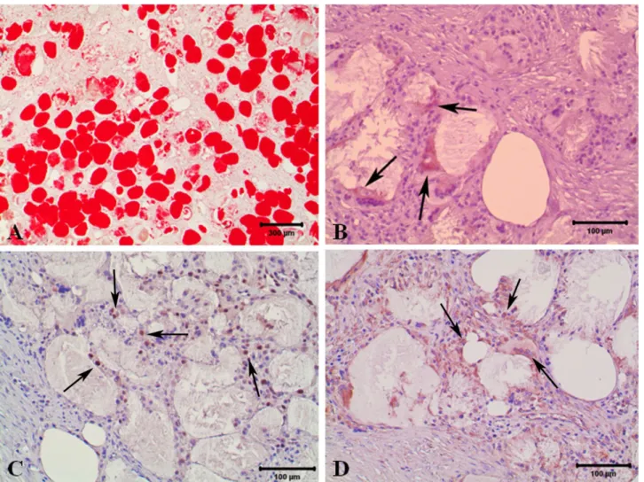
In this case, immunohistochemically CD68, vimentin and PCNA were positive while S-100, SMA and GFAP were negative (Figures 3B-D).
Xanthomas are benign, proliferative lesions that commonly occur in the skin, subcutis, or tendon sheaths of hyperlipidemic patients in humans and animals (Banarjee et al., 2011; Dhanuthai and Torrunruang, 2007; Garner et al., 1999; Chastain and Graham, 1978). They typically form smooth, white to pale yellow nodules and plaques in the skin (Massengale and Nesbitt, 2004). Genital localization of xanthomas was reported in humans but not in animals (Metgud et al., 2008; Ersahin et al., 2005). However, xanthomas are uncommon in animals compared to humans (Massengale and Nesbitt, 2004; Jones et al., 1997; Jackler and Brackmann, 1987), they generally occur in the subcutis of birds (Goldschmidt and Hendrick, 2002). But xanthomas have been rarely reported in other animals (Ozmen and Haligur, 2012; Derakhshanfar and Molaei, 2007; Jones et al., 1997; Jackler and Brackmann, 1987). Extra dermal localizations also reported like as ocular and intestinal areas in animals (Romanucci et al., 2008; Derakhshanfar and Molaei, 2007). There is no report available about vaginal localization of this lesion in cattle.
Microscopically, diffuse, nodular infiltrates of multinucleated giant cells, large macrophages containing abundant foamy cytoplasm and cholesterol clefts are characteristic for xanthoma (Banarjee et al., 2011; Balme
et al., 2009; Dhanuthai and Torrungruang, 2007; Reavill, 2004; Garner et al., 1999; Burgdorf, 1997; Oberman et al., 1992; Chastain and Graham, 1978). Xanthomas are not neoplastic, but can be locally invasive (Banarjee et al., 2011; Hirokawa et al., 2003; Oberman et al., 1992). In this case the mass localized the wall of the vagina and have characteristic histopathological appearance of xanthomas.
Xanthomas must be differentiated from granulomatous inflammation secondary to infectious agents such as fungi and mycobacteria (Goldschmidt and Hendrick, 2002). The histopathological features in this case were therefore highly suggestive of xanthoma. Periodic acid-Schiff (PAS), Ziehl-Neelsen and Gram’s stains failed to detect microorganisms. Frozen tissue from the mass stained positively with Oil Red O. Xanthomas in humans show positive CD68 indicating a macrophage origin and negative S100 protein immunoreactivity (Romanucci et al., 2008; Scott et al., 2001). In this case, CD68 positivity
and absence of S-100 reactivity indicated the histiocytic origin. In addition, the positive vimentin immunostaining seen in our case further indicates a mesenchymal origin. The cells were negative for SMA and GFAP, which would not support muscle or nerve origin. The positive PCNA immunostaining was probably due to increased proliferative activity of the cells in the mass because of the proliferative nature of the xanthomas (Ozmen and Haligur, 2012).
Treatment consists of conservative excisional surgery and recurrence is rare (Burgdorf, 1997). Postoperatively, the cow had no sequelae secondary to the operation on postoperative day 2. At 5 months after surgery, no recurrence observed in present case. Xanthomas are commonly associated with hyperlipidemia in humans, dogs and cats (Scott et al., 2001; Bonhomme et al., 2000; Jackler and Brackmann, 1987). Unfortunately, we were unable to measure blood lipids. The pathogenesis of this atypical xanthoma in this cow remains obscure.
Figure 3. A: Xanthoma, positive staining for lipid found in both the extracellular space and in the cytoplasms of macrophages (Oil Red O, Harris haematoxylin counterstain). B: Brown color CD68 immunopositive cells (arrows); C: Brown color PCNA immunopositive cells (arrows); D: Brown color vimentin immunopositive cells (arrows) of xanthoma (Streptavidin–biotin complex method, haematoxylin counterstain).
Şekil 3. A: Ksantom, hem ektreselüler bölgedeki hemde makrofajlar sitoplazmasındaki lipid için pozitif boyanma (Oil Red O, Harris haematoxylin zıt boyama). B: CD68 için kahverengi immun pozitif hücreler (oklar); C: PCNA için immunopozitif hücreler (oklar) D: Vimentin immunopozitif boyanma (oklar) of xanthoma (Streptavidin–biotin complex method, haematoxylin counterstain).
pathological findings of a vaginal xanthoma in a Holstein cow.
References
1. Balme E, Thuilliez C, Lejeune T, Chateau-Escoffier L, Bernex F (2009): Multiple atypical mucosal xanthomas in
a dog similar to human verruciform xanthoma. J Vet
Diagn Invest, 21, 124-128.
2. Banarjee KH, Orandle MS, Ratterree W, Bauer RW, Gaunt SD (2011): Idiopathic solitary cutaneous xanthoma
in a dog. Vet Clin Pathol 40, 95-98.
3. Bonhomme GR, Loevner LA, Yen DM, Deems DA, Bigelow DC, Mirza N (2000): Extensive intracranial
xanthoma associated with type II hyperlipidemia. Am J
Neuroradiol, 21, 353–355.
4. Burgdorf WHC (1997): The histiocytoses. 591-616. In: D Elder, R Elenitsas, C Jaworsky, B Johnson, (Eds.), Lever’s Histopathology of the Skin. Lippincott-Raven Publishers, New York.
5. Chastain CB, Graham CL (1978): Xanthomatosis secondary
to diabetes mellitus in a dog. JAVMA, 172, 1209-1211.
6. Derakhshanfar A, Molaei MM (2007): Occurrence of
xanthelasma in a cow (the first report). Iranian J Surg, 2,
73-76.
7. Dhanuthai K, Torrungruang K (2007): Verruciform
xanthoma: Report of three cases. Acta Stom Croat, 41,
166-162.
8. Ersahin C, Szpaderska AN, Foreman K, Yong S (2005):
Verucciform xanthoma of the penis not associated with human papillomavirus infection. Arch Path Lab Med, 129, 62–64.
9. Garner MM, Lung NP, Murray S (1999): Xanthomatosis in geckos: five cases. J Zoo Wildlife Med, 30, 443-447. 10. Goldschmidt MH, Hendrick MJ (2002): Tumors of the
skin and soft tissues. 111-113. In: DJ Meuten (Ed.), Tumors
in Domestic Animals, Iowa State Press, Ames, Iowa. 11. Hirokawa M, Takenaka R, Takahashi A, Sugihara K,
Wada H, Tashiro T, Horiguchi H, Wakatsuki S, Sano T (2003): Esophageal xanthoma: Report of two cases and a
review of the literature. J Gastroenterol Hepatol, 18, 1105–
1158.
13. Jones TC, Hunt RD, King NW (1997): Veterinary
Pathology, Williams and Wilkins, Maryland.
14. Massengale WT, Nesbitt Jr LT (2004): Xanthomas. 1447-1454. In: JL Bolognia, JL Jorizzo, RP Rapini (Eds), Dermatology. Mosby, Philadelphia.
15. Metgud M, Malur P, Mallya S (2008): A rare case of
verruciform xanthoma of vulva. J Turkish-German
Gynecol Assoc, 9, 350-352.
16. Muthusamy KA, Azmi K, Rad M, Narayanan P, Rajagopalan R, Rahmen NA, Waran V (2008): Bilateral
temporal bone xanthoma. J Neurosurg, 108, 361–364.
17. Oberman A, Kreisberg RA, Henkin Y (1992): Principles
and Management of Lipid Disorders: A Primary Care Approach. Williams and Wilkins, Baltimore.
18. Ozmen O, Haligur M (2012): A case of xanthoma in a
Saanen goat. Vet Dermatol, 23, 150- e32.
19. Reavill DR (2004): Tumors of pet birds. Vet Clin North America: Exotic Anim Prac. 7, 537-560.
20. Romanucci M, Malatesta D, Guardiani P, Frescura P, Salda LD (2008): Xanthogranulomatous inflammation of
the small bowel in a dog. Vet Pathol, 45, 207-211.
21. Scott DW, Miller WH, Griffin CE (2001): Small Animal
Dermatology. Saunders, Philadelphia.
22. Vogelnest, LJ (2001): Cutaneous xanthomas with concurrent demodicosis and dermatophytosis in a cat. Aust Vet J 79, 470-475.
Geliş tarihi: 13.12.2013 / Kabul tarihi: 15.04.2014
Address for correspondence:
Prof. Dr. Özlem Özmen Mehmet Akif Ersoy University, Faculty of Veterinary Medicine, Department of Pathology,
Istiklal Yerleskesi, 15030 Burdur, Turkey e- mail: [email protected]