Journal Of Ankara University Faculty of Medicine 2011, 64(3)
Ayșegül Öksüzoğlu, Özlem Moraloğlu, Ayla Aktulay, Yaprak Engin-Üstün, Șevki Çelen, Leyla Mollamahmutoğlu 141
Conjoined Twins: A Case Report of Thoraco-Omphalopagus
Yapıșık İkizler; Bir Torako-Omfalofagus Olgu SunumuAyșegül Öksüzoğlu
1, Özlem Moraloğlu
1, Ayla Aktulay
1, Yaprak Engin-Üstün
1,
Șevki Çelen
1, Leyla Mollamahmutoğlu
11 Zekai Tahir Burak WomAn’s Health Education and Research
Hospital, Department of Perinatology, Ankara, Turkey A 29-year old woman, gravida 2, para 1 was referred for routine scan at 12 weeks. Sonographic
examination performed at 12 gestational weeks revealed conjoined twins. The fetal biometry of both twins was consistent with the menstrual age. The upper and lower limbs of both twins were normal. Fetuses were positioned face-to-face and fused from umbilicus to lower thorax. Fetuses were found to share the heart and liver. One fetus had cystic hygroma. The parents opted to terminate the pregnancy but refused autopsy.
Key Words: Conjoined twins, thoracoomphalopagus, monozygotic pregnancies.
29 yașında, gravida 2, parite 1 olan hasta 12. gebelik haftasında ikiz gebelik nedeniyle rutin ultrason taramasında refere edildi. Her iki fetusun fetal biometri ölçümleri gebelik haftası ile uyumluydu. İkizlerin üst ve alt ekstremiteleri normal gözlendi. Fetuslar ultrasonik gözlemde yüz-yüze pozisyondaydılar ve umblikus ve toraksın alt seviyesinde yapıșık oldukları gözlendi. Ultrasonda karaciğer ve kalp tek izlendi. Fetuslardan birinde kistik higroma mevcuttu. Aile bilgilendirildikten sonra gebelik terminasyonunu kabul etti fakat otopsiyi kabul etmedi. Anahtar Sözcükler: Yapıșık ikizler, torako-omfalofagus, monozigotik gebelikler. Conjoined twins are a rare complication
of monozygotic pregnancies. Due to its high mortality and morbidity, early prenatal diagnosis becomes important. Prognosis is related to the vitality of the shared organs and gravity of the accompanying congenital anomalies, as well as to the location and extent of adhesion. Conjoined twins are classified according to the adhesion regions: thoracopagus (thorax), omphalopagus (abdomen), pygopagus (sacrum), ischiopagus (pelvis), craniopagus (cranium), cephalopagus (face), ve rachipagus (dorsal). Thoracopagus is the most frequent type. 75 % of conjoined twins are seen as thoracoomphalopagus. Here we described the prenatal diagnosis
of a case of thoraco-omphalopagus conjoined twin diagnosed during the first trimester with two-dimensional ultrasound at 12 weeks of gestation.
Case report
A 29-year old woman, gravida 2, para 1 was referred for routine scan at 12 weeks. Family history was negative.
Her past medical history was unremarkable. There was no information on drug usage during pregnancy. Her first birth was by cesarean section due to breech presentation. She had no family or personal history of twins. Sonographic examination performed at 12 gestational weeks revealed conjoined twins (Figure 1).
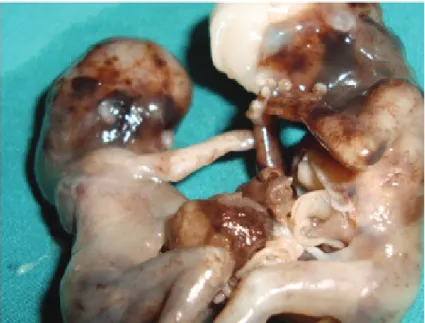
The fetal biometry of both twins was consistent with the menstrual age. The upper and lower limbs of both twins were normal. Fetuses were positioned face-to-face and fused from umbilicus to lower thorax. We had inability to separate fetal bodies. There was no change in the relative positions of the fetuses despite attempts. Fetuses were found to share the heart and liver. One fetus had cystic hygroma. The parents opted to terminate the pregnancy (Figure 2). The family refused autopsy.
Discussion
Conjoined twins are one of the rare congenital malformation /anomalies, Ankara Üniversitesi Tıp Fakültesi Mecmuası 2011, 64 (3)
DOI: 10.1501/Tıpfak_000000799
CERRAHİ BİLİMLER/SURGICAL SCIENCES
Case Report/ Olgu Sunumu
Received: 03.01.2011 • Accepted: 18.10.2011 Corresponding author
Ozlem MORALOGLU, MD
Talatpasa Bulvarı 158/5 Cebeci/Ankara Perinatoloji Anabilim Dalı
Gsm : +905326855157 Fax : 00903123627778
Ankara Üniversitesi Tıp Fakültesi Mecmuası 2011, 64(3)
Conjoined Twins: A Case Report of Thoraco-Omphalopagus 142
seen every 50,000-100,000 births, where 1 % of the monozygotic twins are effected. Two different theories have been suggested on the formation of conjoined twins. According to the “fission” theory, 13-15 days after the fertilization embryonic disc has an incomplete separation where as in the “fusion” theory, two separate monoovulatuar embryonic discs undergo a secondary association. Recently the fusion theory is accepted because it can explain all conjoined twin phenomenon (1). Long-term usage of contraceptive
drugs, abnormal calcium metabolism and extremely underweight woman with ovulatory dysfunctions are suspected in etiology. It is three times more frequent in girls.
In thoracopagus the twins are positioned face-to-face, and very commonly sternum, diaphragm, upper abdominal wall and around 75% the heart are conjoined. Almost always there is an atrial joint and a mutual pericardium. Twins with omphalopagus are conjoined in the umbilical region, often containing the lower thorax.
Liver is around 80 % mutual and this sharing is usually not equally balanced. In cases where duodenum is shared biliary anomalies are frequent. Heart is almost always separate, but a pericardial adhesion may be present. Congenital heart disease will be seen in 30% of the cases. In our report fetuses were found to share the heart and liver. Cardiac defects, congenital diaphragmatic hernia, neural tube defect, cystic hygroma, renal dysplasia, club foot, intestinal atresia are frequently seen in conjoined twins (2). One of the fetuses in our case had cystic hygroma. Developments in the imaging technologies,
widespread utilization of high resolution transvaginal ultrasonography enables prenatal diagnosis of conjoined twins in the first trimester. The earliest reported case of thoracopagus case in literature was in the 7th week of pregnancy (3). The false positive ratio is very high before the 10th week of pregnancy. In the earlier weeks of pregnancy fetal movements are limited and monochorionic twins may be mistaken for conjoined twins (4). Hence the ultrasonography screening should be repeated in the 11th and
12th weeks of pregnancy. 3D USG
can be helpful in early first trimester. An adherent umbilical cord containing varying 2 to 7 blood vessels is present. When monoamniotic twin pregnancy is determined by ultrasonography, the fetuses should be carefully examined for conjointment aspects. Similar positioning of the twins, lack of independent movements, non-detaching fetus bodies during positional changes, a single umbilical cord containing more than three blood vessels, excessive flexion of fetal spinal column, lack of position changes in the twins among examinations performed at different intervals will be helpful for diagnosing conjoined twins. Evaluation of the conjoined twins, the location and extent of the adhesion, presence of fetal anomalies, existent organ sharing will be very difficult to evaluate through ultrasonography in the 2nd and 3rd trimesters.
Figure 1: Sonographic examination performed at 12 gestational weeks .
Journal Of Ankara University Faculty of Medicine 2011, 64(3)
Ayșegül Öksüzoğlu, Özlem Moraloğlu, Ayla Aktulay, Yaprak Engin-Üstün, Șevki Çelen, Leyla Mollamahmutoğlu 143
Magnetic resonance and recently widely used ultrafast MR will assist in prenatal diagnosis. Conjoined twins can be diagnosed in the first trimester. 40 % are stillborn. 75 % of live births are lost in the first 24 hours of life. Treatment is most frequently termination. If a decision for continuation of pregnancy is given, congenital cardiac anomalies should be determined by fetal echo, degrees of organ sharing and congenital anomalies should be
determined by fetal MRI and delivery should take place in a tertiary center. Mode of delivery is cesarean section and a vertical uterine incision is often required. The location and extent of the conjoinment, presence of additional anomalies in the fetuses, whether the vital organs are shared or not, the extent of the shared organs are very important in terms of pregnancy prognosis. They will greatly effect the decisions for surgical separation or termination of
pregnancy. Mortality and morbidity ratios for the conjoined twins are still high despite the developments in the radiological imaging methods and surgical treatment techniques. Once the diagnosis of conjoined twins is established, the family should be informed in detail. Besides the choice of termination, if continuation of pregnancy and a postnatal separation procedure is desired they should be provided with detailed information on prognosis and results.
KAYNAKLAR
1. Spencer R. Theoretical and analytical embryology of conjoined Twins: part I: embryogenesis. Clin Anat. 2000;13:36-53. 2. Chen CP. Thoraco-omphalopagus conjoined twins associated with omphalocele and an umbilical cord cyst. Taiwan J Obstet Gynecol. 2007;46:183-4.
3 Taner MZ, Kurdoglu M, Taskiran C, Kurdoglu Z, Himmetoglu O, Balci S. Early prenatal diagnosis of conjoined twins at 7 weeks and 6 days' gestation with two-dimensional Doppler ultrasound: a case report. Cases J. 2009;2:8330.
4. Pajkrt E, Jauniaux E. First-trimester diagnosis of conjoined twins. Prenat Diagn. 2005;25:820-6.