INTRODUCTION
Dental impression exposed to infected saliva and blood is a challenging factor for cross contamination1-5). Microorganisms can survive on impression surface and can be transferred to the laboratory1,4,6). Therefore impressions have to be disinfected after removal from mouth before pouring cast1-3,7). Disinfection procedure should be proper because it is important for dimensional stability of impression materials8-12).
Reversible and irreversible hydrocolloids, polyethers, and some additional silicone materials are more hydrophilic than other types of impressions and are more suspect to dimensional changes when exposed to solutions13,14). Some studies15-17) have shown that immersion disinfectants clinically have irrelevant effect even on hydrophilic materials however, other studies12,16,18) have indicated that the dimensional stability of hydrophilic materials is adversely affected by immersion. Currently elastomeric impression materials are recommended to be disinfected by immersion1,6,10,13,14) because it ensures that all of the surfaces of the impression come in contact with the disinfectant solution19,20).
Chemical disinfectants can be broadly classified into three categories20): high-level glutaraldehyde (GA), intermediate sodium hypochlorite (NaOCl) and low level chlorhexidin21). GA (2%) and NaOCl (0.5%) are commonly used disinfectants for elastomeric impression materials9-11,19,22,23). Many studies have been carried out to find the effect of disinfection procedures on the dimensional stability of elastomeric impression materials1,5,6,10) and some studies used full arch
casts23-26) while others studied on a die or disc shaped samples1,5,13,27,28). The recommended exposure time for the most surface disinfectants is less than 30 min (for immersion)13) and approximately 10–15 min without affecting the accuracy10,13,14).
Elastomeric impression materials are commonly preferred because of their good physical properties29-32). Two widely used elastomeric impressions are vinyl polysiloxane (also called addition silicone, VPS) and polyether (PE)33,34).
Vinyl polyether silicones (VPES) are introduced as new generation elastomeric impression materials with good mechanical and flow properties and the advantages of improved dimensional accuracy, surface reproduction and hydrophilicity29,33) And also enhancement of hydrophilicity may influence the accuracy of impressions33). These impression materials are combination of PE and VPS29,35,36). The VPES manufacturer data sheet indicates that PE comprises 5% to 20% of the total composition to enhance the hydrophilicity of the material, thus making a final impression more successful where humidity is a concern35,36).
VPS and PE impression materials are used to produce final impressions for edentulous patients. VPS and PE impression materials both have excellent dimensional stability35).
Different measuring techniques used in determining dimensional changes after disinfection. Some studies use travelling microscope19,20,32,37,38) with various accuracy (±0.001 to ±0.005 mm), Pandita et al.29) were utilized 3D laser scanner and Hiraguchi et al.13) were used laser scan micrometer, also digital caliper was also used in the study of Amin et al.5).
3D evaluation of the effect of disinfectants on dimensional accuracy and stability
of two elastomeric impression materials
Gokce SOGANCI1, Duygu CINAR2, Alper CAGLAR2 and Ayberk YAGIZ3
1 Department of Prosthodontics, Yildirim Beyazit University Dental School, Ayvali Mah. Gazze Cad. No:7 Etlik-Kecioren/Ankara, Turkey 2 Department of Prosthodontics, Baskent University Dental School, Yukarı Bahçelievler, 06490 Çankaya/Ankara, Turkey
3 Ay Tasarim Ltd., 3267. Sk Basın Kulup 3 Sitesi A Blok No:13 Yaşamkent, 06810 Çankaya/Ankara, Turkey
Corresponding author, Gokce SOGANCI; E-mail: [email protected]
The aim of this study was to determine and compare the dimensional changes of polyether and vinyl polyether siloxane impression materials under immersion disinfection with two different disinfectants in three time periods. Impressions were obtained from an edentulous master model. Sodium hypochlorite (5.25%) and glutaraldehyde (2%) were used for disinfection and measurements were done 30 min later after making impression before disinfection, after required disinfection period (10 min), and after 24 h storage at room temperature. Impressions were scanned using 3D scanner with 10 microns accuracy and 3D software was used to evaluate the dimensional changes with superimpositioning. Positive and negative deviations were calculated and compared with master model. There was no significant difference between two elastomeric impression materials (p>0.05). It was concluded that dimensional accuracy and stability of two impression materials were excellent and similar.
Keywords: Dimensional stability, Dimensional accuracy, Polyether, Vinyl polyether silicone, 3D scanning
Color figures can be viewed in the online issue, which is avail-able at J-STAGE.
Received Mar 29, 2017: Accepted Nov 17, 2017
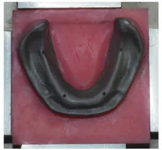
Fig. 1 Master model used for impressions with 4 landmarks (two at the canine areas and two at the molar areas) on alveolar ridge area.
Fig. 2 Assembly prepared for stabilizing the model while making impressions. However all dimensional accuracy measurements
with these measuring techniques are aimed to determine linear dimensional change, the distance between two points. In this study it is intended to measure dimensional change in whole model surface area with 3D software.
The purpose of this study was to determine the effect of two disinfectant agents, 5.25% NaOCl and 2% GA on dimensional accuracy and stability of two elastomeric impression materials, VPES and PE.
The null hypothesis was that there would be no significant difference in dimensional stability and accuracy between VPES and PE impressions treated with two different disinfectants.
MATERIALS AND METHODS
An impression was made from edentulous lower jaw model (Frasaco, Tettnang, Germany) using C-Silicone putty impression paste (Optosil, Heraus Kulzer, Hanau, Germany). Otopolymerizing acrylic resin (Meliodent self curing, Heraeus Kulzer) was poured into the impression and waited for completion of polymerization. Acrylic resin model was sent to the laboratory to construct the edentulous arch area where the impression was making from chromium-cobalt alloy to prevent abrasion and dimensional changes of resin master model due to multiple impressions. Four holes 1.5 mm in diameter were drilled on the top surface of alveolar ridge of the model, two at the canine areas and two at the molar areas, to serve as landmarks (Fig. 1). PE (Impregum Soft Monophase, 3M ESPE, Neuss, Germany) and vinyl polyether siloxane (EXA’lence 370 monophase, GC America, Alsip, IL, USA) impression materials were used. Fifteen impressions were obtained for each impression material for each measurement. At the sum 180 measurements were done.
Custom trays were prepared from light cured acrylic resin (Major Prodotti Dentari, Moncalire, Italy). PE adhesive (3M ESPE) for PE impressions and VPS adhesive for VPES impressions (3M ESPE) were applied inside the custom trays and allowed for dry. An assembly
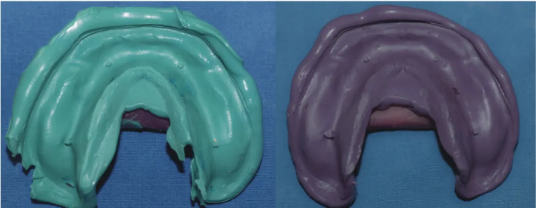
was arranged for stabilizing the model while making impressions. 2.5 kilograms constant weight was seated on the tray to ensure standard pressure for leakage of excess material (Fig. 2). Impression materials were mixed using automatic dispensing and mixing systems. Setting time was two times longer than the time recommended by the manufacturer to compansate for impression fabrication at room temperature, 23°C instead of mouth temperature and all impressions were mixed and stored at 23°C10,13). After setting was completed impressions were removed from model and rinsed 10 s under tap water for simulating the clinical situation for avoiding blood and saliva. Than impressions were remained for air dry (Fig. 3).
Two disinfection solutions; 2% GA (Steranios, Anios Laboratoires, Hellemmes, France) and 5.25% NaOCl (Aklar Kimya, Ankara, Turkey) were used in this study.
Fig. 3 Impressions of polyether and vinyl polyether siloxane.
Immersion technique was applied for 10 min in a tank according to the manufacturers.
Measurement of impressions were completed at 3 intervals; 30 min later after making impression before disinfection (T1: measurement time), after required disinfection period (10 min) (T2), and after 24 h storage at room temperature (T3). Fifteen impressions were repeated from both impression materials for each interval and for two disinfectants. After disinfection impressions were again rinsed 10 s under tap water to remove residues from disinfectants and allow for air dry.
Master model and impressions were scanned by a 3D scanner (SmartOptics Activity 880, smart optics Sensortechnik, Bochum, Germany) with 10 microns accuracy for measurement of dimensional changes. Scanned impressions were transferred to a software (VirtualGrid, Vr-Mesh Studio, Bellevue, WA, USA) in a personal computer.
Comparisons were made with superposing the four holes on 3D images of master model and impression materials. Holes in the master model which were transferred to the impression materials were served as landmarks in canine and molar area on alveolar ridge. These holes were used for superposition of the images accurately. Surface matchings were completed and the deviation area percentages were calculated.
RESULTS
The results were evaluated depends on three variables; impression materials, disinfection solutions and measurement times. Surface area deviations of two impression materials were evaluated according to the master model in software. Positive and negative deviation areas were evaluated at the 60–120, 120– 180, 180–240, 240–300 microns intervals. Deviations at 0–60 microns intervals were not evaluated because dimensional changes of impression materials at these intervals were accepted as similar to master model. Deviation intervals were determined with different colors between red and blue for each interval. Above and
below 300 microns were ignored and defined as yellow in the figures. Red color tones indicated positive deviations toward the tray and marked as (+) sign, blue color tones indicated negative deviation vice versa and indicated as (−) sign.
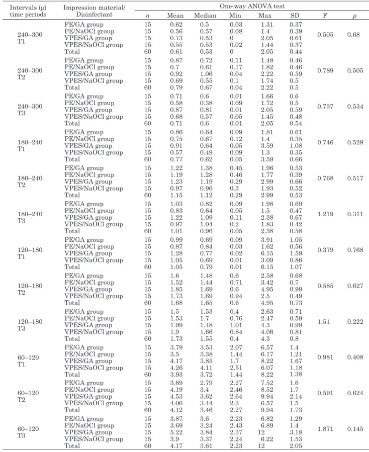
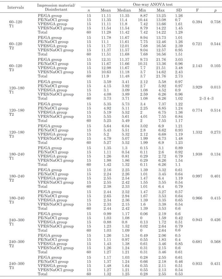
Deviation values of each group at every 60 microns interval were analyzed by statistical method of One-way Analysis of Variance (ANOVA). These deviation values were evaluated at 4 intervals for positive deviations and 4 intervals for negative deviations between different groups and statistical mean ranks were obtained. Values for positive and negative deviations were indicated in Tables 1 and 2, respectively.
According to the statistical analysis it was found that there was not any time dependent differences for both impression materials immersed in two disinfection materials.
However, VPES impression material was found to be more accurate than PE according to the master model in terms of positive deviation before disinfection at 120– 180 micron interval (Table 1).
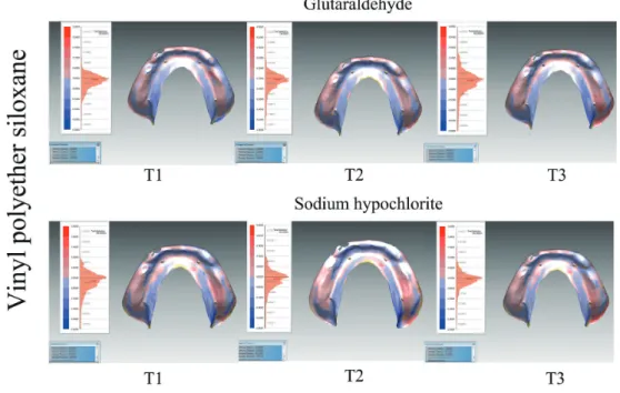
Positive deviations for both PE and VPES impression materials were seen in the posterior alveolar crest region and negative deviations were seen in retromylohyoid area (Figs. 4 and 5).
DISCUSSION
Dimensional accuracy and stability are important for final impression of edentulous arches39,40). Two popular impression materials, PE and VPES were used to evaluate in terms of dimensional change when subjected to the most used disinfection solutions, GA (2%) and NaOCl (5.25%) with immersion method in this study. Area deviation percantages of impression surfaces were compared the developing method of 3D optic scanner with surface superimposition. Scanned master model and impressions with 3 time periods; non disinfected impressions (before disinfection), immersion after 10 min and 24 h later after immersion were measured. Fifteen impressions for both PE and VPES were evaluated.
Table 1 Comparison of VPES and PE impression materials treated with GA and NaOCl disinfection materials under 3 different time periods (T1; 30 min later after making impression before disinfection, T2; after 10 min disinfection period, T3; after 24 h storage at room temperature) and four positive deviation intervals (µ) using One-way ANOVA Intervals (µ)
time periods Impression material/Disinfectant
One-way ANOVA test
n Mean Median Min Max SD F p
240–300 T1 PE/GA group PE/NaOCl group VPES/GA group VPES/NaOCl group Total 15 15 15 15 60 0.62 0.56 0.73 0.55 0.61 0.5 0.57 0.53 0.53 0.53 0.03 0.08 0 0.02 0 1.31 1.4 2.05 1.44 2.05 0.37 0.39 0.61 0.37 0.44 0.505 0.68 240–300 T2 PE/GA group PE/NaOCl group VPES/GA group VPES/NaOCl group Total 15 15 15 15 60 0.87 0.7 0.92 0.69 0.79 0.72 0.61 1.06 0.55 0.67 0.11 0.17 0.04 0.1 0.04 1.48 1.82 2.22 1.74 2.22 0.46 0.46 0.59 0.5 0.5 0.789 0.505 240–300 T3 PE/GA group PE/NaOCl group VPES/GA group VPES/NaOCl group Total 15 15 15 15 60 0.71 0.58 0.87 0.68 0.71 0.6 0.38 0.81 0.57 0.6 0.01 0.09 0.01 0.05 0.01 1.66 1.72 2.05 1.45 2.05 0.6 0.5 0.59 0.48 0.54 0.737 0.534 180–240 T1 PE/GA group PE/NaOCl group VPES/GA group VPES/NaOCl group Total 15 15 15 15 60 0.86 0.75 0.91 0.57 0.77 0.64 0.67 0.64 0.49 0.62 0.09 0.12 0.05 0.09 0.05 1.81 1.4 3.59 1.3 3.59 0.61 0.35 1.08 0.35 0.66 0.746 0.529 180–240 T2 PE/GA group PE/NaOCl group VPES/GA group VPES/NaOCl group Total 15 15 15 15 60 1.22 1.19 1.23 0.97 1.15 1.38 1.28 1.19 0.96 1.12 0.45 0.46 0.29 0.3 0.29 1.96 1.77 2.99 1.93 2.99 0.53 0.39 0.66 0.52 0.53 0.768 0.517 180–240 T3 PE/GA group PE/NaOCl group VPES/GA group VPES/NaOCl group Total 15 15 15 15 60 1.03 0.83 1.22 0.97 1.01 0.82 0.64 1.09 1.04 0.96 0.09 0.05 0.11 0.2 0.05 1.98 1.5 2.38 1.83 2.38 0.69 0.47 0.67 0.42 0.58 1.219 0.311 120–180 T1 PE/GA group PE/NaOCl group VPES/GA group VPES/NaOCl group Total 15 15 15 15 60 0.99 0.87 1.28 1.05 1.05 0.69 0.84 0.77 0.69 0.79 0.09 0.03 0.02 0.01 0.01 3.91 1.62 6.15 3.09 6.15 1.05 0.56 1.59 0.86 1.07 0.379 0.768 120–180 T2 PE/GA group PE/NaOCl group VPES/GA group VPES/NaOCl group Total 15 15 15 15 60 1.6 1.52 1.85 1.73 1.68 1.48 1.44 1.69 1.69 1.65 0.6 0.71 0.6 0.94 0.6 2.58 3.42 4.95 2.5 4.95 0.68 0.7 0.99 0.49 0.73 0.585 0.627 120–180 T3 PE/GA group PE/NaOCl group VPES/GA group VPES/NaOCl group Total 15 15 15 15 60 1.5 1.53 1.99 1.9 1.73 1.53 1.7 1.48 1.66 1.55 0.4 0.76 1.01 0.84 0.4 2.63 2.47 4.3 4.06 4.3 0.71 0.59 0.99 0.81 0.8 1.51 0.222 60–120 T1 PE/GA group PE/NaOCl group VPES/GA group VPES/NaOCl group Total 15 15 15 15 60 3.79 3.5 4.17 4.26 3.93 3.53 3.38 3.85 4.11 3.72 2.07 1.44 1.7 2.51 1.44 6.57 6.17 8.22 6.07 8.22 1.4 1.21 1.67 1.18 1.38 0.981 0.408 60–120 T2 PE/GA group PE/NaOCl group VPES/GA group VPES/NaOCl group Total 15 15 15 15 60 3.69 4.19 4.53 4.06 4.12 2.79 3.4 3.62 3.44 3.46 2.27 2.46 2.64 2.3 2.27 7.52 8.52 9.94 6.57 9.94 1.6 1.7 2.14 1.5 1.73 0.591 0.624 60–120 T3 PE/GA group PE/NaOCl group VPES/GA group VPES/NaOCl group Total 15 15 15 15 60 3.87 3.69 5.22 3.9 4.17 3.6 3.24 3.84 3.37 3.61 2.23 2.43 2.37 2.24 2.23 6.82 6.89 12 6.22 12 1.29 1.4 3.18 1.53 2.05 1.871 0.145
Positive surface deviations of impression materials were compared according to the master model (Statistical significance level, 0.05).
Table 2 Comparison of VPES and PE impression materials treated with GA and NaOCl disinfection materials under 3 different time periods (T1; 30 min later after making impression before disinfection, T2; after 10 min disinfection period, T3; after 24 h storage at room temperature) and four negative deviation intervals (µ) using One-way ANOVA Intervals Impression material/Disinfectant One-way ANOVA test
n Mean Median Min Max SD F p
60–120 T1 PE/GA group PE/NaOCl group VPES/GA group VPES/NaOCl group Total 15 15 15 15 60 11.11 11.35 11.11 11.54 11.28 11.35 11.4 11.8 11.54 11.42 8.87 10.44 7.42 8.79 7.42 13.25 13.08 13.66 14.22 14.22 1.26 0.7 1.61 1.45 1.28 0.394 0.758 60–120 T2 PE/GA group PE/NaOCl group VPES/GA group VPES/NaOCl group Total 15 15 15 15 60 11.78 11.1 11.77 11.37 11.51 11.67 11.61 12.01 11.57 11.68 9.94 7.75 7.68 9.04 7.68 13.73 12.46 16.56 12.57 16.56 1.01 1.26 2.39 0.95 1.51 0.721 0.544 60–120 T3 PE/GA group PE/NaOCl group VPES/GA group VPES/NaOCl group Total 15 15 15 15 60 12.31 11.67 12.98 10.63 11.9 11.37 11.66 11.67 11.18 11.48 9.73 10.31 7.7 3.7 3.7 21.76 13.36 21.51 14.62 21.76 3.03 0.96 3.48 2.43 2.73 2.143 0.105 120–180 T1 PE/GA group PE/NaOCl group VPES/GA group VPES/NaOCl group Total 15 15 15 15 60 3.59 4.15 3.1 4.08 3.73 3.41 3.92 3.09 3.89 3.71 2.12 2.7 1.08 2.59 1.08 5.58 5.92 4.52 6.26 6.26 0.97 0.97 0.9 0.96 1.02 3.929 0.013 2–3 4–3 120–180 T2 PE/GA group PE/NaOCl group VPES/GA group VPES/NaOCl group Total 15 15 15 15 60 5.35 4.92 5.19 5.55 5.25 5.73 5.11 5.52 5.61 5.49 3.4 2.25 2 4.01 2 7.37 6.85 6.75 7.55 7.55 1.22 1.24 1.36 0.84 1.17 0.774 0.514 120–180 T3 PE/GA group PE/NaOCl group VPES/GA group VPES/NaOCl group Total 15 15 15 15 60 5.64 5.43 5.2 4.79 5.27 6.04 5.51 5.32 5.07 5.52 3.02 2.8 2.12 1.99 1.99 6.9 6.62 6.68 6.73 6.9 1.21 0.93 1.19 1.48 1.23 1.332 0.273 180–240 T1 PE/GA group PE/NaOCl group VPES/GA group VPES/NaOCl group Total 15 15 15 15 60 1.35 1.11 1.26 1.98 1.42 1.3 0.54 0.81 1.86 1.32 0.15 0.11 0.29 0.29 0.11 3.1 2.6 2.72 6.26 6.26 0.89 0.92 0.79 1.54 1.1 1.938 0.134 180–240 T2 PE/GA group PE/NaOCl group VPES/GA group VPES/NaOCl group Total 15 15 15 15 60 2.16 2.24 2.55 2.55 2.38 2.25 2.26 2.54 2.42 2.33 1.39 1.01 1.47 1.55 1.01 3.32 3.45 6.4 3.55 6.4 0.61 0.64 1.19 0.54 0.79 0.997 0.401 180–240 T3 PE/GA group PE/NaOCl group VPES/GA group VPES/NaOCl group Total 15 15 15 15 60 2.44 2.65 2.34 2.33 2.44 2.52 2.94 2.36 2.15 2.42 1.47 1.67 1.39 1.6 1.39 3.27 3.53 3.35 3.38 3.53 0.57 0.64 0.65 0.54 0.6 0.966 0.415 240–300 T1 PE/GA group PE/NaOCl group VPES/GA group VPES/NaOCl group Total 15 15 15 15 60 0.99 1.03 0.88 1.23 1.03 1.17 1.08 0.8 1.52 1.09 0.06 0 0.13 0.02 0 2.19 1.59 1.72 2.64 2.64 0.6 0.42 0.51 0.79 0.6 0.943 0.426 240–300 T2 PE/GA group PE/NaOCl group VPES/GA group VPES/NaOCl group Total 15 15 15 15 60 1.26 1.11 1.43 1.26 1.27 1.34 0.97 1.38 1.24 1.21 0.49 0.55 0.61 0.31 0.31 2.06 1.83 3.46 2.15 3.46 0.5 0.4 0.85 0.6 0.61 0.681 0.568 240–300 T3 PE/GA group PE/NaOCl group VPES/GA group VPES/NaOCl group Total 15 15 15 15 60 1.17 1.37 1.48 1.27 1.32 1.03 1.24 1.64 1.21 1.25 0.28 0.66 0.35 0.55 0.28 2.55 2.18 2.11 2.13 2.55 0.61 0.46 0.51 0.54 0.53 0.933 0.431
Negative surface deviations of impression materials were compared according to the master model. (Statistical significance level, 0.05).
Fig. 4 Positive and negative deviations for PE impression material.
T1, T2 and T3 measurement times with hypochlorite and GA disinfection, from left to right, respectively.
Fig. 5 Positive and negative deviations for VPES impression material.
T1, T2 and T3 measurement times with hypochlorite and GA disinfection, from left to right, respectively.
changes with 10 microns accuracy in this study. Technical improvements in 3D imaging procedures enable a direct digitalization of impression negatives38). It is an advantageous method for assessing the dimensional accuracy and stability of impressions than other two dimensional methods36,38,41) because it is a quick,
non-contact procedure with no physical invasion of the object and also time saving42). Dimensional changes occur in three dimensions. So that it might be a more reliable method for measuring dimensional changes and 3D scanning digitizers provide greater precision and 3D analysis. And also software programs help to eliminate
subjective errors depends on the operator. However sharp line angles and undercuts or transparent and reflective materials could be difficult to scan and need to apply matte white coat over the the object. And also it is more expensive than the other mechanical measuring techniques38,41). These may be counted as challenging factors for 3D optic scanners.
Most of the studies5,8,15,32,35) measured the dimensional accuracy between selected points on model especially at canine and first molar regions. The length between selected points were compared among models in two dimensions and concluded as differences between distances or percentages. In the present study, four holes were also created at canine and first molar positions and these holes were used for superposing the whole impression surfaces in 3D software. Percentage of surface area deviation values were obtained using 3D scanning method with superposing the images.
Shah et al.38) and Kurtulmuş-Yılmaz et al.43) were used 3D superimpositional data for measuring dimensional accuracy as similar with this present study.
Master models in studies were varied. Some studies15,20,35,38,44) used metal arch shaped models and the others used disc shaped models or dies28,32,45,46). To simulate the edentulous mandibular ridge, metal arch shaped model was chosen in this study similar with other studies.
Another variable that might influence the dimensional accuracy and stability of impressions is tray selection. Some studies used self cured acrylic resin or light cured acrylic (VLC)15,39) trays47) and others used metal stock trays48). Schaefer et al.34) declared that modified stock trays contribute dimensional accuracy with well fitting and limiting unintended flow of material. Custom trays ensure the uniformity of the impression material so that the dimensional changes due to excess and thick material might be decreased. As a matter of fact that custom trays ensure uniform and optimum material thickness, increase dimensional accuracy and decrease deformation49). Brosky et al.41) declared that the dimensional stability of an elastomeric impression material was optimal when custom-made acrylic resin impression tray was used. In a review of Petropoulos and Rashedi39) it was concluded that custom trays (both acrylic and VLC) were mostly used for final impression of edentulous arches. And also standardized tray positioning with a constant force is clinically advisable but not generally practicable34,48). Guiraldo et al.50) and Carvalhal et al.11) made impressions with a pressure of 2 kgf to allow for leakage of excess material. In this present study, a custom made light cured acrylic resin trays were used and a 2.5 kg constant weight was seated on the tray to ensure standard pressure and uniform distribution of impression materials. Thus, subjective errors might be excluded. On the contrary, it could be argued that metal trays are rigid and dimensionally more stable than acrylic trays48). It is determined that acrylic trays tend to absorb and expand in humidity so that dimensional change of trays should be kept in mind when evaluating the dimensional accuracy and stability
of impression materials8). Piwowarczyk et al.48) declared that deformation of the impression materials was eliminated by using a rigid impression tray and they removed the tray in a specific force in vertical dimension. Custom VLC impression trays were prepared for impressions in this study. The deformation effect of tray might be negligible in this study because all impressions compared together in the same conditions.
Dimensional accuracy and stability could be measured directly from impressions8,28) or from stone casts obtained from impressions. Due to the difficulty to scan or measure the dimensional accuracy from the impression, most of the studies used stone cast in their studies8,43,34,51,52). Measuring impressions may have advantages over measurements done from stone casts8) because besides the dimensional change of impression material, dimensional behavior of dental stone has to be taken into consideration when measurements done from stone cast models. So that comparison of dimensional changes between impression materials might be carried out on impressions. Martin et al.28) evaluated direct measurement of the impression material, without the need to pour a cast. Dimensional changes were evaluated directly from scanning the impression materials in this study.
Dimensional accuracy and stability of impression materials are crucial factors for successive treatment steps48). It is known that dimensional stability of VPS and PE impression materials are excellent35,51,53). However Shah et al.38) and Faria et al.49) declared that PE were more accurate than VPS. And also in the study of Petrie et al.32) even hydrophilic VPS materials were used on moist or wet surfaces, an acceptable impression could not always obtain. VPES materials composed of PE and VPS materials, which protect their properties with the advantages of both materials. PE component counteracted the dimensional contraction of VPES with increasing moisture absorption and reduced silica content8,29,35,45). Due to the greater properties of PE over VPS, PE impression material was compared with a newly introduced impression material VPES in terms of dimensional accuracy and stability under two different disinfection materials in this study.
There are controversions about disinfection impact on impression materials or stone casts. Some of the studies concluded a significant effect of disinfection solution not only depends on water absorption but also chemical interactions. However some studies declared that there is no significant effect on impression dimensional accuracy. Walker et al.45) stated in their study that NaOCl (0.5%) had an adverse effect on PE impression surface. And also it was seen that PE significantly expanded when disinfected with NaOCl. Besides there was not any significant change in VPS dimensional stability after disinfection. Amin et al.5) used 0.5% hypochlorite and it caused smaller dimensional change than that of 2% GA in the additional silicone impression material. Previous investigations showed that the least dimensional change was occurred when GA used1,10) Sinobad et al.10) were used GA and 5.25%
NaOCl for immersion of impression materials to evaluated the dimensional accuracy and stability of additional silicones and condensation silicones on impressions. It was concluded that 5.25% concentration of NaOCl caused significant dimensional changes for both impression materials.
In this present study, dimensional stability of VPES and PE impression materials were found similar under immersion with 2% GA and 5.25% NaOCl disinfection materials.
Dimensional changes according to the disinfection were also evaluated in terms of measurement times in different studies. Nassar et al.35) were evaluated the dimensional stability of PE, VPS and VPES impression materials with immersion disinfection of GA (2.5%) at four pouring time periods (immediate, 1 day, 1 week and 2 weeks). Measurements were done on stone casts and distances between selected areas (two canine and two first molar) using digital micrometer and found that VPS or PE measurements were similar with VPES impression. For all measuring times dimensional stability was found similar and it was indicating that the VPES material was accurate and dimensionally stable. Kronström et al.44) were studied on dimensional accuracy of three elastomeric impression materials under two disinfection methods and different times (disinfection by spray for 10 min and disinfection by immersion for 90 min) and results of cross arch and anteroposterior die measurements using measuring microscope were found similar. They reported that neither disinfection method nor the disinfection time affected the dimensional stability and accuracy. Five different brands of addition-type silicone were immersed in two different disinfectants for 30 min and 24 h in the study of Hiraguchi et al.13) and diameter of the casts were measured using a laser scan micrometer. They concluded that the type of disinfectant did not affect the dimensional changes in immersion disinfection. Carvalhal et al.11) evaluated the immersion times on different elastomeric impression materials. It was observed that VPS and PE impression materials showed no significant differences when immersed with 2% GA and 0.5% hypochlorite solutions at different immersion time intervals.
Using 2% GA can be safely recommended for 10 min of immersion disinfection without affecting the wettability of PE. In contrast, 5.25% phenol and 0.5% NaOCl should not be considered for immersion disinfection because it adversely affects the wettability of PE materials54). However, Amin et al.5) recommended 10 min immersion disinfection with 0,5% NaOCl for additional cured silicones.
In the present study, immersion time was selected as 10 min both for NaOCl and GA disinfection materials, which recommended in the literatures and it was seen that this short immersion time did not affect the dimensional accuracy and stability of PE and VPES impression materials. However, Thouati et al.46) used additional silicones and PEs disinfected with 5.25% NaOCl disinfectant solution for 30 min and found that there were statistically significant dimensional
variations.
Measurements were done using 3D method dissimilar with the studies above done with 2D measurements. Impression surfaces directly scanned with optic scanner and compared with master model. Dimensional changes were determined at every 60 microns intervals because optic scanner used in this study had 10 microns standard deviation. Deviations between impression materials under two disinfectants at every interval were compared in itself. Deviations at 0–60 micron intervals were not evaluated because dimensional changes of impression materials were so close to master model at these intervals.
Impression dimensional accuracy under changing conditions such as angulated implant analogs, different impression materials and techniques were evaluated and it was concluded that there was a discrepancy of 50 microns even if impression was good44).
Statistically there were no dimensional changes for impression materials under two disinfectants at different time periods. However, small differences were recorded between PE and VPES at 120–180 micron intervals. VPES were found more accurate than PE comparing with the master model before disinfection. This difference was ignored when thought that the other intervals were statistically similar to each other.
Independent from the measuring method the results of this study were similar with the results of other studies about elastomeric impression materials.
The limitations of this study were about making impressions and their removal which were not the same as impressions in clinical practice. Master model and conditions were not resembled the resiliency of oral tissues. Saliva and soft tissues were eliminated with using metal model and neglected in this study.
CONCLUSION
According to the results of this research dimensional accuracy and stability of two elastomeric impression materials PE and VPES are similar when immersed in two different disinfectants. Short measurement time period (maximum 24 h) for impression materials may not cause the dimensional changes. It can be concluded that both impression materials are excellent and similar.
Further clinical studies need to carry out to simulate oral environment.
REFERENCES
1) Melilli D, Rallo A, Cassaro A, Pizzo G. The effect of immersion disinfection procedures on dimensional stability of two elastomeric impression materials. J Oral Sci 2008; 50: 441-446.
2) Choi YR, Kim KN, Kim KM. The disinfection of impression materials by using microwave irradiation and hydrogen peroxide. J Prosthet Dent 2014; 112: 981-987.
3) Bhasin A, Vinod V, Bhasin V, Mathew X, Sajjan S, Ahmed ST. Evaluation of effectiveness of microwave irradiation for disinfection of silicone elastomeric impression material. J
Indian Prosthodont Soc 2013; 13: 89-94.
4) Goel K, Gupta R, Solanki J, Nayak M. A comparative study between microwave irradiation and sodium hypochlorite chemical disinfection: a prosthodontic view. J Clin Diagn Res 2014; 8: 42-46.
5) Amin WM, Al-Ali MH, Al Tarawneh SK, Taha ST, Saleh MW, Ereifij N. The effects of disinfectants on dimensional accuracy and surface quality of impression materials and gypsum casts. J Clin Med Res 2009; 1: 81-89.
6) Kalantari MH, Malekzadeh A, Emami A. The effect of disinfection with sodium hypochlorite 0.5% on dimensional stability of condensation silicone impression materials of speedex and irasil. J Dent (Shiraz) 2014; 15: 98-103. 7) Blalock JS, Cooper JR, Rueggeberg FA. The effect of
chlorine-based disinfectant on wettability of a vinyl polysiloxane impression material. J Prosthet Dent 2010; 104: 333-341. 8) Kotsiomiti E, Tzialla A, Hatjivasiliou K. Accuracy and stability
of impression materials subjected to chemical disinfection —a literature review. J Oral Rehabil 2008; 35: 291-299.
9) Bock JJ, Fuhrmann RA, Setz J. The influence of different disinfectants on primary impression materials. Quintessence Int 2008; 39: 93-98.
10) Sinobad T, Obradović-Djuricić K, Nikolić Z, Dodić S, Lazić V, Sinobad V, Jesenko-Rokvić A. The effect of disinfectants on dimensional stability of addition and condensation silicone impressions. Vojnosanit Pregl 2014; 71: 251-258.
11) Carvalhal CI, Mello JA, Sobrinho LC, Correr AB, Sinhoreti MA. Dimensional change of elastomeric materials after immersion in disinfectant solutions for different times. J Contemp Dent Pract 2011; 12: 252-258.
12) Saleh Saber F, Abolfazli N, Kohsoltani M. The effect of disinfection by spray atomization on dimensional accuracy of condensation silicone impressions. J Dent Res Dent Clin Dent Prospects 2010; 4: 124-129.
13) Hiraguchi H, Kaketani M, Hirose H, Kikuchi H, Yoneyama T. Dimensional changes in stone casts resulting from long-term immersion of addition-type silicone rubber impressions in disinfectant solutions. Dent Mater J 2013; 32: 361-366. 14) Estafanous EW, Palenik CJ, Platt JA. Disinfection of
bacterially contaminated hydrophilic PVS impression materials. J Prosthodont 2012; 21: 16-21.
15) Jagger DC, Vowles RW, McNally L, Davis F, O’Sullivan DJ. The effect of a range of disinfectants on the dimensional accuracy and stability of some impression materials. Eur J Prosthodont Restor Dent 2007; 15: 23-28.
16) Lepe X, Johnson GH, Berg JC, Aw TC, Stroh GS. Wettability, imbibition, and mass change of disinfected low-viscosity impression materials. J Prosthet Dent 2002; 88: 268-276. 17) Al-Omari WM, Jones JC, Wood DJ. The effect of disinfecting
alginate and addition cured silicone rubber impression materials on the physical properties of impressions and resultant casts. Eur J Prosthodont Restor Dent 1998; 6: 103-110.
18) Johnson GH, Chellis KD, Gordon GE, Lepe X. Dimensional stability and detail reproduction of irreversible hydrocolloid and elastomeric impressions disinfected by immersion. J Prosthet Dent 1998; 79: 446-453.
19) Suprono MS, Kattadiyil MT, Goodacre CJ, Winer MS. Effect of disinfection on irreversible hydrocolloid and alternative impression materials and the resultant gypsum casts. J Prosthet Dent 2012; 108: 250-258.
20) Rentzia A, Coleman DC, O’Donnell MJ, Dowling AH, O’Sullivan M. Disinfection procedures: their efficacy and effect on dimensional accuracy and surface quality of an irreversible hydrocolloid impression material. J Dent 2011; 39: 133-140.
21) McDonnell G, Russell AD. Antiseptics and disinfectants: activity, action, and resistance. Clin Microbiol Rev 1999; 12: 147-179.
22) Al Kheraif AA. Surface roughness of polyvinyl siloxane impression materials following chemical disinfection, autoclave and microwave sterilization. J Contemp Dent Pract 2013; 14: 483-487.
23) Pal PK, Kamble SS, Chaurasia RR, Chaurasia VR, Tiwari S, Bansal D. Evaluation of different disinfactants on dimensional accuracy and surface quality of type IV gypsum casts retrieved from elastomeric impression materials. J Int Oral Health 2014; 6: 77-81.
24) Lepe X, Johnson GH. Accuracy of polyether and addition silicone after long-term immersion disinfection. J Prosthet Dent 1997; 78: 245-249.
25) Hiraguchi H, Kaketani M, Hirose H, Yoneyama T. The influence of storing alginate impressions sprayed with disinfectant on dimensional accuracy and deformation of maxillary edentulous stone models. Dent Mater J 2010; 29: 309-315.
26) Godbole SR, Dahane TM, Patidar NA, Nimonkar SV. “Evaluation of the effect of ultraviolet disinfection on dimensional stability of the polyvinyl silioxane impressions.” an in-vitro study. J Clin Diagn Res 2014; 8: 73-76.
27) Thota KK, Jasthi S, Ravuri R, Tella S. A comparative evaluation of the dimensional stability of three different elastomeric impression materials after autoclaving —an invitro study. J Clin Diagn Res 2014; 8: 48-50.
28) Martin N, Martin MV, Jedynakiewicz NM. The dimensional stability of dental impression materials following immersion in disinfecting solutions. Dent Mater 2007; 23: 760-768. 29) Pandita A, Jain T, Yadav NS, Feroz SM, Pradeep, Diwedi A.
Evaluation and comparison of dimensional accuracy of newly introduced elastomeric impression material using 3D laser scanners: an in vitro study. J Contemp Dent Pract 2013; 14: 265-268.
30) Pande NA, Parkhedkar RD. An evaluation of dimensional accuracy of one-step and two-step impression technique using addition silicone impression material: an in vitro study. J Indian Prosthodont Soc 2013; 13: 254-259.
31) Kambhampati S, Subhash V, Vijay C, Das A. Effect of temperature changes on the dimensional stability of elastomeric impression materials. J Int Oral Health 2014; 6: 12-19.
32) Petrie CS, Walker MP, O’mahony AM, Spencer P. Dimensional accuracy and surface detail reproduction of two hydrophilic vinyl polysiloxane impression materials tested under dry, moist, and wet conditions. J Prosthet Dent 2003; 90: 365-372.
33) Stober T, Johnson GH, Schmitter M. Accuracy of the newly formulated vinyl siloxanether elastomeric impression material. J Prosthet Dent 2010; 103: 228-239.
34) Schaefer O, Schmidt M, Goebel R, Kuepper H. Qualitative and quantitative three-dimensional accuracy of a single tooth captured by elastomeric impression materials: an in vitro study. J Prosthet Dent 2012; 108: 165-712.
35) Nassar U, Oko A, Adeeb S, El-Rich M, Flores-Mir C. An in vitro study on the dimensional stability of a vinyl polyether silicone impression material over a prolonged storage period. J Prosthet Dent 2013; 109: 172-178.
36) Nassar U, Chow AK. Surface detail reproduction and effect of disinfectant and long-term storage on the dimensional stability of a novel vinyl polyether silicone ımpression material. J Prosthodont 2015; 24: 494-498.
37) Tan E, Chai J, Wozniak WT. Working times of elastomeric impression materials determined by dimensional accuracy. Int J Prosthodont 1996; 9: 188-196.
38) Shah S, Sundaram G, Bartlett D, Sherriff M. The use of a 3D laser scanner using superimpositional software to assess the accuracy of impression techniques. J Dent 2004; 32: 653-658. 39) Petropoulos VC, Rashedi B. Current concepts and techniques
Prosthodont 2003; 12: 280-287.
40) Petrie CS, Walker MP, Williams K. A survey of U.S. prosthodontists and dental schools on the current materials and methods for final impressions for complete denture prosthodontics. J Prosthodont 2005; 14: 253-262.
41) Brosky ME, Pesun IJ, Lowder PD, Delong R, Hodges JS. Laser digitization of casts to determine the effect of tray selection and cast formation technique on accuracy. J Prosthet Dent 2002; 87: 204-209.
42) Rignon-Bret C, Dupuis R, Gaudy JF. Application of a 3-dimensional measurement system to complete denture impressions. J Prosthet Dent 2002; 87: 603-612.
43) Kurtulmus-Yilmaz S, Ozan O, Ozcelik TB, Yagiz A. Digital evaluation of the accuracy of impression techniques and materials in angulated implants. J Dent 2014; 42: 1551-1559.
44) Kronström MH, Johnson GH, Hompesch RW. Accuracy of a new ring-opening metathesis elastomeric dental impression material with spray and immersion disinfection. J Prosthet Dent 2010; 103: 23-30.
45) Walker MP, Rondeau M, Petrie C, Tasca A, Williams K. Surface quality and long-term dimensional stability of current elastomeric impression materials after disinfection. J Prosthodont 2007; 16: 343-351.
46) Thouati A, Deveaux E, Iost A, Behin P. Dimensional stability of seven elastomeric impression materials immersed in disinfectants. J Prosthet Dent 1996; 76: 8-14.
47) Corso M, Abanomy A, Di Canzio J, Zurakowski D, Morgano SM. The effect of temperature changes on the dimensional
stability of polyvinyl siloxane and polyether impression materials. J Prosthet Dent 1998; 79: 626-631.
48) Piwowarczyk A, Ottl P, Büchler A, Lauer HC, Hoffmann A. In vitro study on the dimensional accuracy of selected materials for monophase elastic impression making. Int J Prosthodont 2002; 15: 168-174.
49) Faria AC, Rodrigues RC, Macedo AP, Mattos Mda G, Ribeiro RF. Accuracy of stone casts obtained by different impression materials. Braz Oral Res 2008; 22: 293-298.
50) Guiraldo RD, Borsato TT, Berger SB, Lopes MB, Gonini-Jr A, Sinhoreti MA. Surface detail reproduction and dimensional accuracy of stone models: influence of disinfectant solutions and alginate impression materials. Braz Dent J 2012; 23: 417-421.
51) Wadhwani CP, Johnson GH, Lepe X, Raigrodski AJ. Accuracy of newly formulated fast-setting elastomeric impression materials. J Prosthet Dent 2005; 93: 530-539.
52) Taylor RL, Wright PS, Maryan C. Disinfection procedures: their effect on the dimensional accuracy and surface quality of irreversible hydrocolloid impression materials and gypsum casts. Dent Mater 2002; 18: 103-110.
53) Johnson GH, Mancl LA, Schwedhelm ER, Verhoef DR, Lepe X. Clinical trial investigating success rates for polyether and vinyl polysiloxane impressions made with full-arch and dual-arch plastic trays. J Prosthet Dent 2010; 103: 13-22. 54) Hussein LA. Accuracy of optimized rubber base ımpression
materials (linear and surface analysis). Aust J Basic Appl Sci 2014; 8: 543-551.