DEVELOPMENT OF PEPTIDE BASED MATERIALS AS A
SYNTHETIC SCAFFOLD TO MIMIC EXTRACELLULAR MATRIX
A DISSERTATION SUBMITTED TO
THE GRADUATE SCHOOL OF ENGINEERING AND SCIENCE OF BILKENT UNIVERSITY
IN PARTIAL FULFILLMENT OF THE REQUIREMENTS FOR THE DEGREE OF
DOCTOR OF PHILOSOPHY IN
MATERIALS SCIENCE AND NANOTECHNOLOGY
By
MELİKE SEVER July 2017
ii
DEVELOPMENT OF PEPTIDE BASED MATERIALS AS A SYNTHETIC SCAFFOLD TO MIMIC EXTRACELLULAR MATRIX
By Melike Sever July 2017
We certify that we have read this dissertation and that in our opinion it is fully adequate, in scope and in quality, as a dissertation for the degree of Doctor of Philosophy.
Ayşe Begüm Tekinay (Advisor)
Çağlar Elbüken
Yusuf Şükrü Çağlar
Nülüfer Tülün Güray
Michelle Marie Adams
Approved for the Graduate School of Engineering and Science:
Ezhan Karaşan
iii ABSTRACT
DEVELOPMENT OF PEPTIDE BASED MATERIALS AS A SYNTHETIC SCAFFOLD TO MIMIC EXTRACELLULAR MATRIX
Melike Sever
Ph.D. in Materials Science and Nanotechnology Advisor: Ayşe Begüm Tekinay
July, 2017
Biomaterials obtained through self-assembling process of peptide amphiphile (PA) molecules provide great potential to introduce new therapeutic approaches in regenerative medicine through mimicking the natural environments of different types of tissues. The ability of self-assembled PA nanofibers to mimic natural extracellular matrix (ECM) renders them attractive for regenerative medicine applications. The materials-cell interactions can be modulated through the surface modification of the materials such as introducing the bioactivity via short bioactive peptide sequences derived from natural ECM proteins, which regulate cell behavior through controlling of cellular activities such as proliferation and differentiation.
Herein, I described my studies on the development of PA nanofibers in order to mimic natural ECM with differentiation and regeneration purposes. Heparan sulfate mimetic and laminin mimetic PA nanofibers were used as a potential therapeutic approach in Parkinson's disease (PD). These bioactive PA nanofibers were found to reduce the progressive cell loss in SH-SY5Y cells caused by 6-hydroxydopamine treatment in
iv
rats and provide a promising new strategy for treatment of PD. These nanofibers also proved to be effective in enhancing the viability of Schwann cells and increase nerve growth factor (NGF) release from these cells in vitro. Since NGF has a crucial role in nerve injury repair and myelination in the regenerating nerve, the bioactive epitopes used in this study present also a promising approach as guidance cues for regenerating axons.
Tenascin-C is another multifunctional ECM glycoprotein common in both nerve and bone tissue. By decorating peptide nanofibers with tenascin-C derived epitope and using in three-dimensional (3D) system, this tenascin-C mimetic 3D cell culture system was found to provide both the biochemical and physical aspects of the native environment of neural cells, thereby filling the gap between 2D cell culture models and in vivo environments and contributing to more tissue-like structure and more predictive approaches to organogenesis and tissue morphology.
Within the scope of this thesis, tenascin-C mimetic nanofibers were also used for osteogenic differentiation of mesenchymal stem cells (MSCs). They were found to significantly enhance the attachment, proliferation, and osteogenic differentiation of MSCs even in the absence of any external bioactive factors and regardless of the suitable stiff mechanical properties normally required for osteogenic differentiation.
Keywords: Peptide nanofibers, extracellular matrix, Parkinson’s disease, neural
v ÖZET
HÜCRELER ARASI MATRİSİN TAKLİT EDİLMESİ İÇİN SENTETİK İSKELE OLARAK PEPTİT TABANLI MALZEMELERİN
GELİŞTİRİLMESİ
Melike Sever
Malzeme Bilimi ve Nanoteknoloji, Doktora Tez Danışmanı: Ayşe Begüm Tekinay
Temmuz, 2017
Peptid amfifil (PA) moleküllerinin kendiliğinden bir araya gelmesiyle elde edilen biyomalzemeler, farklı doku türlerinin doğal ortamlarını taklit ederek rejeneratif tıpta yeni terapötik yaklaşımlar getirme potansiyeli taşımaktadır. Kendiliğinden oluşan PA nanofiberlerin doğal hücre dışı matrisi (HDM) taklit etme yeteneği onları rejeneratif tıp uygulamaları için çekici kılmaktadır. Biyoaktif sinyaller taşıyan ve kendiliğinden oluşan peptit nano yapılar, sentetik HDM malzemeleri olarak yaygın bir şekilde kullanılmaktadır. Malzeme-hücre etkileşimlerini geliştirmek amacıyla proliferasyon ve farklılaşma gibi hücresel aktivitelerin modülasyonu yoluyla hücre davranışını düzenleyen doğal HDM proteinlerinden türetilen kısa biyoaktif peptit sekansları materyallerin yüzeylerini biyoaktifleştirmede kullanılmıştır.
Bu tezde, farklılaşma ve rejenerasyon amaçlarıyla doğal hücre dışı matrisi taklit edebilmek için PA nanofiberlerin gelişimi üzerine uygulamalar anlatılmıştır. Parkinson hastalığında (PH) potansiyel bir tedavi yaklaşımı olarak heparan sülfat ve laminin taklidi PA nanofiber kullanılmıştır. Bu biyoaktif PA nanofiberlerin, in vitro
vi
olarak 6-hidroksidopaminin neden olduğu SH-SY5Y hücrelerindeki ilerleyici hücre kaybını azalttığı ve sıçanlarda PH'nin nörokimyasal ve davranışsal sonuçlarını düzelttiği ve PH'nin tedavisinde umut verici yeni bir strateji sağladığı bulunmuştur. Bu nanofiberlerin ayrıca Schwann hücrelerinin canlılığı üzerinde etkili oldukları ve in
vitro olarak bu hücrelerden sinir büyüme faktörü salımını arttırdığı gösterilmiştir. Sinir
büyüme faktörünün yenilenen sinirde, sinir hasarının onarımı ve miyelinasyonunda önemli rolü olduğu için, bu çalışmada kullanılan biyoaktif epitoplar, yenilenen aksonların yol gösterici ipuçları olarak umut verici bir yaklaşım sunmaktadır.
Tenascin-C, hem sinir hem de kemik dokusunda ortak olan, çok işlevli bir hücre dışı matris glikoproteinidir. Tenascin-C türevli epitop ile peptit nanofiberlerin biyoaktif hale getirilmesi ve üç boyutlu (3B) sistemde kullanılmasıyla, tenascin-C mimetik 3B hücre kültürü sisteminin sinir hücrelerinin doğal ortamını hem biyokimyasal hem de fiziksel yönleriyle taklit ettiği ve böylece iki boyutlu hücre kültürü modelleriyle in vivo ortamlar arasındaki boşluğu doldurduğu ve organogeneze ve doku morfolojisine daha akıllı yaklaşımlar getirdiği gösterilmiştir.
Bu tez kapsamında tenascin-C mimetik nanofiberler mezenkimal kök hücrelerin (MKH) osteojenik farklılaşması için de kullanılmıştır. Bu nanofiberlerin, normal olarak osteojenik farklılaşma için gerekli olan dış biyoaktif faktörlerin yokluğunda ve yumuşak mekanik özelliklere bakılmaksızın MKH'lerin bağlanma, çoğalma ve osteojenik farklılaşmasını önemli ölçüde arttırdığı bulunmuştur.
Anahtar kelimeler: Peptid nanofiberler, hücre dışı matris, Parkinson hastalığı, sinirsel
vii
Acknowledgement
I would like to express my deepest appreciation to my thesis advisors Prof. Ayşe Begüm Tekinay and for their scientific knowledge, guidance, encouragement and support throughout my PhD thesis studies. In addition to guiding my PhD studies, she has preened me as a scientist, and fully supported my professional development in any way she could. I would like to thank Prof. Mustafa Özgür Güler for his scientific support during my studies. I would also like to acknowledge my jury members for their contributions to my thesis.
I would like to express my special thanks to Prof. Dr. Mehmet Cansev, Dr. Büşra Mammadov, Mesut Türkyılmaz and Gökhan Günay for their support and great collaboration.
I would like to acknowledge The Scientific and Technological Research Council of Turkey (TÜBİTAK BIDEB 2211-E) for the financial support. Also, our experimental studies were supported by TÜBİTAK projects 111M410, 113S038 and 113S538.
I would like to acknowledge my previous and present lab members Dr. Gözde Uzunallı, Öncay Yaşa, Yasin Tümtaş, Ceren Garip Yaşa, Didem Mumcuoğlu, Elif Arslan, Dr. Hakan Ceylan, Dr. Göksu Çınar, Dr. Melis Şardan, Dr. Berna Şentürk, Seren Hamsici, Egemen Deniz Eren, Ayşegül Dede, Çağla Eren, Mustafa Beter, Nuray Gündüz, İdil Uyan, Zeynep Orhan, İbrahim Çelik, Canelif Yılmaz, Özüm Şehnaz Günel, Dr. Gülcihan Gülseren, Melis Göktaş, Özge Uysal, Dr. M. Aref Khalily, Nurcan Haştar, Fatih Yergöz, Merve Şen, Oğuz Tuncay, Begüm Dikeçoğlu, Alper Özkan, Ahmet Emin Topal, Aygül Zengin, Meryem Hatip, Dr. Ruslan Garifullin, Dr.
viii
Rashad Mammadov, Dr. Özlem Erol, Dr. Ashif Shaikh, Hatice Kübra Kara, Hepi Hari Susapto, Şehmus Tohumeken, Burak Demircan, Dr. Seher Yaylacı and Dr. Hilal Ünal Gülsüner for creating such a warm working environment. I would like to thank Dr. Gülistan Tansık and Dr. Canan Kurşungöz for their friendships and support during my PhD studies at UNAM. Also, I would like to thank Zeynep Erdoğan and Mustafa Guler for their technical contribution to my thesis.
I would like to extend my special thanks to İsmail Cem Yılmaz, İrem Sevgi Öztürk, Eren Öztürk, Sinan Tarık Türesay, Latif Serkan Atabey, Beril Özbalcı, Utku Aslan and Orbay Tuğ who are always with me during those years and creating unforgettable memories with me. I also thank to Oya Gül and Ahu Durmuşçelebi for their endless friendship, help and support. I also thank to Temiz İş Band for their perfect sound and being my models to improve my photography skills.
I would like to thank Bahçekapılı, Kazan and Metinbaş families for their love and support. I especially thank to Ayşen Kazan, my new little sister, for making all the times enjoyable with her emotional support and cheerfulness.
I would like to express my most sincere gratitude to my parents, my mother Birsen Sever and my father Saffet Sever for their endless love, motivation, support and encouragement. I would not be where I am today without their help and support. My special thanks for my life partner, my love Oytun Bahçekapılı. We were right by each other side through all the highs and lows. Thank you so much for being a huge part of my life. Last but not least, I would like to thank to my little brother. He is my best friend and always supports me whenever I need. I am so lucky to have a brother like him. I dedicate this thesis to my beloved brother, Oğuz Sever.
ix
Contents
ABSTRACT ... iii
ÖZET ... v
Acknowledgement ... vii
List of Tables ... xix
Abbreviations ... xx
Chapter 1 ... 1
1. Introduction ... 1
1.1 Neurodegenerative Diseases ... 1
1.2 Extracellular Matrix (ECM) of the Nervous System ... 1
1.2.1 Components of ECM of the Nervous System ... 2
1.2.2 Challenges to Nerve Regeneration ... 3
1.3 Approaches in the Design of ECM-Mimetic Scaffolds for Neural Differentiation and Regeneration ... 4
1.3.1 Biochemical Functionalization of the Scaffolds for Neural Differentiation and Regeneration ... 5
1.3.2 Physical Functionalization of the Scaffolds for Neural Differentiation and Regeneration ... 8
Chapter 2 ... 12
2. Regenerative Effects of Peptide Nanofibers in an Experimental Model of Parkinson’s Disease ... 12
2.1 Introduction ... 12
2.2 Experimental Section ... 16
x
2.2.2 Synthesis of PA Molecules ... 16
2.2.3 Scanning Electron Microscopy (SEM) Imaging of PA Nanofibers ... 18
2.2.4 Scanning Transmission Electron Microscopy (STEM) Imaging of PA Nanofibers ... 18
2.2.5 Secondary Structure Analysis ... 18
2.2.6 Oscillatory Rheology ... 19
2.2.7 Cell Culture and Maintenance ... 19
2.2.8 Drug Treatment and Viability Assay ... 20
2.2.9 Flow Cytometry Analysis of Apoptosis ... 20
2.2.10 In Vivo Surgery ... 21
2.2.10.1 Animals ... 21
2.2.10.2 Surgical Procedure ... 21
2.2.10.3 Intrastriatal lesioning with 6-OHDA and PA Injection... 22
2.2.11 Behavioral Analysis ... 23
Rotation Test ... 23
Cylinder Test ... 24
Stepping Test ... 24
2.2.12 Animal Perfusion and Tissue Acquisition... 24
2.2.13 Histological Analyses... 25
2.2.14 Dopamine Analyses ... 25
2.2.15 Western Blot Analyses ... 26
2.2.16 Statistical Analyses ... 26
2.3 Results and Discussion ... 27
xi
2.3.2 In vitro Studies ... 33
2.3.3 Effect of Peptide Nanofibers on Behavioral Functions ... 36
2.3.4 Histological Assessments ... 41
2.4 Conclusion ... 46
Chapter 3 ... 48
3. Effect of Schwann Cell Activity in Sciatic Nerve Regeneration by Glycosaminoglycan and Laminin Mimetic Peptide Nanofibers ... 48
3.1 Introduction ... 48
3.2 Experimental Section ... 49
3.2.1 Materials ... 49
3.2.2 Synthesis of PA Molecules ... 50
3.2.3 Schwann Cell Isolation ... 54
3.2.4 Viability Assay ... 57
3.2.5 SEM imaging of Schwann cells on PA nanofiber and PLL coated surfaces ... 57
3.2.6 Immunocytochemistry... 58
3.2.7 Analysis of NGF secretion by Schwann cells ... 58
3.2.8 Statistical Analysis ... 59
3.3 Results and Discussion ... 59
3.3.1 Cell Behavior and Viability of Schwann Cells on Peptide Nanofibers 59 3.3.2 NGF Secretion from Schwann Cells on Peptide Nanofibers ... 62
3.4 Conclusion ... 63
xii
4. Tenascin-C Derived Signaling for Neural Differentiation in Three-Dimensional
Nanofiber System ... 65
4.1 Introduction ... 65
4.2 Experimental Section ... 67
4.2.1 Materials ... 67
4.2.2 Synthesis and Characterization of PA Molecules ... 67
4.2.3 SEM Imaging of PA Nanofibers ... 68
4.2.4 Secondary Structure Analysis ... 69
4.2.5 Oscillatory Rheology ... 69
4.2.6 Cell Culture and Maintenance ... 70
4.2.7 3D Cell Cultures ... 70
4.2.8 Flow Cytometry Analysis of Viability ... 71
4.2.9 PC-12 Neurite Extension Assay ... 72
4.2.10 SEM Imaging of PC-12 Cells on 2D or within 3D PA Nanofibers .... 73
4.2.11 Gene Expression Analysis... 73
4.2.12 Western Blot Analysis... 74
4.2.13 Statistical Analysis ... 75
4.3 Results and Discussion ... 75
4.3.1 Design and Characterization of Peptide Nanofibers ... 75
4.3.2 Effect of TN-C Mimetic Peptide Nanofibers on Cell Viability ... 80
4.3.3 Effect of TN-C Mimetic Peptide Nanofibers on Neurite Outgrowth .... 81
4.3.4 Effect of TN-C Mimetic Peptide Nanofibers on Neural Gene Expressions ... 83
xiii
4.3.5 Effect of TN-C Mimetic Peptide Nanofibers on Neural Protein
Expressions ... 86
4.4 Conclusion ... 88
Chapter 5 ... 90
5. Tenascin-C mimetic peptide nanofibers for stem cell differentiation into osteogenic lineage ... 90
5.1 Introduction ... 90
5.2 Experimental Section ... 93
Materials ... 93
Synthesis of PA Molecules ... 94
SEM Imaging of PA Nanofiber Network ... 95
STEM Imaging of PA Nanofiber Matrices ... 95
Secondary Structure Analysis ... 95
Oscillatory Rheology ... 96
Cell Culture and Maintenance ... 96
Viability Assay ... 97
SEM Imaging of MSCs on PA Nanofiber Coated Surfaces ... 97
Gene Expression Analysis... 98
ALP Activity Assay ... 100
Imaging Mineral Deposition by Alizarin Red Staining ... 100
Statistical Analysis ... 101
5.3 Results and Discussion ... 101
xiv
5.3.2 Effect of TN-C Mimetic Nanofibers on Cell Adhesion and Spreading
... 106
5.3.3 Effect of TN-C Mimetic Peptide Nanofibers on Gene Expressions of Osteogenic Markers ... 108
5.3.4 Effect of TN-C Mimetic Peptide Nanofibers on ALP Activity and Mineralization ... 112
5.4 Conclusion ... 115
Chapter 6 ... 117
6. Conclusion and Future Prospects ... 117
xv
List of Figures
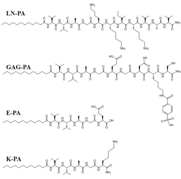
Figure 2. 1 Chemical structures of PA molecules. ... 28
Figure 2. 2 LC-MS of LN-PA (a,b), GAG-PA (c,d), K-PA (e,f) and E-PA (g,h). .... 29
Figure 2. 3 SEM and STEM images of LN-PA/GAG-PA (a), LN-PA/E-PA (b), K-PA/GAG-PA (c) and K-PA/E-PA (d) ... 31
Figure 2. 4 Characterization of secondary structure of peptide nanostructures by CD. ... 32
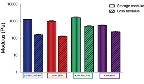
Figure 2. 5 Mechanical properties of PA gels measured by oscillatory rheology ... 33
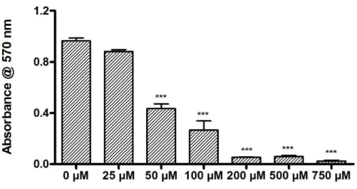
Figure 2. 6 Concentration-dependent toxicity of 6-OHDA in SH-SY5Y cells determined by Alamar blue assay.. ... 34
Figure 2. 7 Viability of SH-SY5Y cells when cultured on peptide nanofibers in the presence or absence of 50 µM 6-OHDA for 24 h analyzed by calcein/ethidium homodimer live-dead assay. ... 35
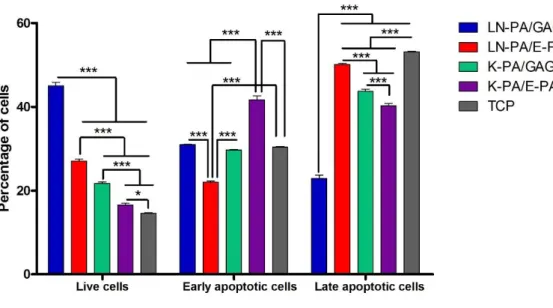
Figure 2. 8 Cell apoptosis on different PA combinations and TCP after 24 h of culture tested by flow cytometry analysis ... 36
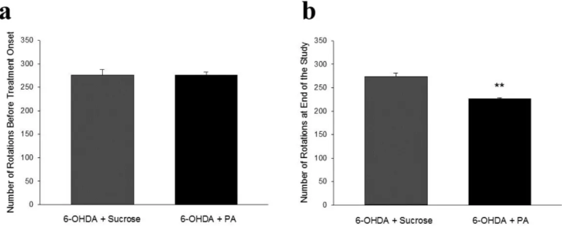
Figure 2. 9 Changes in rotational behavior evaluated by rotameter ... 37
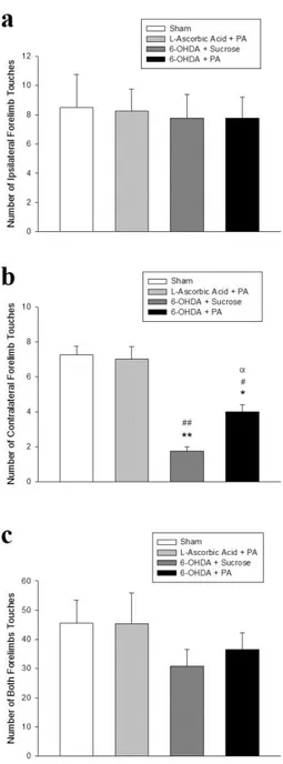
Figure 2. 10 Changes in forelimb asymmetry evaluated by cylinder test. ... 38
Figure 2. 11 Changes in forelimb akinesia evaluated by stepping test ... 40
Figure 2. 12 H&E staining of striata of rat brain sections at postoperative week 6... 41
Figure 2. 13 Immunohistochemistry against Iba-1 protein for rat brain sections at postoperative week 6. ... 42
Figure 2. 14 Levels of dopamine in lesioned striatum. ... 43
Figure 2. 15 Levels of TH analyzed by Western blot in lesioned striatum. ... 44
xvi
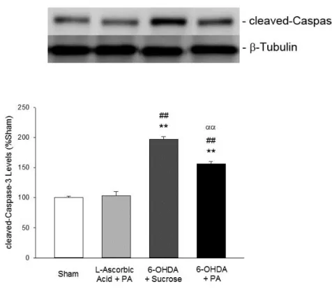
Figure 2. 17 Levels of cleaved-Caspase-3 analyzed by Western blot... 46
Figure 3. 1 Chemical structures of PA molecules used in the study. ... 51
Figure 3. 2 LC-MS of LN-PA (a,b), GAG-PA (c,d), K-PA (e,f) and E-PA (g,h). .... 53
Figure 3. 3 Images of rat sciatic nerve (a), white stripes of nerve trunk under the epineurial connective tissue (b), stripped epineurial connective tissue and nerve fascicles (c), white stripes of nerve fascicle after the removal of connective tissue (d) and a number of nerve fibers after being teased (e) ... 55
Figure 3. 4 Light microscopy images illustrating Schwann cell cultures at day 10 (A) and day 20 (B) and after the first passage (C). ... 56
Figure 3. 5 Viability of Schwann cells on PLL coated surface (a), K-PA/E-PA scaffold (b) and LN-PA/GAG-PA scaffold (c) analyzed by live–dead assay after 48 h of incubation ... 60
Figure 3. 6 PA substrates are biocompatible and support adhesion of Schwann cells. SEM images of Schwann cells cultured on PLL (a,d), K-PA/E-PA (b,e) and LN-PA/GAG-PA (c,f) gels ... 61
Figure 3. 7 Immunostaining against S100 ... 62
Figure 3. 8 NGF concentration in culture medium of Schwann cells cultured on LN-PA/GAG-PA, K-PA/E-PA and PLL coated surfaces after 48 h of incubation. ... 63
Figure 4. 1 In vitro experimental design. ... 71
Figure 4. 2 Chemical structures of PA molecules. ... 76
Figure 4. 3 LC-MS of TC-PA (A), KK-PA (B) and EE-PA (C). ... 77
Figure 4. 4 SEM images of TC-PA/EE-PA and KK-PA/EE-PA. ... 78
xvii
Figure 4. 6 Equilibrium storage and loss modulus of TC-PA/EE-PA and
KK-PA/EE-PA in water. ... 79
Figure 4. 7 Viability of PC-12 cells seeded on 2D or within 3D peptide nanofibers which was tested by flow cytometry analysis. ... 81
Figure 4. 8 PC-12 cells cultured on TC-PA/EE-PA and KK-PA/EE-PA nanofibers and quantification of neurite length and percentage of neurite bearing cells. ... 82
Figure 4. 9 SEM images of PC-12 cells cultured on 2D TC-PA/EE-PA, EE-PA/KK-PA nanofibers, and 3D TC-EE-PA/KK-PA/EE-EE-PA/KK-PA, EE-EE-PA/KK-PA/KK-EE-PA/KK-PA gels on day 7. ... 83
Figure 4. 10 Gene expression analyses of β-III tubulin and SYN1 on day 7. ... 85
Figure 4. 11 Western blot analysis of βIII-tubulin and SYN1 protein expression of PC-12 cells seeded in 3D TC-PA/EE-PA or KK-PA/EE-PA hydrogels with NGF induction. ... 87
Figure 4. 12 Western blot analysis of ERK phosphorylation (p ERK) and total ERK expression ... 88
Figure 5. 1 Schematic diagram of the study. ... 93
Figure 5. 2 Self-assembled PA nanofibers.. ... 102
Figure 5. 3 LC and MS of PA molecules used ... 103
Figure 5. 4 STEM images of the TC-PA/E-PA and K-PA/E-PA nanofibers formed at pH 7.4. ... 104
Figure 5. 5 SEM images of TC-PA/E-PA and K-PA/E-PA gels reveal the ECM-like morphology of PA scaffolds. ... 105
Figure 5. 6 Characterization of secondary structure of peptide nanostructures by CD spectroscopy. ... 105
xviii
Figure 5. 8 Viability of rMSCs when cultured on peptide nanofibers and TCP analyzed by calcein ethidium homodimer live−dead assay. ... 107 Figure 5. 9 SEM images of rMSCs cultured on TC-PA/E-PA, E-PA/K-PA gels, and TCP at 7 days and 12 days after cell seeding. ... 108 Figure 5. 10 Gene expression analyse of (a) MSC markers and (b) osteogenic markers.. ... 110 Figure 5. 11 Gene expression levels of different differentiation markers in rMSCs at week 2 on TC-PA/E-PA substrate and bare surface ... 111 Figure 5. 12 ALP activity of rMSCs on days 3 and 7. ... 113 Figure 5. 13 Deposition of calcium on peptide coated substrates and TCP on days 7 and 12 as demonstrated by alizarin red staining. ... 114 Figure 5. 14 Quantification of relative calcium deposition on days 7 and 12. ... 114
xix
List of Tables
Table 4. 1 Experimental Groups ... 71
Table 4. 2 Primers Used for qRT-PCR Expression Analysis... 74 Table 5. 1 Primers Used for qRT-PCR Expression Analysis... 99
xx
Abbreviations
6-OHDA 6-hydroxydopamine
ALP Alkaline phosphatase
ANOVA Analysis of variance
BSA Bovine serum albumin
CD Circular dichroism
CNS Central nervous system
DAB 3,3′-diaminobenzidine
DCM Dichloromethane
DIEA N,N-diisopropylethylamine
DIT Digital integration time
DMEM Dulbecco's modified Eagle's medium DMF N,N-Dimethylformamide
ECM Extracellular matrix
ETD Everhart–Thornley detector
FBS Fetal bovine serum
Fmoc 9-Fluorenylmethoxycarbonyl
GAPDH Glyceraldehyde 3-phosphate dehydrogenase HBTU N,N,N′,N′-Tetramethyl-O-(1H-benzotriazole-1-yl)
uronium hexafluorophosphate
HCl Hydrochloric acid
H&E Hematoxylin and eosin
HPLC High pressure liquid chromatography
HRP Horseradish peroxidase
HS Horse serum
HSPG Heparan sulfate proteoglycans
i.p. intraperitoneally
LC-MS Liquid chromatography-mass spectroscopy
MEM Minimum essential medium
MSC Mesenchymal stem cell
xxi
NGF Nerve growth factor
NSC Neural stem cell
PA Peptide amphiphile
PBS Phosphate buffered saline
PD Parkinson’s disease
PLL Poly-L-lysine
PNS Peripheral nervous system
PVDF Polyvinylidene fluoride
P/S Penicillin/streptomycin
rMSC Rat mesenchymal stem cell
RPMI Roswell Park Memorial Institute medium
qRT-PCR Quantitative real-time polymerase chain reaction
sem Standard error of mean
SEM Scanning electron microscopy
STEM Scanning transmission electron microscopy
SYN1 Synaptophysin I
TBST Tris-buffered saline and Tween 20
TCP Tissue culture plate
TFA Trifluoroacetic acid
TH Tyrosine hydroxylase
TIS Triisopropyl silane
TN Tenascin
1
Chapter 1
1. Introduction
1.1 Neurodegenerative Diseases
Neurodegenerative diseases are characterized by progressive neuronal degeneration, intracellular or extracellular protein aggregation, and motor and cognitive dysfunctions. The prevalence of most of the neurodegenerative diseases increases with age. For instance, ~15 million people are affected worldwide by Alzheimer disease, which is a common neurodegenerative disease. Since there are currently no available therapeutic approaches in order to prevent the progression of the disease, it is predicted that this will increase to 13.2 million and 16.2 million in the United States and Europe by 2050, respectively [1, 2]. Therefore, development of effective strategies for prevention or treatment of neurogenerative diseases are essential in order to deal with social and financial costs of neurodegeneration.
1.2 Extracellular Matrix (ECM) of the Nervous System
ECM is the noncellular collection of extracellular molecules secreted by the cells, and provides physical support for the cells and affects the intracellular signaling cascades through biomechanical and biochemical cues that are essential for cell fate including proliferation, adhesion, differentiation and cell death. ECM components show synergy with the signals coming from growth factors and hormones and are involved in tissue-specific gene expression control via transduction mechanisms [3, 4]. Also, ECM displays a dynamic structure by being remodeled by the cells and constantly altering its composition and structure during development, remodeling, repair and aging [5].
2
1.2.1 Components of ECM of the Nervous System
Although some components of ECM such as water, proteins and polysaccharides are common in all tissues, ECM of each tissue has a specific structure in terms of physical, topological, and biochemical composition. ECM is mainly composed of two types of macromolecules which are fibrous proteins and proteoglycans [6, 7]. Collagens, elastins, laminins and fibronectins are the main proteins found in ECM. The ECM shows different degrees of stiffness, which varies several orders of magnitude from brain tissue to bone tissue. This variation occurs as a result of concentration of some ECM components such as collagen and elastin. Collagen is the most abundant fibrous protein of ECM and has different functions including providing tensile strength, regulation of cell adhesion, support for migration, and tissue development [8]. Elastin, which is another protein, provides recoil for the tissue during repeated stretching while fibronectin directs the ECM organization and has an important role in cell attachment [9]. As a non-fibrous protein, laminin plays a role in tissue structure and cell function including cell adhesion, migration, and differentiation [10]. Proteoglycans are the glycosylated proteins which form the majority of the extracellular space and display different functions [6]. While some proteoglycans are specific to different tissue types, some of them are common and distributed widely. For instance, aggrecan is the major type of proteoglycan found in cartilage tissue while perlecan is a heparan sulfate proteoglycan (HSPG) present in basement membrane [11]. Composition of ECM also varies according to the developmental stage and aging. For instance, levels of some junctional proteins such as cadherin decrease during aging and gaps occur between the epithelial cells, which affects the junctional integrity [12]. Therefore, composition and
3
dynamic structure of ECM must be taken into consideration while designing synthetic materials.
1.2.2 Challenges to Nerve Regeneration
Nervous system is the most complex and highly organized system in the body and is composed of a complex network of nerves. Since the nervous system has very limited regeneration capacity, any damage in this system induced by either physical injuries or neurological disorders can cause degeneration and neuronal cell death due to loss of communications between the healthy cells. Following the injuries or traumas in the nervous system, patients generally suffer from the loss of sensory or motor function, and neuropathic pains.
Although the current therapeutic approaches provide regeneration up to a certain point, they are not quite effective. For the development of new therapies for nervous system, it is important to understand the nature and requirements of the cells, including physical, chemical and biological signals, and to induce regeneration process while preventing the formation of glial scars, which makes it a complex process. Peripheral nervous system (PNS) and central nervous system (CNS) respond to injury differently. In the PNS, a series of pathophysiological events occur after the Wallerian degeneration in the distal end. The distal part of the nerve is degenerated and the remaining cellular debris is removed by the macrophages and monocytes [13]. In PNS injuries, direct end-to-end reconnection through surgical sutures is a common method for the treatment of small injury gaps. For larger nerve defects, autografts are used as a gold standard to bridge the gaps. However, there are still problems which limit the use of autografts such as loss of function at donor sites and limited number of donor
4
grafts. Allografts and xenografts may be considered [14, 15], but they also have problems including transfer of several diseases and immunological rejections. CNS has much lower regeneration capacity compared to PNS. Following the injury in CNS, glial scar formation and release of inhibitory molecules occur at the injury site. Removal of cell debris is slower in CNS than PNS due to the limited infiltration levels of macrophages through the brain-spinal cord barrier [16]. Because of these reasons, the scaffolds constructed by tissue engineering strategies may offer an alternative strategy to facilitate neural repair. The studies about neural regeneration generally focus on the inhibitory nature of the nervous system after the injuries and combination of multiple cues in order to increase regeneration capacity of nervous system. In nervous system, regeneration occurs through mechanisms to recover the function of degenerated cells. The limited regeneration capacity of nervous system is aimed to be improved by using biomaterials designed for stem cell culture, differentiation and in
vivo regeneration. The strategies to construct biomaterials for nerve repair should
include the removal of inhibitory environment after the damage, induction of axon guidance, managing the cell signaling, increasing the local concentration of neurotrophic factors and providing an artificial microenvironment by mimicking the native ECM of the cells in order to fill the gap originating from the injury [17].
1.3 Approaches in the Design of ECM-Mimetic Scaffolds for Neural Differentiation and Regeneration
Biomaterials, that are generated as a result of self-assembling process and include short peptides and peptide derivatives, have significant potential in regenerative medicine applications. Mimicking native ECMs which have regulatory functions in tissue
5
formation and regeneration is a promising strategy for the design of synthetic biomaterials. Synthetic materials are commonly used in tissue engineering applications because of their specific properties such as modifiable elasticity, high water content and ability for encapsulation of cells. Cell adhesion, proliferation and differentiation are other important significant criteria to be considered when designing a nanomaterial for neural tissue engineering. The main goal of regenerative medicine is to enhance tissue regeneration and healing after injury or disease leading to degeneration of the tissue of interest. Guidance of cell behavior by the features of a material at cell-biomaterial interfaces has significant importance, and developing novel biomaterials with certain surface modifications to induce controlled cell function would be beneficial for improving therapeutic potential of current regenerative medicine protocols. The biodegradability and biocompatibility are the most important features of synthetic biomaterials. These synthetic materials must also be multifunctional in order to mimic the natural ECM both biochemically and biophysically.
1.3.1 Biochemical Functionalization of the Scaffolds for Neural Differentiation and Regeneration
Mimicking the cytoarchitecture of native ECM of neural cells has become a common approach in tissue engineering applications. ECM is the non-cellular component of organisms which provides vital physical support and presents the biological cues for tissue development and differentiation [6]. ECM-mimetic scaffolds can be decorated with a number of different peptide motifs derived from the components of ECM at very high density in order to introduce bioactivity to the system and direct the
6
biochemical signaling cascade. Especially cell-binding epitopes of fibrous proteins of ECM including RGDS, YIGSR, and IKVAV, are widely used in the literature since they have both structural and adhesive roles [18]. In addition to bioactive moieties for initial attachments of the cells, further proliferation, migration and differentiation signals or degradable sites such as hydrolysable ester linkages [19] and enzyme-mediated degradable sequences [20] may also be incorporated into the scaffolds. Among these scaffolds, peptide amphiphiles (PA), which can self-assemble into nanofibers through non-covalent interactions and introduce bioactive epitopes on their surface, have great potential to mimic the regulatory characteristics of natural environment of the cells for biological studies as well as therapeutic applications. PA molecules can form self-organized biocompatible fibers at nanoscale and their surface can be tailored in order to induce biochemical responses in a desired way [21, 22]. Moreover, two or more PAs can be used to form nanofibers with multiple cues in order to obtain cumulative effects [23].
When compared to other systems in the body, nervous system has a very unique structure due to different compositions of ECM and its mechanical properties. While designing a bioactive scaffold for neural regeneration purposes, it is important to consider its biological and chemical properties as well as its physical structure. In order to introduce bioactivity into the system, bioactive epitopes of ECM proteins can be incorporated into the scaffold. Within the concept of biomaterials, use of peptide nanofibers has become extensive in neural regeneration applications. Decorating the peptide molecules with different bioactive epitopes derived from ECM proteins such as, isoleucine-lysine-valine-alanine-valine (IKVAV) and
tyrosine-isoleucine-glycine-7
serine-arginine (YIGSR) derived from laminin, arginine-glycine-aspartate (RGD) derived from fibronectin and valine-phenylalanine-aspartate-asparagine-phenylalanine-valine-leucine-lysine (VFDNFVLK) peptide derived from tenascin-C (TN-C) makes the scaffolds biofunctional and has been shown to induce neural differentiation, attachment and migration [23-26]. These peptides have become favorable for regenerative medicine applications, since they are more stable and easy to synthesize than the bulk proteins.
Incorporation of the bioactive epitopes into PA molecules and self-assembly of these molecules into nanofibrous scaffolds through non-covalent interactions can provide an artificial microenvironment which is similar to the natural ECM [27, 28]. Arginine– alanine–aspartate (RAD)16-I and RAD16-II were used in the form of self-assembled peptide nanofibrous scaffolds for neural cell cultures. RAD16 peptide scaffolds functionalized with RGD and laminin-derived motifs, GFLGFPT and BMHP were used for differentiation of neural stem cells (NSCs) [27]. IKVAV sequence, which is known to promote and direct neurite outgrowth, was used in different studies as a part of self-assembling peptide nanofibers. Silva et al. cultured neural progenitor cells on IKVAV-bearing scaffolds to study cellular differentiation of these cells in vitro, and they selectively induced neuronal differentiation while they suppressed differentiation to astrocytes [29]. In addition to in vitro studies, animal models have been used in order to investigate the potential of self-assembling peptide nanofibers for repair and regeneration of nervous system in several studies. Tysseling-Mattiace et al. inhibited the glial scar formation, promoted axon elongation and facilitated regeneration with significant behavioral improvement after spinal cord injury using IKVAV-bearing
8
peptide nanofibers [30]. Also, self-assembling peptide nanofibers functionalized with IKVAV sequence were used to encapsulate NSCs and enhance stem cell survival as well as to reduce formation of glial astrocytes in a rat brain surgery model to demonstrate the damage in cerebral neocortex/neopallium loss [31].
1.3.2 Physical Functionalization of the Scaffolds for Neural Differentiation and Regeneration
When designing biomaterials for tissue-engineering applications, physical properties of a material should also be taken into account in addition to biological and chemical properties. Physical characteristics including stiffness, dimensionality, substrate topography and electrical conductivity are important parameters for scaffold functionalization to induce neural differentiation.
Mechanical properties of the microenvironment are important regulators of cellular characteristics including morphology [32] and motility [33] of the cells as well as differentiation [34]. Normally, brain and spinal cord are the softest tissues in the body with elastic moduli of about 2000 Pa. However, when glial scar forms as a result of injury, it increases the stiffness of this area which leads to problems in neurite extension and neural regeneration. Soft materials have become more favorable for regeneration studies in CNS, because neurons and astrocytes give different responses to matrix stiffness. When designing a scaffold for neural differentiation, the mechanical properties of the scaffold should be similar to that of brain tissue, which is below 1 kPa [35]. It was shown that stiff substrates having elastic moduli between 1,000 and 10,000 Pa caused the differentiation of adult NSCs into glial cells, while soft substrates with elastic moduli between 100-500 Pa induced primarily neuronal
9
differentiation [36]. Also, spinal cord and cortical brain neurons prefer soft materials in order to be able to extend their neurites [36, 37] while astrocytes form stress fibers and favor the surfaces with high elastic modulus [35]. The effect of stiffness was also studied on human mesenchymal stem cells. This effect was investigated by using three-dimensional (3D) porous scaffolds generated by type I collagen and hyaluronic acid. Using different concentration of 1-ethyl-3(3-dimethylaminopropyl) carbodiimide as a crosslinking agent, the elastic modulus of the 3D substrates was modified and the stiffness was controlled in the range of 1–10 kPa. Results showed that mesenchymal stem cells (MSCs) were likely to differentiate into neurons at 1 kPa while they preferred glial lineage in the scaffold at 10 kPa stiffness verified with experiments focused on up-regulation of neuronal mid- and late protein markers after longer mechanical induction time for 21 days [38].
In vitro models are important tools to study cell behavior and fate in a
highly-controlled manner. Although two-dimensional (2D) cell cultures are commonly used in differentiation studies, three-dimensional cell cultures are important in vitro models to fill the gap between 2D cell culture experiments and in vivo studies. To study regeneration of neural tissue which has a very low regeneration capacity, use of 3D models is important. They better mimic the ECM of neural cells in terms of providing support and enhanced diffusion of oxygen and nutrients. There are fundamental differences between cells grown on a monolayer surface and in 3D manner. The 3D cultures have been shown to display longer neurite outgrowth, higher levels of survival as well as different patterns of differentiation when compared to 2D monolayers [39, 40]. Also, hippocampal neurons cultured on a 3D aragonite matrix displayed higher survival as compared to 2D counterparts with higher-density network formation [41].
10
In another study, 3D cell culture systems with the functional motifs of RGD (Arg-Gly-Asp), bone marrow homing peptide 1 and 2 were developed for the culture of adult NSCs. The proliferation and differentiation of NSCs were supported by these scaffolds by allowing a satisfactory supply of nutrients and oxygen [42].
Nanotopography is an important tool to guide cell differentiation and it can be modified in size and shape according to desired application. Interaction of the cells with nanotopographies can induce different responses including changes in cell morphology [43], adhesion [44], proliferation [45] and gene regulation [46]. It is important to understand that neurite growth along topographical patterns is important for tissue engineering applications in neurology. In one study, nanotopographic control of neuronal polarity was studied and the interaction between focal adhesions and topographic features were exploited to design a set of scaffolds made of cyclic olefin copolymer yielding control over neuronal polarity establishment and neurite pathfinding. By this way, specific neuronal polarity states were selectively favored by a set of biocompatible textured scaffolds and it was demonstrated that this selection can be tailored through varying the topographical constraint applied to the maturation of focal adhesions during neuritogenesis [47].
Neurons are electrically excitable cells and as a result of their inherent nature, they are capable of transmitting electrochemical signals throughout the nervous system making them highly sensitive to electrical stimuli. The action potential generated at the synapse is the key component of neural communication, so electrical conductivity is an important physical property to enhance neural cell activity [48]. Providing electrical conductivity to the scaffolds and electrical stimulation of the cells cultured on these scaffolds might be useful to improve synaptic connections. One of the new approaches
11
in scaffold generation is the design of conductive substrates which provide external electrical stimuli to the stem cells, because these stimuli can affect the cell behavior including proliferation, migration and differentiation [49, 50]. Electrical conductivity can be provided to the scaffold through utilization of conductive polymers and carbon based materials which include carbon nanotubes, graphite and graphene [51]. Combining the conductivity of carbon nanotubes with the alignment of the poly(lactic acid) nanofibers supports and increases the neural differentiation of mouse embryonic stem cells which was detected by enhanced expression of mature neuronal markers even in the absence of direct electrical stimulation [52].
Overall, self-assembled nanofibrous scaffolds provide different perspectives in neural regeneration processes. The fibrous structure is quite similar to natural ECM, and tailoring the biochemical and physical properties of the scaffolds enables the manipulation of cellular functions depending of the type of the injury or degeneration. Altogether, these bioactive nanofibrous scaffolds hold great potential for neural regeneration both after injury and for neurodenerative diseases.
12
Chapter 2
2. Regenerative Effects of Peptide Nanofibers in an
Experimental Model of Parkinson’s Disease
This chapter of thesis was published in the following article [53]; Reproduced from “Regenerative effects of peptide nanofibers in an experimental model of Parkinson’s disease”; Sever, M.; Turkyilmaz, M.; Sevinc, C.; Cakir, A.; Ocalan, B.; Cansev, M.; Guler, M. O.; Tekinay, A. B., Acta Biomaterialia, 2016, 46, 79-90, with permission from Elsevier.
2.1 Introduction
Neurodegenerative diseases caused by infection, stroke and acute trauma are the fourth leading cause of death in the world after cardiovascular diseases, cancer and stroke [54]. Among the neurodegenerative diseases, Parkinson’s disease (PD) is the second most common neurodegenerative disease after Alzheimer’s disease. PD is a severe, chronic and progressive disease associated with symptoms such as tremor, postural instability, rigidity and bradykinesia [55]. PD is characterized by diminished levels of striatal dopamine as a consequence of dopaminergic neuron loss in the substantia nigra pars compacta. Beside the degeneration of dopaminergic neurons, Lewy body formation can be included as pathologic hallmarks of PD [56]. In addition, several studies have shown that activation of apoptosis is one of the mechanisms that underlie progressive striatal neurodegeneration [57-60]. A recent study has provided strong evidence for this hypothesis by showing that in vivo suppression of Caspase-3, an apoptotic marker, by RNA interference in a rat model of PD reduced striatal
13
dopaminergic cell loss and improved locomotor activity [61]. Millions of people in US and Europe suffer from PD, but the pharmaceutical agents used for PD treatment including levodopa, Monoamine oxidase-B inhibitors (selegiline and rasagiline), Catechol-O-methyl transferase inhibitors (tolcapone and entacapone) and dopamine agonists (pramipexole and ropinirole) just relieve and modify the symptoms. In addition, since they are administered orally, they can cause systemic toxicity with different adverse effects [62]. There are no therapies yet available for PD to slow down the degeneration process in the brain and recover the lost function. Besides these current strategies, there are various experimental approaches to improve the efficacy of currently available treatment strategies. Uses of adenosine A2A receptor antagonists, glutamate receptor antagonists, monoamine oxidase inhibitors, anti-apoptotic agents, antioxidants and coenzyme Q10 are among the emerging pharmacotherapies at different stages of preclinical and clinical trials [63]. Also, there are several non-pharmacological approaches offering alternative strategies for the treatment of PD. Viral vectors were studied to silence the over-expressed defective genes considered as risk factors in PD or to transfer the genes such as glutamic acid decarboxylase gene [64]. Stem cell transplantation is another promising approach for the replacement of dopaminergic neurons progressively lost in PD. Induced pluripotent stem cells, NSCs and MSCs are the most commonly studied resources to generate dopaminergic neurons as a treatment strategy for PD [65]. However, there are some drawbacks for this technique such as selecting tumor-free cell type for transplantation, which limits the potential therapeutic benefits. Additionally, some surgical procedures are within the emerging techniques for PD treatment, including deep brain stimulation, pallidotomy and thalamotomy, especially for PD patients having severe disabling
14
problems and not responding to traditional treatment options. However, these procedures also have high risks and require close post-operative follow-up period [63].
Although repairing the damaged area in the brain is extremely challenging, recent advances in regenerative medicine and tissue engineering provide new therapeutic approaches for neurodegenerative disease treatment. Biomaterials, which are designed to interact with biological systems provide new platforms to replace the damaged neurons or slow down the progression of the diseases [66]. Biomaterial scaffolds can be modified with physical or/and chemical cues depending on the aim of the study. Cell attachment, adhesion, migration and spreading as well as cell differentiation into specific lineages can be achieved through physical or chemical modification of the surface of the scaffold, including stiffness, topography, charge and interactions with ECM proteins or cells [67].
Laminins are heterotrimeric proteins and the major non-collagenous components of the basal lamina. They bind to cell membrane through interaction with integrin receptors on the cell surface, and by this way, they can influence diverse biological activities including cell adhesion, migration and differentiation [68]. Moreover, laminin has a fundamental role in axonal growth and myelination [69] and functions as a neurite-outgrowth promoting factor for peripheral and central neurons [70]. Laminin also interacts with other matrix elements such as HSPG through noncovalent interactions, and laminin-HSPG complex was found to be involved in neurite outgrowth [71, 72]. Heparan sulfates are highly sulfated glycosaminoglycans and function as cell–ECM interface to modify cell signaling. They also interact with
15
various ECM molecules such as growth factors. They act as a reservoir of such growth factors and increase their local concentration [73-76].
The self-assembled PA nanofibers provide suitable platforms to mimic ECM. As a result of the hydrophobic interaction between alkyl tails and β-sheet formation between peptide segments, they self-assemble into nanofibers in aqueous environment. Also, biochemical signals provided by a specific protein can be introduced into the nanofiber system through addition of bioactive epitopes into individual PA molecules instead of use of bulk protein [77]. The peptide sequences Ile-Lys-Val-Ala-Val (IKVAV) of cell-binding domain of laminin was discovered and found to facilitate neurite extension [78]. After the discovery of this small peptide sequence, IKVAV-carrying PAs were used in both in vitro and in vivo studies for neural differentiation [29] and spinal cord regeneration [30, 79], respectively. We previously reported that combination of IKVAV-carrying PAs along with heparan-sulfate-mimicking PA nanofibers displayed dual bioactivity and promoted much longer neurite outgrowth compared to the scaffold with laminin-derived signals alone, even in the presence of inhibitory conditions provided by chondroitin sulfate proteoglycans [23]. Although bioactive PA nanofibers were shown to enhance in vitro neurite extension or peripheral nerve regeneration, there is no study on the therapeutic effects of bioactive PA nanofibers on PD.
Here we investigated whether these heparan sulfate and laminin mimetic PA nanofibers have potential therapeutic effect for both protection of SH-SY5Y cells against 6-hydroxydopamine (6-OHDA)-induced apoptosis in in vitro studies and for reducing striatal injury and enhancing dopaminergic nerve regeneration in
16
experimental PD model. Six weeks following treatment with bioactive PA nanofibers, rats with 6-OHDA-induced Parkinsonism displayed improvements in behavioral functions, i.e., reductions in forelimb asymmetry, contralateral forelimb akinesia and d-amphetamine-induced rotational behavior in cylinder, stepping and rotation tests, respectively. Moreover, brain dopamine content and tyrosine hydroxylase (TH) levels increased, while cleaved-Caspase-3 levels decreased in rats treated with PA nanofibers compared to sucrose control. Histological assessment also showed that PA injection to the striatum provided better tissue integrity by reducing the progressive cell loss caused by 6-OHDA toxicity, which makes this bioactive nanofiber system a promising new platform for PD treatment.
2.2 Experimental Section 2.2.1 Materials
All protected amino acids, lauric acid, 4-[α-(2′,4′-dimethoxyphenyl) 9-Fluorenylmethoxycarbonyl (Fmoc)-aminomethylphenoxyacetomidonorleucyl-MBHA resin (Rink amide (Fmoc)-aminomethylphenoxyacetomidonorleucyl-MBHA resin), N,N,N′,N′-Tetramethyl-O-(1H-benzotriazole-1-yl) uronium hexafluorophosphate (HBTU) and
N,N-diisopropylethylamine (DIEA) were purchased from Nova-Biochem, ABCR, or Sigma-Aldrich. 6-OHDA was purchased from Sigma Aldrich. Alamar Blue, viability assay reagents and other cell culture materials were purchased from Invitrogen. Apoptosis assay reagents were purchased from Biotium. All other chemicals and materials used in this study were purchased from Thermo Scientific or Sigma Aldrich. 2.2.2 Synthesis of PA Molecules
PA molecules were synthesized on Rink Amide MBHA Resin or Fmoc-Glu(OtBu)-Wang Resin by using Fmoc-protected solid phase peptide synthesis method. Amino
17
acid couplings were performed with 2 equivalents of amino acids activated with 1.95 equivalents of HBTU and 3 equivalents of DIEA for 2 h. Fmoc removal was performed with 20% piperidine–N,N-dimethylformamide (DMF) solution for 20 min. 10% acetic anhydride–DMF solution was used to permanently acetylate the unreacted amine groups after each coupling step. DMF and dichloromethane (DCM) were used as washing solvents after each step. p-Sulfobenzoic acid was coupled to the side chain of lysine to synthesize sulfonated PAs. A lysine residue with 4-methytrityl (Mtt) side chain protection was used for selective deprotection of amine groups. Mtt removal was performed by shaking resins for 5 min with trifluoroacetic acid (TFA):triisopropyl silane (TIS):H2O:DCM in the ratio of 5:2.5:2.5:90. Cleavage of the PAs and protection groups from the resin was carried out with a mixture of TFA:TIS:H2O in the ratio of 95:2.5:2.5 for 3 h. Excess TFA removal was carried out by rotary evaporation. PAs in the remaining solution were precipitated in ice-cold diethyl ether overnight. The precipitate was collected by centrifugation next day and dissolved in ultrapure water. This solution was frozen at -80 °C for 4 h and then lyophilized for 4–5 days. PAs were characterized by liquid chromatography–mass spectrometry (LC–MS). Mass spectrum was obtained with Agilent LC-MS equipped with Agilent 6530 Q-TOF with an ESI source and Zorbax Extend-C18 2.1 × 50 mm column for basic conditions and Zorbax SB-C8 4.6 × 100 mm column for acidic conditions. A gradient of water (0.1% formic acid or 0.1% NH4OH) and acetonitrile (0.1% formic acid or 0.1% NH4OH) was used. In order to remove residual TFA, positively-charged PAs were treated with 0.1 M hydrochloric acid (HCl) solution and lyophilized. To purify the peptides, Agilent preparative reverse-phase high pressure liquid chromatography (HPLC) system equipped with Zorbax Extend-C18 21.2 × 150 mm column was used for basic
18
conditions and Zorbax SB-C8 21.2 × 150 mm column was used for acidic conditions. A gradient of water (0.1% TFA or 0.1% NH4OH) and acetonitrile (0.1% TFA or 0.1% NH4OH) was used. All peptide batches were freeze-dried and reconstituted in ultrapure water at pH 7.4 before use.
2.2.3 Scanning Electron Microscopy (SEM) Imaging of PA Nanofibers
PA nanofiber networks were observed by SEM imaging. Oppositely charged PA solutions (1 wt%) were mixed in appropriate volume ratio (final volume being 30 μL) to produce gels with neutral charge. Gels were formed on silicon wafer and dehydrated by transferring to 20%, 40%, 60%, 80% and 100% v/v ethanol, sequentially. They were critical point-dried afterwards by using Autosamdri 815B equipment from Tousimis. Dried PA gels were coated with 4 nm Au/Pd and SEM (FEI Quanta 200 FEG) images were taken by using an Everhart–Thornley Detector (ETD) at high vacuum mode at 5 keV beam energy.
2.2.4 Scanning Transmission Electron Microscopy (STEM) Imaging of PA Nanofibers
Samples for STEM imaging were prepared by mixing equal volumes of negatively and positively charged PA molecules with appropriate concentrations for charge neutralization and placing them on a 200-mesh carbon TEM grid for 10 min followed by 2 wt% uranyl acetate staining for 2 min and drying. STEM images at HAADF mode were acquired with FEI Tecnai G2 F30 TEM at 300 kV.
2.2.5 Secondary Structure Analysis
A JASCO J815 circular dichroism (CD) spectrometer was used at room temperature. Oppositely charged 2.5 × 10⁻⁴ M PA solutions were mixed in appropriate volume
19
ratios (final volume being 500 μL) to produce nanofibers with net neutral charge. Measurements were carried out from 300 nm to 190 nm; data interval and data pitch being 0.1 nm, and scanning speed being 100 nm min−1. All measurements were performed with three accumulations. Digital Integration Time (DIT) was selected as 1 s, band width as 1 nm, and the sensitivity was standard.
2.2.6 Oscillatory Rheology
Oscillatory rheology measurements were performed with Anton Paar Physica RM301 Rheometer operating with a 25 mm parallel plate configuration at 25 °C. 250 μL total volume with 1 wt% of each PA component was carefully loaded onto the center of the lower plate and incubated for 10 min for gelation before measurement. After equilibration, the upper plate was lowered to a gap distance of 0.5 mm. Storage moduli (G′) and loss moduli (G″) values were scanned from 100 rad s−1
to 0.1 rad s−1 of angular
frequency, with a 0.5% shear strain. Three samples were measured for each PA gel.
2.2.7 Cell Culture and Maintenance
SH-SY5Y cells (kindly provided by Prof. Dr. Fikrettin Sahin, Yeditepe University, Istanbul, Turkey) were used in cell culture experiments. They were cultured in 75 cm2 flasks at 37 °C in a humidified incubator and supplied with 5% CO2. Cells were maintained in 1:1 mixture of Eagle’s minimum essential medium and F12 medium supplemented with 10% fetal bovine serum (FBS) and 1% penicillin/streptomycin (P/S). All cell experiments were carried out after reaching 90% confluency. The culture medium was changed every 3–4 days.
20 2.2.8 Drug Treatment and Viability Assay
6-OHDA was dissolved in 0.3% l-ascorbic acid/0.9% NaCl solution to create a stock concentration of 5 mM and was used at a final concentration of 25, 50, 100, 200 and 500 μM. SH-SY5Y cells were seeded at a density of 2 × 10⁴ cells/well on 96-well plate and incubated for 24 h. Then, they were treated with 6-OHDA (25, 50, 100, 200 and 500 μM) for 24 h. Medium was discarded after 24 h of incubation and replaced with medium containing 10% Alamar blue. Blank group contained only Alamar blue medium without cells. After 3 h incubation at 37 °C, absorbance measurement was performed by using Spectramax M5 microplate reader at 570 and 600 nm as reference. After determining the toxic dose, the viability test of SH-SY5Y cells was performed after 24 h of 6-OHDA treatment by using calcein-AM/ethidium homodimer 1 (EthD-1) staining. In brief, cells were incubated on PA-coated and uncoated 96 well-tissue culture plates (TCPs) at a density of 5 × 10³ cells/well. After 24 h, the cells were exposed to 50 μM 6-OHDA dissolved in 0.3% l-ascorbic acid/saline solution or vehicle (0.3% L-ascorbic acid/saline solution). After 24 h of incubation, cell medium was discarded; cells were washed with phosphate buffered saline (PBS) and then incubated with 2 μM calcein-AM and 2 μM EthD-1 in PBS for 30 min at room temperature. Finally, five random images were taken at 100x magnification from each well for both qualitative and quantitative analysis by using Zeiss Axioscope fluorescence microscope. Cells were counted with Image J system for analyzing proliferation.
2.2.9 Flow Cytometry Analysis of Apoptosis
Apoptosis in SH-SY5Y cells seeded on different PA combinations was measured using the Annexin V-FITC Apoptosis Detection kit. After 24 h incubation in 6-well plates
21
at a density of 5 × 10⁵ cells/well, the cells were exposed to 50 μM 6-OHDA dissolved in 0.3% l-ascorbic acid/saline solution. After 24 h exposure to 6-OHDA, flow cytometry protocol for Annexin V and propidium iodide was performed. Medium was discarded; cells were washed with cold PBS and resuspended in 1X annexin-binding buffer. 100 μL of 1X annexin-binding buffer per assay was added with 5 μL Alexa Fluor® 488 annexin V and 1 μL of 100 μg/mL propidium iodide. Cells were incubated at room temperature for 15 min. The stained cells were analyzed by flow cytometry, measuring the fluorescence emission at 530 nm (FL1 channel) and >575 nm (FL3
channel). 2.2.10 In Vivo Surgery
2.2.10.1 Animals
A total of 32 male Sprague Dawley rats (weighing 300–350 g) were housed individually in each cage with free access to food and water in a 12/12 h light/dark cycle. Experimental procedures conformed to the National Institute of Health Guide for the Care and Use of Laboratory Animals and were approved by the Local Ethics Committee on Experimental Animal Research of Uludag University, Bursa, Turkey (Approval ID: 2012-10/1).
2.2.10.2 Surgical Procedure
Rats (n = 24) anesthetized with ketamine and xylazine (80 and 10 mg/kg, respectively) were placed into the stereotaxic apparatus (David Kopf Instruments, Tujunga, CA, USA). Their heads were shaved and a 2-cm incision was made on the skull under aseptic conditions ensured with 70% alcohol. After visualization of the bregma, two burr holes were drilled and hand-made guide cannulas (each 4 mm long) were placed (first one +0.48 mm anterior and second one -0.40 mm posterior to bregma). The
22
cannulas were plugged and rats were returned to their cages after injection with a single dose of analgesic (buprenorphine; 0.05 mg/kg; s.c.). A group of rats (n = 8) underwent the same operation without placement of guide cannulas in order to serve as sham controls.
2.2.10.3 Intrastriatal lesioning with 6-OHDA and PA Injection
24 h after the placement of guide cannulas, two injection apparatus were placed into the right striata of rats through the guide cannulas at the following coordinates: first at anteroposterior: +0.48 mm, mediolateral: −2.2 mm and vertical: −4.6 mm and second at anteroposterior: −0.40 mm, mediolateral: −4.0 mm and vertical: −6.0 mm according to the Rat Brain Atlas [80]. Freely-moving rats were injected intrastriatally through polyethylene tubing (PE 20; Becton Dickinson, Franklin Lakes, NJ, USA) attached to the apparatus at the given coordinates with either 8 μg of 6-OHDA (dissolved in saline containing 0.3% L-ascorbic acid, n = 16) or its solvent 0.3% L-ascorbic acid (n = 8) at a volume of 2 μL using an injection pump with a flow rate of 1 μL/min. The cannulas were plugged and rats were followed up in individual cages with free access to food and water.
The dose and stereotaxic coordinates for 6-OHDA injections were selected from previous studies [81, 82] which showed that this protocol is adequate for consistent depletion of approximately 70–80% of striatal dopamine and induce Parkinsonism in rats resembling the early phase of PD in clinical setting.
One week later, rats receiving intrastriatal 6-OHDA injections were tested for d-amphetamine-induced rotational behavior. Rats were injected intraperitoneally (i.p.) with d-amphetamine (5 mg/kg) and ipsilateral rotations between 15 and 45 min were
23
recorded using an automated Rotameter system (TSE Systems, Germany). Initial behavioral task was performed in order to ensure that the unilateral striatal 6-OHDA lesion was established and the data were used to randomize the 6-OHDA-lesioned rats into two treatment groups (i.e., 6-OHDA + Sucrose [n = 8] and 6-OHDA + PA [n = 8] groups) with almost equal mean number of ipsilateral rotations (275 ± 11 and 275 ± 6, respectively) in this task. No rotational behavior was observed in rats in “Sham” and “L-Ascorbic Acid + PA” groups.
Rats were followed-up in their cages for three days after the initial rotational behavior task in order to allow for wash-out of d-amphetamine from the brain and then allocated into treatment groups as follows: (i) Sham group, (ii) L-Ascorbic Acid + PA group, (iii) 6-OHDA + Sucrose group and, (iv) 6-OHDA + PA group.
In rats receiving intrastriatal 6-OHDA or L-ascorbic acid injections, PA (1% aqueous solutions) or sucrose (0.25 M) injections were made using the same coordinates of the right striata through the existing guide cannulas. The 1% solutions dissolved in sucrose of both the heparan sulfate-mimicking (GAG) and laminin-derived (LN) PA nanofibers were injected consecutively by the help of the polyethylene tubing attached to the injection apparatus with a 5 min interval in between two injections. After injection, rats were followed-up for a period of 6 weeks in individual cages with free access to food and water.
2.2.11 Behavioral Analysis Rotation Test
24 Cylinder Test
Cylinder test was performed in order to evaluate the forelimb asymmetry in rats using a modified version [83] of the initially-described procedure [84]. Briefly, rats were put individually in a glass cylinder (21 cm diameter, 34 cm height) and video recorded for 5 min without prior habituation. To stimulate rats that showed little or no tendency to explore, the following methods were used in the given order: (i) turning the lights in the room on and off 2 ± 3 times; (ii) mildly shaking the cylinder for 2 ± 3 s; (iii) taking the rat out of the cylinder for approximately 30 s and then putting it back, as described previously [85]. Ipsilateral or contralateral forelimb touches to the cylinder wall were counted by a blinded observer from the video recordings.
Stepping Test
Stepping test (Adjusting Steps Test) was performed in order to evaluate forelimb akinesia three times on the same day with 30 min intervals in between tests according to the procedure described previously [86]. Briefly, rats were held by the experimenter with one hand fixing the hindlimbs and slightly raising the hind part above the surface of a table with a width of 100 cm. The other hand fixed the forelimb not to be monitored. The rats were moved sideways with one paw touching the table at a speed of 100 cm/5 s first in the forehand and then in the backhand direction. The number of adjusting steps was counted for both paws in both directions by a blinded observer from the video recordings.
2.2.12 Animal Perfusion and Tissue Acquisition
Three days after the behavioral tests to allow for the wash-out of d-amphetamine, rats in all groups were sacrificed either with or without transcardiac perfusion using 4% paraformaldehyde solution under ketamine and xylazine anesthesia. Brains were
25
obtained and striata were excised in rats sacrificed without perfusion. Striata were homogenized using 0.4 N HCl, and homogenates were kept for future analyses of dopamine, TH and cleaved-Caspase-3. Brains of rats obtained following paraformaldehyde perfusion were sectioned and processed for immunohistochemical analyses.
2.2.13 Histological Analyses
Sections were deparaffinized in xylene and rehydrated in serial ethanol series for hematoxylin & eosin (H&E) staining according to the standard protocol. For immunohistochemistry experiments, sections were stained with anti-TH (1:250; Millipore AB152) and anti-Iba1 (1:2000 Sigma ab178846) antibodies. After primary antibody staining, horseradish peroxidase (HRP) conjugated goat anti-rabbit secondary antibody (1:500; Millipore) was used followed by 3,3′-diaminobenzidine (DAB) staining. All samples were mounted onto glass slides using xylene based mounting medium. Digital images were acquired via Zeiss Axio Scope A1. Images were acquired by using 10x and 20x objectives.
2.2.14 Dopamine Analyses
Dopamine contents of striata were analyzed using HPLC coupled with an electrochemical detector and an analytical column as described previously [81]. The samples were run at a rate of 1 mL/min using a mobile phase containing 0.15 M Na2HPO4, 0.5 mM sodium octasulfate and 0.1 mM Na2EDTA dissolved in 10% methanol solution.
![Figure 2. 2 LC-MS of LN-PA (a,b), GAG-PA (c,d), K-PA (e,f) and E-PA (g,h). Mass spectrometry of LN-PA; [M+H] + (calculated): 1292.93, [M+H] + (observed): 1293.01, [M+2H] +2 /2 (calculated): 646.96, [M+2H] +2 /2(observed): 647.01, [M+3H] +3 /3 (ca](https://thumb-eu.123doks.com/thumbv2/9libnet/5671273.113565/50.892.185.791.134.760/figure-gag-mass-spectrometry-calculated-observed-calculated-observed.webp)