Aims: Conservative treatment of jaw cysts establishes low surgical complication risk and protects vital anatomical structures such as inferior alveolar nerve, maxillary sinus and permanent tooth germs. Marsupialization and decompression have been widely used in children. The aim of this study was to evaluate the results of the conservative treatment of jaw cysts in five children. Materials and Methods: This article presents case series of marsupialization in jaw cysts associated with impacted teeth in five children. A total of nine impacted teeth within the cystic lesions were observed. Results: Complete resolution of all cystic lesions and simultaneous eruption of six impacted teeth within the cyst were managed. The other two teeth were erupted orthodontically and one had to be extracted. Conclusions: Marsupialization is effective for the treatment of cystic lesions in growing patients as it preserves vital anatomical structures and enables eruption of the impacted teeth within the cyst.
Keywords: Children, conservative treatment, cyst treatment, marsupialization
Conservative Surgical Treatment of the Jaw Cysts in Children: Case
Study of Five Patients
G Gurler, S Yilmaz, C Delilbasi, E Dilaver, E Yuzbasioglu1, A Patir-Munevveroglu2
Address for correspondence: Dr. G Gurler, Department of Oral and Maxillofacial Surgery Istanbul Medipol University School of Dentistry, Atatürk Bulvarı No. 27, Unkapani, Istanbul, Turkey. E-mail: [email protected] In this case series, we present outcomes of conservative surgical approach to jaw cysts in children.
c
AseP
resentAtIonsFive children (three girls and two boys) whose age ranged 9–12 years with no systemic disease, with a mean of 10 years, referred to our clinic for dental treatment. Lesions suspected of cysts were detected in routine panoramic radiographs [Figure 1A]. In clinical examination, three lesions (one mandible, two maxilla) showed buccal expansion but no sign of infection was found in any patient. Cone beam computerized tomography (CBCT) was taken for the evaluation of lesion borders and anatomic structures (tooth roots, inferior alveolar nerve, etc.).
Nine impacted permanent teeth were detected in six lesion cavities (two maxilla, three mandible). (Table 1)
I
ntroductIonC
onservative surgical approach in children is the most preferred way to treat cysts and tumors in children to decrease morbidity and protect neighboring vital structures. Furthermore, thanks to conservative surgery, growth and development of the organs may not be adversely affected. Marsupialization is a conservative surgical treatment in which cyst wall is sutured to oral mucosa and contents of cyst are drained. As a result, intraluminal pressure decreases and bone regeneration is promoted around the cyst cavity. In decompression, a small window on the cyst wall is created and an obturator is inserted to this opening to establish a continuity between cystic cavity and oral mucosa.[1,2] Both marsupialization and decompression treatments preserve vital structures such as roots of the teeth, the maxillary sinus, or inferior alveolar nerve from surgical damage.[3] They also promote the spontaneous eruption of the involved tooth within the cyst when sufficient space for eruption exists. Some parameters such as patient’s age, gender, cusp depth, tooth axis, tooth angulation, interdental space, and impacted teeth size are used to predict possibility of eruption of the impacted tooth.[4] Departments of Oral andMaxillofacial Surgery, 1Prosthodontics and 2Pediatric Dentistry, Istanbul Medipol University School of Dentistry, Unkapani, Istanbul, Turkey
A
bstr
A
ct
This is an open access article distributed under the terms of the Creative Commons Attribution-NonCommercial-ShareAlike 3.0 License, which allows others to remix, tweak, and build upon the work non-commercially, as long as the author is credited and the new creations are licensed under the identical terms.
For reprints contact: [email protected]
How to cite this article: Gurler G, Yilmaz S, Delilbasi C, Dilaver E, Yuzbasioglu E, Patir-Munevveroglu A. Conservative surgical treatment of the jaw cysts in children: Case study of five patients. Niger J Clin Pract 2017;20:1216-20.
Date of Acceptance: 18-Jul-2016
Access this article online Quick Response Code:
Website: www.njcponline.com
DOI: 10.4103/1119-3077.217244
A removable acrylic obturator was prepared and adjusted to the cyst window 2 weeks after the procedure [Figure 2A,2B]. Patients and their families were informed about how to rinse the lumen of the cyst with sterile saline solution twice a day through the opening of the cyst wall. Patients were recalled every 2 weeks for follow-up and for adjustment of the obturator. Eruption of the impacted teeth and resolution of the cyst lesions were evaluated with panoramic radiograph monthly. By using preoperative and follow-up panoramic radiographs the following criteria were assessed: resolution of the cystic lesion, eruption of the impacted teeth, the relation between angulation of the impacted teeth and eruption, the relation between depth of the impacted teeth and eruption.
Radiographic landmarks were recorded as Fuji et al.[4] described: cusp depth (the distance of the central cusp tip of the cyst-associated tooth from the line that passes the cement–enamel junction of the adjacent teeth) [Figure 3A], angulation (the angle between the impacted tooth axis and the bisector of both adjacent tooth axes) [Figure 3B], cyst size (the area size of the cyst in mm2) [Figure 3C].
Complete marsupialization period ranged 20 weeks to 60 weeks with a mean of 44.2 weeks. (Table 2) Gradual resolution of cystic lesions and complete or partial eruptions of the impacted teeth at the end of the treatment were observed [Figure 4]. Six permanent teeth in five cases erupted uneventfully. Eruption of the two impacted teeth was managed by orthodontic treatment. One tooth in cystic lesion had to be extracted. We have not encountered any complications except in one case, the tooth within the cystic cavity had to be extracted during marsupialization period.
Two cases needed orthodontic alignment after eruption of impacted teeth. While the mean cusp depth of spontaneously erupted teeth was 5.9 mm, the mean cusp depth of teeth which were erupted orthodontically was 4.2 mm. Mean angulation of spontaneous erupted teeth and orthodontically erupted teeth 19.8° and 33°. [Table 3] In all our cases, we used obturators which were prepared One patient had bilateral cystic lesion in mandible. The
inclination and depth of impacted teeth in alveolar bone were measured on CBCT images. The angle between impacted tooth axis and bisector of adjacent teeth was recorded as “angulation” of the impacted tooth. The vertical distance of cusp tip of impacted permanent tooth to alveolar ridge was recorded as “cusp depth.” The lesion size was measured by In Vivo Dental Program (Anatomage Dental, San Jose, CA) by using Dicom images obtained from CBCT scan.
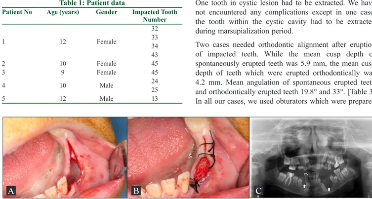
Surgical treatments were carried out at Department of Oral and Maxillofacial Surgery, Istanbul Medipol University School of Dentistry. Informed consent was obtained from parents of the children. Marsupializations were performed under local anesthesia and conscious sedation. Following the extraction of deciduous teeth, widening of the extraction socket to create a window for the insertion of the obturator was carried out. A biopsy sample was incised from the epithelial lining surrounding the cyst cavity and processed for histopathologic evaluation. Cyst cavity was irrigated with sterile saline solution and packed with nitrofurazone pomade immersed gauze [Figure 1B,1C]. All surgical procedures were completed without complication and postoperative follow-up was uneventful. Patients and their families were recalled twice a week for controlling and replacing a new gauze. Histopathological examination revealed dentigerous cyst in four patients and bilateral odontogenic keratocyst in one patient.
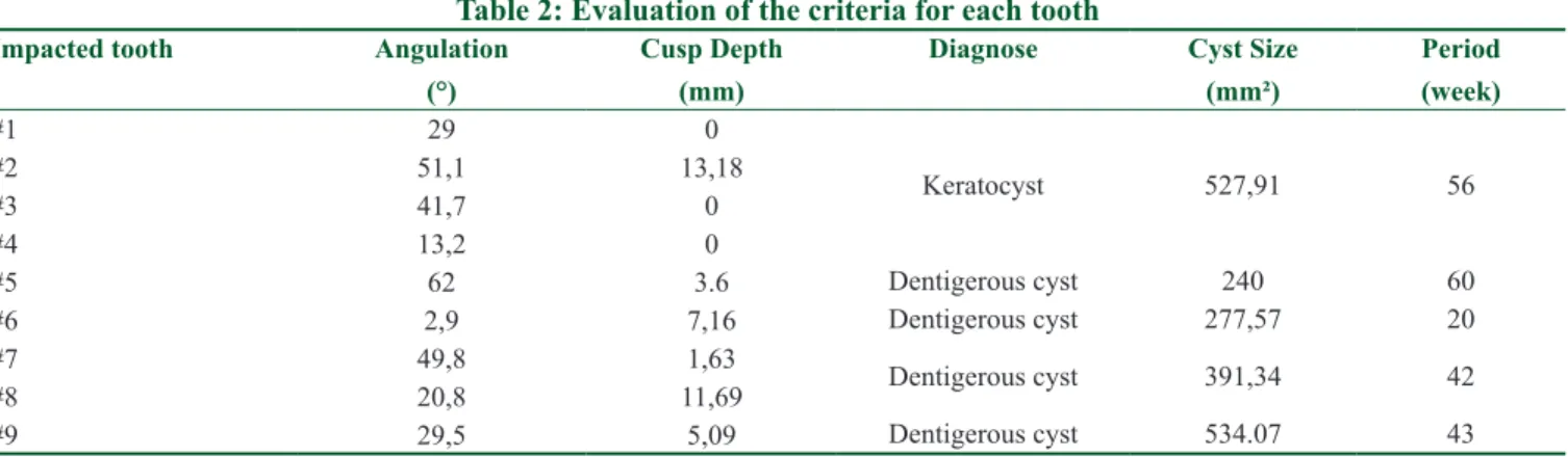
Table 1: Patient data
Patient No Age (years) Gender Impacted Tooth Number 1 12 Female 32 33 34 43 2 10 Female 45 3 9 Female 45 4 10 Male 2425 5 12 Male 13
Figure 1: (A) Radiographic appearance of bilateral impacted teeth within the cystic lesion; (B) marsupialization of the cystic cavity; (C) insertion of
antibiotic immersed gauze through the cyst cavity
from acrylic. All obturators permitted tooth eruption and enabled the reduction of the cyst cavity. However, the extension of the acrylic plates in the cyst cavity needed adjustment for tooth eruption and cavity shrinkage.
d
IscussIonMarsupialization and decompression are conservative surgical techniques and have been widely performed
for the treatment of jaw cysts.[5] Advantages of marsupialization are: it reduces the cyst size; minimizes the risk of damage to important anatomical structures such as inferior alveolar nerve and maxillary sinus; minimizes the damage to bone tissue; stimulates osteogenesis; and promotes the eruption of the involved teeth.[6,7] On the contrary, the disadvantages are: patient discomfort which is prominent at the early stages of Figure 3: (A) Cusp depth; the distance of the central cusp tip of the cyst-associated tooth from the line that passes the
cement-enamel junction of the adjacent teeth; (B) angulation of the impacted tooth to axis of adjacent teeth; (C) measurement of cyst size
A
B
C
Figure 2: (A) Intraoral appearance of marsupialization cavity in left mandible after two weeks; (B) adaptation of the obturator to the orifice
A
B
Table 2: Evaluation of the criteria for each tooth
Impacted tooth Angulation (°)
Cusp Depth (mm)
Diagnose Cyst Size (mm²) Period (week) #1 29 0 Keratocyst 527,91 56 #2 51,1 13,18 #3 41,7 0 #4 13,2 0 #5 62 3.6 Dentigerous cyst 240 60 #6 2,9 7,16 Dentigerous cyst 277,57 20 #7 49,8 1,63 Dentigerous cyst 391,34 42 #8 20,8 11,69 #9 29,5 5,09 Dentigerous cyst 534.07 43
marsupialization; it requires cooperation of patients which plays a major role in the success of such a long treatment period, longer follow-up period is required for some cases to control eruption of involved teeth and pathological tissue remains in situ.[4,8]
Dentigerous cysts are the most commonly encountered among all developmental odontogenic cysts of the jaws. Different treatment options for dentigerous cysts are enucleation, curettage, decompression, marsupialization, and resection with or without continuity defect. Preservation of the impacted teeth and prevention of the vital tissues from surgical damage are desired in young patients, hence marsupialization can be the first choice of treatment. Koca et al.[9] and Ziccardi VB et al.[10] stated that marsupialization is more efficient than other treatment options particularly in young patients by preserving tooth in cyst cavity and by promoting tooth eruption. However, Hyamoto et al. reported that 30% impacted premolars associated with dentigerous cyst did not erupt spontaneously by enucleation and these teeth had to be removed surgically.[2,11] Koca et al. and Miyawaki et. al. stated that impacted teeth associated with dentigerous cyst can be erupted orthodontically when sufficient space exist.[2,9,12] Complete resolution of the cystic lesions were achieved in all of our cases, six of nine impacted teeth within the cystic lesions erupted spontaneously, two of them were erupted orthodontically, and one had to be extracted. These three teeth had severe angulation.
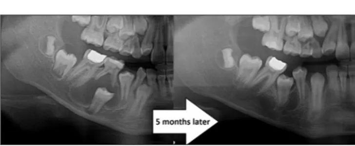
There is a dilemma whether odontogenic keratocyst [keratocystic odontogenic tumor (KCOT)] can be treated with marsupialization. Although some authors advocate enucleation with curettage, the others state that odontogenic keratocyst can completely disappear Figure 4: Resolution of the cystic lesion and eruption of the impacted
tooth after marsupialization
Table 3: Angulation and cusp depth of impacted teeth
Mean Angulation (°) Mean Cusp Depth (mm) Erupted tooth 19.8 5.9 Orthodontically assisted 33 4.2 Extracted 62 3.6
after marsupialization alone.[7] Tabrizi et. al. evaluated 13 patients with keratocyst who were treated with marsupialization.[13] They followed up patients for 60 months. During this period, they declared no sign of recurrence. Johnson et al. reviewed 2736 published articles regarding management and recurrence of KCOT.[14] They found that recurrence rate of enucleation alone, enucleation with adjunctive measures (other than Carnoy solution), and enucleation with Carnoy solution were 25.6%, 30.3%, and 7.9%, respectively. Furthermore, recurrence rate of marsupialization alone and marsupialization with adjunctive therapies were 4.8% and 15.8%, respectively. As a conclusion, they recommended that large KCOT is best treated with a two-stage approach being marsupialization followed by enucleation and adjunctive measures. In this present report, keratocyst was diagnosed in only one patient and managed successfully with marsupialization without performing second stage surgery. During the first-year follow-up, no sign of recurrence was observed.
Fuji et al. carried out a study on prediction of the eruption of impacted teeth associated with dentigerous cyst. They found that as angulation and cusp depth increase, the success rate decreases.[4] In our case series, teeth that did not erupt had higher angulation degrees than the spontaneous erupted teeth. However, the depth of the tooth in the alveolar bone did not affect eruption. Several kinds of obturators and appliances are being used in decompression of the cysts. In all our cases, we used removable acrylic obturators. All obturators permitted tooth eruption and enabled the reduction of the cyst cavity. The extension of the acrylic obturator enables tooth eruption and cavity shrinkage.
c
onclusIonChildren have high capacity of bone regeneration; therefore, conservative surgical treatment enables spontaneous tooth eruption. Several factors should be considered to predict eruption and close follow-up is mandatory to decide the need for orthodontic treatment. Financial support and sponsorship
Nil.
Conflicts of interest
There are no conflicts of interest.
r
eferences1. Serra E, Silva FM, Sawazaki R, de Moraes M. Eruption of teeth associated with a dentigerous cyst by only marsupialization treatment: A case report. J Dent Child (Chic) 2007;74:228. 2. Qian WT, Ma ZG, Xie QY, Cai XY, Zhang Y, Yang C.
cyst-associated mandibular premolars in preadolescent patients. J Oral Maxillofac Surg 2013;71:1825-32.
3. Pogrel MA. Treatment of keratocysts: The case for decompression and marsupialization. J Oral Maxillofac Surg 2005;63:1667-73. 4. Fujii R, Kawakami M, Hyomoto M, Ishida J, Kirita T. Panoramic
findings for predicting eruption of mandibular premolars associated with dentigerous cyst after marsupialization. J Oral Maxillofac Surg 2008;66:272-6.
5. Torres-Lagares D, Segura-Egea JJ, Rodriguez-Caballero A, Liamas-Carreras JM. Gutierrez-Perez JL, Treatment of a large maxillary cyst with marsupialization, decompression, surgical endodontic therapy and enucleation. J Can Dent Assoc 2011;77:b87.
6. Thoma KH, Oral surgery. 3rd ed St. Louis: Mosby; 1958. p. 1033-6.
7. Archer WH, editor. Textbook of oral surgery. 4th ed. Philadelphia: Saunders; 1968. pp. 442-69.
8. Zhao YF, Wei JX, Wang SP. Treatment of odontogenic keratocysts: A follow-up of 255 Chinese patients. Oral Surg Oral Med Oral Pathol Oral Radiol Endod 2002;94:151-6.
9. Koca H, Esin A, Aycan K. Outcome of dentigerous cysts treated
with marsupialization. J Clin Pediatr Dent 2009;34:165-8. 10. Ziccardi VB, Eggleston TE, Schnider RE. Using fenestration
technique to treat a large dentigerous cyst. J Am Dent Assoc 1997;128:201-5.
11. Hyomoto M, Kawakami M, Inoue M, Kirita T. Clinical conditions for eruption of maxillary canines and mandibular premolars associated with dentigerous cysts. Am J Orthod Dentofacial Orthop 2003;124:515.
12. Miyawaki S, Hyomoto M, Tsubouchi J, Kirita T, Sugimura M. Eruption speed and rate of angulation change of a cyst-associated mandibular second premolar after marsupialization of a dentigerous cyst. Am J Orthod Dentofacial Orthop 1999;116:578-84.
13. Tabrizi R, Özkan BT, Dehgani A, Langner NJ. Marsupialization as a treatment option for the odontogenic keratocyst. J Craniofac Surg 2012;23:459-61.
14. Johnson NR, Batstone MD, Savage NW. Management and recurrence of keratocystic odontogenic tumor: A systematic review. Oral Surg Oral Med Oral Pathol Oral Radiol 2013;116:271-6.