73
Research Article
Our Clinical Experience in Upper Lumbar Hernia: Retrospective Evaluation of 47 Patients
Güray Bulut1 ORCID: 0000-0002-9318-4800
1Department of Neurosurgery, Nisa Hospital, Medipol University, Istanbul, Turkey
Submitted: October 17, 2019; Accepted: February 26, 2020
Abstract: Clinical and radiological examination of upper level lumbar disc hernias (L1-2, L2-3, L3-4) and evaluation of surgical results. 47 patients with upper level lumbar disc hernia (ULLDH) among 282 lumbar disc hernias (LDH) performed in our clinic between April 2015 and April 2017. Age, physical examination, disc distances, radiological findings, preoperative and postoperative findings, complications, recurrence, patient satisfaction were evaluated retrospectively according to Prolo scale (via two 5-point Likert-type scales). Maximum resection principle was applied in the operations. All patients were operated with direct lumbar radiographs and lumbar magnetic resonance imaging (MRI). Lumbar computer tomography and electromyography (EMG) were performed and the diagnoses were supported when necessary. 23 (48.9%) of the cases were male and 24 (51.1%) were female. The average age was 49.9 (25-70). The average period between occurrence of symptoms and attendance to clinic is 3.7 months. The occurrence of L1-2: 3 (6.4%) patients, L2-3: 8 (17%) patients and L3-4: 32 (68.1%) in ULLDH cases (16.6%). Four patients (8.5%) with L2-3 and L3-4 were present. The first operation was not recurred during the 2-year follow-up in our clinic. Spondylodiscitis developed in 1 patient (2.1%) and was improved with medical treatment. In 1 patient (2.1%) preoperative dural injury primer was also repaired. In the early postoperative period, leg pain was disappeared in all cases. According to the Prolo follow-up scale, 31.9% were good and 65.96% were excellent. No bad results had been recorded. The incidence of ULLDH is increasing with the widespread use of MRI. ULLDH, with careful microsurgical technique and maximum disc resection, if operated, surgical success rate increases and complication rate decreases. Keywords: Upper; disc; hernia; lumbar; microdiscectomy
Address of Correspondence: Güray Bulut- [email protected] Tel: +90 (212)4544400,
Department of Neurosurgery, Nisa Hospital, Medipol University, Çobançeşme Mahallesi, Fatih Caddesi, Okul Sok. No: 2-4, 34196, Istanbul, Turkey
1. Introduction
Disc herniation is the most common cause of disabling back pain. The upper limits of the tight L4-5, L5-S1. Only 1-11% of disc herniations occur at L1-2, L2-3, L3-4 levels. Disc herniations at this level are called upper level lumbar disc herniations (Hsu et al, 1990; Nadler et al, 1998).
74
Clinical findings of ULLDH, computer-assisted discs are of particular importance (Nadler et al, 1998). The clinical and radiological findings of ULLDH were discussed in our study.
2. Materials and Methods
In this study, 47 upper-level lumbar disc herniations (16.67%) among 282 lumbar disc herniations operated between April 2015 and April 2017 were reviewed retrospectively.
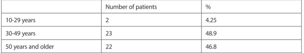
Cases were divided into 3 groups according to age; group 1, 10-29 years of age (4,25%), group 2, 30-49 years of age (48,9%), group 3, 50 years of age and above (46,8%) (Table 1). The cases were evaluated according to their complaints and durations at the time of application, the previous treatments, and the history of physical trauma.
Table 1. Distribution of patients by age ranges
Number of patients %
10-29 years 2 4.25
30-49 years 23 48.9
50 years and older 22 46.8
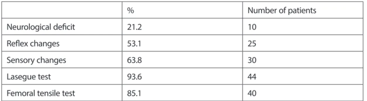
With preoperative neurological examination, muscle strength, loss of sensation and reflex loss were evaluated and femoral stretching test and Lasegue test were performed. In the preoperative radiological examination, lumbosacral radiographs, MRI (Figure 1) and computed tomography (CT) were used. EMG was performed was performed in some cases for neurophysiological examination. All patients underwent microdiscectomy with microsurgical technique. All patients underwent postoperative week, first-month, and third-month follow-up examinations. Prolo follow-up scale was used to determine patient satisfaction.
75 Figure 1. Preoperative and postoperative MRI images of L3-4 right disc herniation
3. Results
In our study, the number of cases with ULLDH was 47 and it accounted for 16.67% of all lumbar disc herniations. Of these, 23 (48.9%) were male and 24 (51.1%) were female (Table 2). Their ages ranged from 25 to 70 years, with a mean of 49.9 years. Twenty-four (51.1%) of the cases were active. We have no cases of acute trauma. There were 6 (12.8%) patients who were traumatized in previous times.
The period between onset of complaints and admission ranged from 7 days to 24 months (4.6 months mean). One patient presented with low back pain, 18 patients with back and right leg pain, 20 patients with waist and left leg pain, 5 patients with back and lower leg pain, and 2 patients with weakness (Table 3). All of the cases had received medical treatment before and 3 (6.4%) received physical therapy. In these cases, 68.1% were at L3-4, 17% at L2-3, 6.4% at L1-2 and 8.5% at two levels (L2-3, L3-4) (Table 4). As a result of neurological examination, 21.2% motor deficit, 63.8% sensory deficit, 53.1% reflex deficits were detected. Lasegue test was found 93.6% and femoral stretching test was positive as 85.1% (Table 5). Lumbosacral X-ray and Lumbosacral MRI were performed directly in all cases. EMG was performed in 5 (10.6%) cases. One of the cases was operated in a different clinic and re-operated at the same level. None of the cases were reoperated due to relapse. There was 1 patient in L1-2 and 3 patients in L3-4. All patients received one dose of prophylactic antibiotic. The skin was brushed with antiseptic solutions for 5 minutes. Distance was determined by peroperative scopy. All patients were operated by microsurgical technique and maximum disc resection was planned using an operation microscope.
76
According to the Prolo follow-up scale, 31.9% and 65.96% were excellent results in the postoperative period (Table 6). In the postoperative early period, none of the patients had leg pain. In the late postoperative period, there was significant improvement in motor, sensory deficits and loss of reflex.
Table 2. Sex
Sex % Number of patients
Male 48.9 23
Female 51.1 24
Table 3. Symptoms
% Number of patients
Low back pain 2.13 1
Low back and right leg pain 38.3 18 Low back and left leg pain 42.55 20 Low back and pain in both legs 10.64 5
Weakness 4.26 2
Table 4. Levels of discectomy performed
% Number of Patients
L1-2 6.4 3
L2-3 17 8
L3-4 68.1 32
Two level 8 4
Table 5. Physical examination findings of patients before surgery
% Number of patients
Neurological deficit 21.2 10
Reflex changes 53.1 25
Sensory changes 63.8 30
Lasegue test 93.6 44
77 Table 6. Clinical results according to Prolo follow-up criteria
% Number of patients Excellent 65.96 31 Good 31.90 15 Intermediate 2.13 1 Bad - -4. Discussion
The incidence of ULLDH has been reported as 3.1% to 10.4% (Albert et al, 1993; Demirbas, 2000; Gutterman and Shenkin, 1982). While the frequency of ULLDH was reported as 5% in the series, Albert et al. reported this rate as 10.4% (Albert et al, 1993). In our series, the incidence of ULLDH was found to be 16.6%, which is higher than in the literature. We can explain this especially with routine use and spread of lumbar MRI.
The majority of the cases with ULLDH are male (Eugene and Kim, 1997; Kotilainen et al, 1993; Rastecchini, 1991; Yasuma et al, 1993). In our series, in contrast to the general literature, it is slightly higher in women (51.1%) than men.
ULLDHs are often seen in the 3rd, 4th and 5th decades as disc hernias at the other level. It is thought that is because of being more active in these age groups (Eugene and Kim, 1997; Rastecchini, 1991; Yasuma et al, 1993). In our series, the most common was in the 31-50 years of age group (48.9%). This result is in parallel with the literature. Approximately half of our cases (48,9%) were in this age group.
Trauma is an important predisposing factor in the formation of lumbar disc herniation. The incidence of trauma cases in large series has been reported as 50-70% (Demirbas, 2000). In our series, there were no cases of acute trauma and 12.8% of cases had previously been traumatized. In the Ken Hsu study, the rate of LDH in active workers was 67% (Hsu et al, 1990). In our series, this ratio is 51.1%.
It has been stated that the symptoms are very variable in ULLDH (Albert et al, 1993; Kotilainen et al, 1993). Therefore, there are some difficulties in early diagnosis of ULLDH. Gutterman reported that the rate of pain spreading to the anterior aspect of the leg was 59% (Gutterman and Shenkin, 1982). In our series, this rate is 80.85%. This rate is higher than Gutterman’s series. It is necessary and useful to question the pain radiating to the front of the leg, as it is particularly distinctive.
Cauda-equina syndrome is the most important and urgent surgical procedure in lumbar disc hernias. While it is expected to be seen in ULLDH due to anatomical neighborhood, it is reported that it is mostly seen in L4-5 level (Hsu et al, 1990; Kostuik et al,1986; Shapiro, 1993). However, lumbar disc herniation is seen more frequently at L4-5 level (Demirbas, 2000). In our series, there were no patients presenting with Cauda-equina syndrome. We believe that early diagnosis and extensive use of MRI are effective.
78
The tests used in the first plan for neurological examination are the Lasegue test and the femoral stretching test. The flat leg lifting test is more positive in the upper lumbar disc hernias because it shows the irritation of the roots at the lower lumbar level. The femoral stretching test is more frequent in ULLDH (Jung and Drovak, 1995). Lumbar disc herniations were found to be positive in 85-90% of the patients. The positivity of the Khe Shu femoral stretching test was reported to be 70%. In our series, this rate is higher than the literature (85.1%). Femoral tensile test positivity is not a pathognomonic finding but a useful test in diagnosis (Estridge et al, 1982).
Reflex examination is an objective criterion for LDH examination. The effect of psychosocial status of the patients is not expected in reflex examination. Dysfunction of the patellar reflex is also expected in ULLDH. Kotilainen found the patella reflex to be 70% hypoactive or areflexia (Kotilainen et al, 1993). In our series, reflex deficits rate was found to be 53.2%. 25 cases and 60% (15 cases) of these cases were patella reflex deficits. This rate was lower than in the literature. The reason for this was the early presentation of the cases and the widespread use of MRI.
Motor dysfunction in ULLDHs is higher than lower LDHs (Paszlar and Szervas, 1981). This suggests that a dynamic and rapid approach is needed in diagnosis and treatment of ULLDH patients. In our series, motor deficits rate was found to be 21.2%. The sensory deficit was more subjective than reflex and motor deficits and was reported as high rates in the literature (Hsu et al, 1990; Paszlar and Szervas, 1981). In our study, sensory deficit was found to be 63.8%. It was thought that the low incidence of our values according to the literature could be the early application of the cases and the widespread use of MRI.
While the incidence of ULLDH was reported as 1% in the pre-MRI period, these rates increased to 10% with the routine use of MRI (Hsu et al, 1990). Because of the superiority of MRI in the correct diagnosis, we think that it is the first advanced examination to be requested after a good neurological examination. If necessary, this should be supported by CT and EMG.
Williams argued that only free disc fragments were sufficient to avoid damaging the healthy disc. Reported a recurrence rate of 9% in the series (Williams, 1977). Rogers also reported a recurrence rate of only 11% in cases of disc fragmentation (Rogers, 1988). Yaşargil and Caspar argued on the maximization of the disc resection (Caspar et al, 1991; Conrad et al, 1992; Yaşargil, 1977). The recurrence rate is 4%. Maximum resection was performed in our series and no recurrence was observed during follow-up. Based on these results we highly recommend the maximum disc resection.
Prolo follow-up scale is widely used in the evaluation of postoperative recovery of the patients (Demirbas, 2000; Prolo et al, 1986). Good results ranged from 74-93% in various series and no difference was found between levels in ULLDH (Demirbas, 2000). In our series, 31.9% good and 65.96% excellent results were obtained in postoperative controls (Table 5). Improvements in motor and reflex deficits, especially in various publications, can be extended up to 1 year (Gutterman and Shenkin, 1982; Kostuik et al,1986; Paszlar and Szervas, 1981). In our study, there was no significant distribution of the cases according to the levels. Complications of dural injury, infection, root injury and epidural fibrosis are the most common complications of ULLDH (Carlson, 1991; Caspar et al, 1991; Gutterman and Shenkin, 1982; Yasargil, 1977). Dura injury and
79 wound infection are observed below 5% (Gutterman and Shenkin, 1982). In our series, spondylodiscitis was
seen in 1 patient (2.1%). After medical treatment, the patient recovered. One patient had dura mater injury (2.1%). It was repaired preoperatively. These results were evaluated as better than the literature. Minimal surgical trauma, excessive laminectomy and avoidance of facetectomy, providing good hemostasis without cauterizing the epidural veins as much as possible, protecting the epidural adipose tissue, exercise in the postoperative period and applying the recommendations made are important (Carlson, 1991; Caspar et al, 1991; Gutterman and Shenkin, 1982; Yasargil, 1977).
Conclusion
The incidence of ULLDHs increases with the widespread use of MRI. Because of the superiority of the MRI in the correct diagnosis, we think that it is the first advanced examination to be requested after a good neurological examination. If necessary, it should be combined with CT and supported by EMG.
Contrary to the literature, the rate of women in our series was higher than men. In our series, ULLDHs are more common in the 3rd and 5th decades than in the literature. The history of trauma and active work were not seen as important factors in the patients in our series. Leg pain, especially pain and weakness on the front of the thighs are the most common symptoms of ULLDH. As reflex deficits, we most commonly observed a decrease or absence of patellar reflex. Although Cauda-equina syndrome is not present in our series, it is a very serious neurological condition and requires urgent treatment. After successful surgery, leg pain needs to be relieved in the early postoperative period. Due to the lack of recurrent disc herniation in our series, we recommend maximum disc resection in the operation. Again, we believe that low complication rates in our cases are due to compliance with microsurgical principles.
Ethical Rules
All procedures performed in studies involving human participants were in accordance with the ethical standards and with the 1964 Helsinki Declaration and its later amendments or comparable ethical standards.
Informed Consent
Informed consent was obtained from all individual participants included in the study.
Funding
No funding was received for this research.
Conflict of Interest
80
References
Albert, T. J., Balderston, R. A., Heller, J. G., Herkowitz, H. N., Garfin, S. R., Tomany, K., An, H. S., Simeone, F. A. (1993). Upper lumbar disc herniations. J Spinal Disord, 6(4), 351-359.
Carlson, G., Abitbol, J. J., Garfin, S. R. (1991). Prevention of complications in surgical management of back pain and sciatica. Orthopedics Clinics of North America, 22(2), 345-351.
Caspar, W., Campbell, B., Barbier, D. D., Kretschmmer, R., Gotfried, Y. (1991). The Caspar microsurgical discectomy and comparison with a conventional standart lumbar disc prosedure. Neurosurgery, 28(1), 78-86. Conrad, T., Pappas, E., Harrington, T., Sonntag, H. (1992). Outcome analysis in 654 surgically treated lumbar disc herniations. Neurosurgery, 30(6), 862-866.
Demirbaş, M. A. (2000). To evaluate the incidence, clinical features, diagnosis and treatment methods in upper-level lumbar disc hernias (Specialization thesis: Haydarpasa Numune Education and Research Hospital).
Estridge, M. N., Rouke, S. A., Johnston, N. G. (1982). The femoral stretching test. J Neurosurgery, 57, 813-816. Eugene, J., Kim, D. H. (1997). A prospective analysis of magnetic resonance imaging findings in patients with sciatica and LDH. Spine, 22(15), 1650-1660.
Gutterman, P., Shenkin, H. A. (1982). Syndromes associated with protrusion of upper lumbar intervertebral discs. J Neurosurgery, 57, 813-816.
Hsu, K., Zucherman, J., Shea, W. (1990). High lumbar disc degeneration: incidence and etiology. Spine, 15, 679-682.
Junge, A., Drovak, J. (1995). Predictors of bad and good outcomes of lumbar disc surgery. Spine, 20(4), 460-468.
Kostuik, J. P., Harington, J., Alexander, D., Rond, W. (1986). Cauda equina syndrome and lumbar disc herniation. The Journal of Bone and Joint Surgery, 68A(3), 386-390.
Kotilainen, E., Valtonen, S., Carison, C. A. (1993). Microsurgical treatment of lumbar disc herniation: follow up of 237 patients. Acta Neurochir (Wien), 120, 143-149.
Nadler, S. F., Campagnolo, D. I., Tomaio, A. C., Stitik, T. P. (1998). High lumbar disc: diagnostic and treatment dilemma. Am J Physical Medicine & Rehabilitation, 77(6), 538-544.
Paszlar, E., Szervas, I. (1981). Herniation of the upper lumbar discs. Neurosurg Rev, 4(3), 151-157.
Prolo, D. J., Oklund, S. A., Butcher, M. (1986). Toward uniformity in evaluating results of lumbar spine operations. Spine, 11(6), 601-606.
81 Rastecchini, F. (1991). Results of surgery compared with conservative management for lumbar disc
herniations. Spine, 21(11), 1383-1387.
Rogers, L. A. (1988). Experience with limited versus extensive disc removal in patients undergoing microsurgical operations for ruptured lumbar disc. Neurosurgery, 22(1), 82-85.
Shapiro, S. (1993). Cauda-equina syndrome secondary to lumbar disc herniation. Neurosurgery, 32(5), 743- 746.
Williams, R. W. (1977). Microdiscectomy-myth, mania or milestone? An 18 years surgical adventure. Adv Neurosurgery, 4, 81- 87.
Yasuma, T., Arai, K., Yamauchi. Y. (1993). The histology of lumbar intervertebral disc herniations. Spine, 18(13), 1761- 1765.