ORIGINAL ARTICLE
Risk factors for skeletal-related events (SREs) and factors
affecting SRE-free survival for nonsmall cell lung cancer patients
with bone metastases
Arife Ulas1&Ahmet Bilici2&Ayse Durnali3&Saadet Tokluoglu3&Sema Akinci4& Kamile Silay5&Berna Oksuzoglu3&Necati Alkis3
Received: 7 July 2015 / Accepted: 5 August 2015 / Published online: 15 August 2015 # International Society of Oncology and BioMarkers (ISOBM) 2015
Abstract Skeletal-related events (SREs) for nonsmall cell lung cancer (NSCLC) patients with bone metastasis lead to serious morbidity. The aim of this study was to determine risk factors for SREs in NSCLC patients with bone metastasis and the factors influencing SRE-free survival and overall survival (OS). From 2000 to 2012, we evaluated retrospectively 835 NSCLC patients. Three hundred and thirty-five of them with bone metastasis were included in the study. SREs and the other prognostic factors were evaluated by univariate and mul-tivariate analysis for SRE-free survival and OS. SREs were detected in 244 patients (72.8 %). The most common SREs were the need for radiotherapy (43.2 %) and malignant hyper-calcemia (17.6 %). The median time to first SRE was 3.5 months at the median follow-up of 17 months. A multi-variate analysis showed that the presence of bone metastasis at diagnosis (p < 0.001), the number of bone metastasis (p = 0.001), baseline hypercalcemia (p=0.004), and the presence of palliative radiotherapy (p=0.04) were independent prog-nostic factors for SRE-free survival. A logistic regression
analysis identified that the presence of bone metastasis at di-agnosis [odds ratio (OR), 12.6], number of bone metastasis (OR, 3.05), and baseline hypercalcemia (OR, 0.33) were found to be predictive factors in the developing of SRE. The median OS time for patients with SRE was worse than that for patients without SRE (7 vs 12 months, respectively). For OS, male gender, ECOG performance status (PS), high lactate de-hydrogenase (LDH) level, hypoalbuminemia, the presence of bone metastasis at diagnosis, the number of bone metastasis, the presence of SREs, the presence of bisphosphonate therapy, and palliative radiotherapy were independent prognostic indi-cators for OS by the multivariate analysis. Our results indicat-ed that the frequency of SREs was high and the presence of bone metastasis at the time of diagnosis, baseline hypercalce-mia, and multiple bone metastases were significant factors predicting the occurrence of SREs. If bone metastases diag-nose earlier, treatments for the prevention of SREs may be initiated earlier; thus, the deterioration of quality of life may be preserved.
Keywords Skeletal-free events . SRE-free survival . Nonsmall cell lung cancer . Bone metastases
Introduction
Lung cancer is the most common tumor worldwide, and it is responsible for the 20.6 % of cancer mortality [1,2]. Bone metastasis is the most common complication in advanced can-cer patients, especially in breast, prostate, and lung cancan-cer. It has been diagnosed at the rate of 30–40 % in advanced nonsmall cell lung cancer (NSCLC) [3]. With the develop-ment of new screening methods such as fluorine-18 deoxyglucose positron emission tomography (PET-CT) scan, the detection of bone metastasis in NSCLC increasingly rises * Arife Ulas
1
Department of Medical Oncology, Ankara Ataturk Training and Research Hospital, Ankara, Turkey
2 Department of Medical Oncology, Medical Faculty,
Istanbul Medipol University, TEM Otoyolu Goztepe Cikisi, Bagcilar, Istanbul 34214, Turkey
3
Department of Medical Oncology, Ankara Oncology Teaching and Research Hospital, Ankara, Turkey
4
Department of Hematology, Ankara Ataturk Training and Research Hospital, Ankara, Turkey
5 Department of Internal Medicine and Geriatrics, Faculty of
Medicine, Ataturk Research and Training Hospital, Yildirim Beyazit University, Ankara, Turkey
[4]. The reason why metastases have an affinity for bone is that there is abundant blood flow to bone marrow and high expression of adhesion molecules on tumor cells, making them adhere to stromal cells of bone marrow and bone matrix and of growth factors. Release of growth factors from bone matrix during osteoclast-mediated osteolysis contributes to the development of metastatic lesions [5].
Skeletal-related events (SREs) developing in patients with bone metastasis result in serious morbidity. In addition, it is a term used to describe a collection of adverse events associated with bone metastases and include pathological fractures, the requirement for surgery or radiotherapy, spinal cord compres-sion, and malignant hypercalcemia [3,6,7]. A prospective study demonstrated that in over 40 % of NSCLC patients with bone metastasis, SRE developed at least once, and this SRE emerged within the first 5 months [8]. In patients with NSCLC, the diagnosis of bone lesions is frequently missed unless serious severe or SREs develop. Furthermore, it seems that the treatment of patients with NSCLC who have bone metastasis is neglected due to short life expectation. However, new treatment modalities have been posed significant im-provement in median overall survival (OS) [9,10]; thus, as survival improves, increase in the prevalence of SREs is likely.
There is limited guiding information as regards to skeleton-associated morbidity and treatment and monitorization of bone metastases for patients with NSCLC. Bone metastasis may de-crease the quality of life and give rise to high financial cost in the terminal stage of life particularly after the development of SRE [11]. Conventional treatments such as surgical stabilization or radiotherapy provide only local control for palliation. In addi-tion, it has been reported that bisphosphonates may delay the onset of SREs [12].
Factors predicting SRE should be well known in order that SREs can be prevented in lung cancer patients with bone me-tastasis. Risk factors for SRE in patients with advanced NSCLC have been reported previously [13]. A previous study reported that increase in N-telopeptide of type I collagen (NTX) levels, which is a sensitive marker of osteolysis, correlates with in-creased risk of SREs [14]. SREs may not occur at a constant rate, and the incidence of these complications may be varied depending on the duration of survival [7,8,15]. In the present study, the determination of the frequency of SREs and its risk factors was aimed. Moreover, clinical factors influencing SRE-free survival and OS were also evaluated.
Material and methods
Patients and data collection
From 2000 to 2012, 835 NSCLC patients followed who were followed up at the department of medical oncology in Ankara
Oncology Training and Research Hospital were evaluated ret-rospectively. Totally, 335 patients of them with bone metasta-ses were included for analysis. All patients had a histological or cytological diagnosis of NSCLC and stage IV disease or postoperative recurrence with distant metastases. However, patients with insufficient disease information and postopera-tive local recurrence without distant metastases were excluded from data analysis. The primary tumor was staged according to the American Joint Committee on Cancer (AJCC) TNM 7th edition staging classification for lung cancer [16]. The Local Ethics Committee of our hospital approved the study, and the informed written consents were obtained from each patients or their relatives.
Clinical information about age, gender, ECOG perfor-mance status (PS), histopathology, history of radiotherapy to the bone, type of chemotherapy and radiation therapy, the presence of bisphosphonate treatment, the presence and num-ber of bone metastases, and baseline biochemical parameters such as albumin, serum calcium level, lactate dehydrogenase (LDH), and alkaline phosphatase (ALP) levels were obtained from patients’ charts. The cutoff value for each biological baseline parameter was defined as follows: hypoalbuminemia, a serum albumin level of less than 3.0 g/dL; ALP level and LDH level, above normal level, according to standard labora-tory norms; and hypercalcemia, a serum calcium level, >10.5 g/dL. Skeletal metastases were diagnosed with positive results on more than two modalities including bone scan, PET-CT, X-ray, and magnetic resonance imaging (MRI). Thereaf-ter, SREs were categorized as (1) pathologic fracture, (2) spi-nal cord compression, (3) required for radiation therapy to the bone, (4) requirement for surgery to the bone or requirement for radiological intervention to the bone, and (5) hypercalce-mia related with malignancy that was either fatal or required emergency treatment.
Statistical analysis
Statistical analyses were performed using SPSS 17.0 (SPSS Inc., Chicago, IL, USA) software.
The relationship between the presence of SRE and the oth-er clinicopathological factors was analyzed by the chi-square test and Fisher’s exact test. The survival analyses and curves were done with the Kaplan-Meier method and compared with the log-rank test. OS was described as the time from diagnosis to the date of the patient’s death or lost in follow-up. SRE-free survival was defined as the time from the start of initial treat-ment to the first SRE developing during or after treattreat-ment. The significance of the clinicopathological indicators as prog-nostic factor were evaluated by univariate analyses for SRE-free survival and OS. All the significant variables observed in univariate analysis were included into the multivariate surviv-al ansurviv-alysis using Cox proportionsurviv-al hazards model for survivsurviv-al. Predictive factors related to SRE were evaluated by logistic
regression analysis. Multivariate p values were used to char-acterize the independence of these factors. The 95 % confi-dence interval (CI) was used to quantify the relationship be-tween survival time and each independent factor. All tests were done using two-sided, and p values <0.05 were accepted statistically significant.
Results
Two hundred and seventy-four (81.8 %) patients were male and 61 (18.2 %) were female. The median age was 58 ranging from 22 to 84 years. One hundred and ninety-one patients were younger than 60 years (55 %) while 141 patients were aged 60 years or older (45 %). Based on the histopathological evaluation, 169 (50.4 %) patients were classified as adenocar-cinoma, 90 (26.9 %) as nonadenocaradenocar-cinoma, and 76 (22.7 %) as not otherwise specified (NOS). Of the study group, 266 (79.4 %) patients had bone metastases at the diagnosis and 69 (20.6 %) had no bone metastases. On the other hand, there were multiple bone metastases in 57.9 % of patients. The most common metastatic bones were vertebrates, pelvic bones, and femur, respectively. The majority of patients (54.3 %) had ECOG PS >2.
First-line platine-based chemotherapy was administered to 246 (73.4 %) at palliative setting. The most common first-line chemotherapy regimens were cisplatin/gemcitabine (n=90), cisplatin/etoposide (n = 43), cisplatin/vinorelbine (n = 30), cisplatin/docetaxel (n=27), and carboplatin/vinorelbine (n= 20). The median number of chemotherapy cycle was 4 (range 2–7) among patients who received chemotherapy as first line, and overall response rate was 52.5 % (stable disease 20.8 % and partial response 31.7 %). EGFR mutation could be eval-uated in 41 patients, and it was detected as positive in 18 patients. These patients were administered Erlotinib, a target-specific agent.
Totally, 244 (72.8 %) patients developed SREs at the diag-nosis (n=105, 43.3 %) or during treatment of disease (n=139, 65.2 %). Of these, 145 (43.3 %) required radiotherapy to the bone, 59 (17.6 %) developed malignant hypercalcemia, 21 (6.3 %) developed pathological fracture, 14 (4.2 %) developed spinal cord compression, and 5 (1.5 %) required surgical treat-ment for the bone. The number of patients who had at least two SREs was 46 (43.3 %). There was no difference according to the metastatic bone sites (p>0.05). The median time to first SRE was 3.5 months at the median follow-up of 17 months (range 7–132 months).
The presence or absence of SRE was closely associated with the number of bone metastasis (p=0.002), smoking his-tory (p=0.02), baseline hypercalcemia (p=0.003), the pres-ence of palliative radiotherapy (p<0.001), and the prespres-ence of bisphosphonate therapy (p<0.001). Patients with multiple bone metastasis had significantly increased SRE when
compared to patients with single bone metastasis. Further-more, palliative radiotherapy and bisphosphonate treatment were given at a significantly higher rate to patients with SRE. The history of smoking was seen at a higher rate in patients with SREs. The relationship between patients with or without SRE based on clinicopathological factors is sum-marized in Table1.
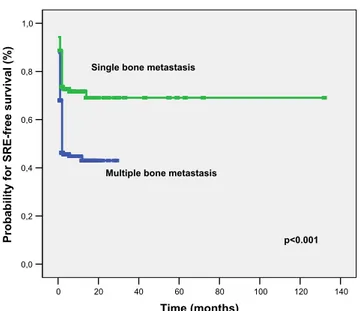
No difference was present with respect to the median follow-up time between patients with or without SRE groups (12.5 vs 14.5 months, respectively, p = 0.86). Uni-variate analysis showed that ECOG PS (p = 0.003), base-line hypercalcemia (p < 0.001), the presence of bone tastasis at diagnosis (p < 0.001), the number of bone me-tastasis (p < 0.001), the presence of palliative radiotherapy (p < 0.001), and bisphosphonate therapy (p = 0.02) were significant prognostic factors for SRE-free survival (Table 2). Patients with single bone metastasis had a bet-ter median SRE-free survival compared to patients with multiple bone metastasis (7 vs 2 months, respectively, Fig. 1). After that, multivariate analysis with Cox regres-sion method in order to further evaluate the prognostic significance of SREs and the other clinicopathological factors was performed. In the multivariate analysis, the presence of bone metastasis at diagnosis [p < 0.001, hazard ratio (HR)1.97], the number of bone metastasis (p = 0.001, HR 1.68), baseline hypercalcemia (HR 0.48, p = 0.004), and the presence of palliative radiotherapy (p = 0.04, HR 1.43) were independent prognostic indicators for SRE-free survival. Table2 shows the results of both univariate and multivariate analysis for SRE-free survival. Then, to determine all of the significant prognostic factors that might be predictive of SRE, a logistic regression analysis was carried out. It indicated that the presence of bone metastasis at diagnosis [p < 0.001, odds ratio (OR) 12.6], the number of bone metastasis (p < 0.001 and OR 3.05), and baseline hypercalcemia (p = 0.007, OR 0.33) were in-dependent factors for predicting the occurrence of SRE. Table 3 demonstrates the results of logistic regression analysis for SRE.
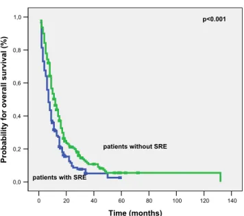
For OS, univariate analysis revealed that male gender (p= 0.01), ECOG PS (p<0.001), high LDH level (p<0.001), high ALP level (p=0.04), hypercalcemia (p=0.003), hypoalbumin-emia (p<0.001), the presence of bone metastasis at diagnosis (p <0.001), the number of bone metastasis (p=0.006), the presence of SREs (p<0.001), the presence of palliative radio-therapy (p = 0.03), and bisphosphonate radio-therapy (p < 0.001) were significant prognostic indicators (Table4). The median OS time for patients with SRE was worse than that for patients without SRE (7 vs 12 months, respectively, Fig.2). Patients without bone metastasis at diagnosis had a better median OS compared with patient bone metastasis at the diagnosis (18 vs 8 months, respectively, Fig.3). In addition, male gender (p= 0.001, HR 0.56), ECOG PS (p=0.008, HR 1.53), high LDH
level (p=0.002, HR 0.55), hypoalbuminemia (p=0.015, HR 0.67), the presence of bone metastasis at diagnosis (p<0.001, HR 2.10), the number of bone metastasis (p=0.016, HR 1.50), the presence of SREs (p=0.035, HR 1.19), the presence of
bisphosphonate therapy (p=0.003, HR 0.60), and the presence of palliative radiotherapy (p=0.001, HR 0.51) were found to be independent prognostic factors for OS in the multivariate analysis (Table4).
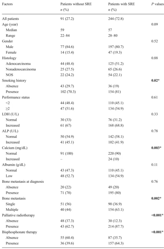
Table 1 Clinicopathological characteristics of patients with or without skeletal-related events (SRE)
Factors Patients without SRE
n (%)
Patients with SRE n (%) P values All patients 91 (27.2) 244 (72.8) Age (year) 0.09 Median 59 57 Range 22–84 28–80 Gender 0.52 Male 77 (84.6) 197 (80.7) Female 14 (15.4) 47 (19.3) Histology 0.88 Adenocarcinoma 44 (48.4) 125 (51.2) Nonadenocarcinoma 25 (27.5) 65 (26.6) NOS 22 (24.2) 54 (22.1) Smoking history 0.02* Absence 43 (29.7) 36 (19) Presence 102 (70.3) 154 (81) Performance status 0.61 <2 44 (48.4) 110 (45.1) ≥2 47 (51.6) 134 (54.9) LDH (U/L) 0.33 Normal 30 (33) 76 (31.2) Increased 61 (67) 168 (68.8) ALP (U/L) 0.78 Normal 50 (54.9) 142 (58.1) Increased 41 (45.1) 102 (41.9) Calcium (mg/dL) 0.003* Normal 91 (100) 220 (90) Increased – 24 (10) Albumin (g/dL) 0.11 Normal 43 (47.3) 110 (45.1) Low 48 (52.7) 134 (54.9)
Bone metastasis at diagnosis 0.76
Absence 20 (22) 49 (20) Presence 71 (78) 195 (80) Bone metastasis 0.002* Single 51 (56) 90 (36.9) Multiple 40 (44) 154 (63.1) Palliative radiotherapy <0.001* Absence 48 (37.3) 30 (12.3) Presence 43 (62.7) 214 (87.7) Bisphosphonate therapy <0.001* Absence 55 (60.4) 87 (35.7) Presence 36 (39.6) 157 (64.3)
NOS not otherwise specified, LDH lactate dehydrogenase, ALP alkaline phosphatase *Statistically significant
Discussion
In the present study, the presence of bone metastasis at the time of diagnosis, multiple bone metastases at diagnosis, and
high baseline serum calcium levels were found to be predic-tive factors of the occurrence of SREs. It was also determined that these three factors also had prognostic significance for SRE-free survival. Moreover, in patients with NSCLC with Table 2 Univariate and
multivariate analysis of patients for SRE-free survival according to clinicopathological factors
Factors Median SRS
(months)
Univariate
p values Multivariatep values HR (95 % CI)
Age (year) 0.89 <60 4.0 ≥60 3.0 Gender 0.53 Male 4.0 Female 2.0 Histology 0.62 Adenocarcinoma 2.0 Nonadenocarcinoma 4.0 NOS 5.0 Smoking history 0.60 Absence 3.0 Presence 5.0 Performance status 0.003* 0.23 1.2 (0.89–1.61) <2 6.0 ≥2 2.0 LDH (U/L) 0.09 Normal 4.0 Increased 3.0 ALP (U/L) 0.17 Normal 5.0 Increased 2.0 Calcium (mg/dL) <0.001* 0.004* 0.48 (0.29–0.78) Normal 4.0 Increased 1.0 Albumin (g/dL) 0.92 Normal 4.0 Low 2.0
Bone metastasis at diagnosis <0.001* <0.001* 1.97 (1.36–2.86)
Absence 12.0 Presence 2.0 Bone metastasis 7.0 <0.001* 0.001* 1.68 (1.22–2.30) Single 2.0 Multiple Bisphosphonate therapy 0.02* 0.40 1.14 (0.83–1.57) Absence 6.0 Presence 2.0 Palliative radiotherapy <0.001* 0.04* 1.43 (0.94–2.19) Absence 7.0 Presence 2.0
HR hazards ratio, CI confidence interval, SRS skeletal-related event (SRE)-free survival, LDH lactate dehydro-genase, ALP alkaline phosphatase
bone metastases, male sex, ECOG PS, high LDH, hypoalbu-minemia, the presence of bone metastasis at diagnosis, multi-ple bone metastases, the presence of SREs at diagnosis, biphosphonate treatment, and palliative radiotherapy were in-dependent prognostic factors for OS.
We detected development of SRE at the diagnosis or during treatment of disease in 72.8 % of patients, and time to devel-opment of first SRE was 6.9 months. In a recent study carried out by Bae et al., including 196 patients presenting with bone metastasis, overall rate of SRE development that was found to be 67.8 and 44 % of the patients had SRE as presenting symp-tom [13]. In the present study, 43.3 % of the patients presented with SRE, and in 13.7 %, at least two SREs were detected. Our results were thus compatible with literature [13].
Tsuya et al. showed that overall rate of SRE was found to be 50 % and median OS was 6.2 months for patients with any SRE versus 12.2 months in patients without an SRE [17]. In our study, median OS was markedly worse in patients with SREs compared to those without SREs (7 vs 12 months). In addition, we established that in patients presenting with bone metastasis, SRE-free survival was shortened by 1.96-fold. The presence of multiple bone metastases at presentation was also evaluated as a risk factor for the development of SREs. The
rate of SRE development was 36.9 % in patients with a single metastasis, while it was 63.1 % in those with multiple metas-tases. SRE-free-survival was 7 months in those with single metastasis, while it was as short as 2 months in those with multiple metastases. In a previous study, for patients with advanced NSCLC undergoing chemotherapy, male sex, a poor ECOG PS, and the presence of multiple bone metastases were found to be the most important risk factors for poor SRE-free survival and development of SREs [18].
A recent Korean study demonstrated that long-term smoking, nonadenocarcinoma tumors, poor ECOG PS, and no history of treatment with EGFR TKIs were predictors for SREs [19]. In the current study, baseline ECOG PS was found to be significant in the univariate analysis for SRE-free sur-vival, but lost its significance in the multivariate analysis. Moreover, male sex and ECOG PS were not found to have any prognostic significance for either occurrence of SREs or SRE-free survival. This might be related to the relatively het-erogeneous sample size and short follow-up interval of the study. However, these two factors were detected to affect only OS. Bae et al., in lung cancer patients with bone metastasis at diagnosis, reported that ECOG PS and the number of bone metastases were found to be prognostic factors for OS [13], as similar to our results.
In a retrospective analysis of 135 stage IV NSCLC patients performed by Tsuya et al., the most common SREs were ra-diotherapy for bone in 34.3 % and hypercalcemia in 20 % [17]. Our rate of common SREs was compatible with the literature [17,20]. In another prospective study for 250 pa-tients with bone metastasis, 120 of whom had NSCLC, radi-ation to bone in 32 %, pathological fracture in 21 %, surgery to bone in 4 %, spinal cord compression in 4 %, and hypercal-cemia in 3 % of patients who were mostly found as SREs [7]. On the other hand, the most common SREs were radiotherapy (67 %) followed by spinal cord compression (21 %) in a study including 1146 lung cancer patients with bone metastasis [21]. In the present study, spinal cord compression was detected in 4.2 % of the patients. This might be related to the relatively small sample size with SREs. Palliative radiotherapy is one of the most important treatment modalities in bone metastasis [22]. In the present study, 43.3 % of patients presented with SRE at diagnosis, and they received palliative radiotherapy to 140 120 100 80 60 40 20 0 Time (months) 1,0 0,8 0,6 0,4 0,2 0,0
Probability for SRE-free survival (%)
Multiple bone metastasis Single bone metastasis
p<0.001
Fig. 1 SRE-free survival of patients according to number of bone metastasis
Table 3 Logistic regression analysis of the predictive factors for SRE for patients with metastatic nonsmall cell lung cancer
Factors p OR 95 % CI
Performance status (<2 vs≥2) 0.42 1.27 0.70–2.31
Bone metastasis at diagnosis <0.001 12.6 4.37–36.7
The number of bone metastasis (single vs multiple) <0.001 3.05 1.67–5.57
Bisphosphonate therapy 0.08 1.73 0.93–3.23
Paliative radiotherapy 0.48 1.29 0.62–2.69
Baseline hypercalcemia (mg/dL) 0.007 0.33 0.13–0.69
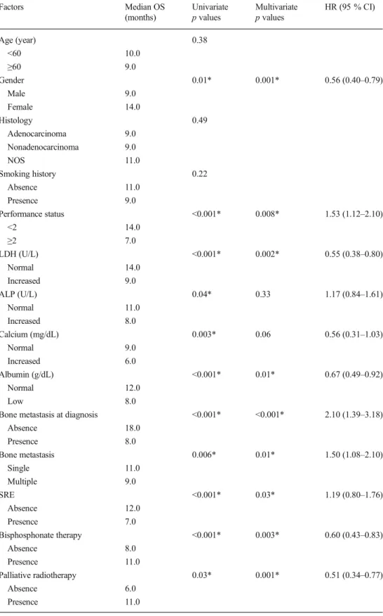
Table 4 Univariate and multivariate analysis of patients for overall survival according to clinicopathological factors
Factors Median OS
(months)
Univariate
p values Multivariatep values HR (95 % CI)
Age (year) 0.38 <60 10.0 ≥60 9.0 Gender 0.01* 0.001* 0.56 (0.40–0.79) Male 9.0 Female 14.0 Histology 0.49 Adenocarcinoma 9.0 Nonadenocarcinoma 9.0 NOS 11.0 Smoking history 0.22 Absence 11.0 Presence 9.0 Performance status <0.001* 0.008* 1.53 (1.12–2.10) <2 14.0 ≥2 7.0 LDH (U/L) <0.001* 0.002* 0.55 (0.38–0.80) Normal 14.0 Increased 9.0 ALP (U/L) 0.04* 0.33 1.17 (0.84–1.61) Normal 11.0 Increased 8.0 Calcium (mg/dL) 0.003* 0.06 0.56 (0.31–1.03) Normal 9.0 Increased 6.0 Albumin (g/dL) <0.001* 0.01* 0.67 (0.49–0.92) Normal 12.0 Low 8.0
Bone metastasis at diagnosis <0.001* <0.001* 2.10 (1.39–3.18)
Absence 18.0 Presence 8.0 Bone metastasis 0.006* 0.01* 1.50 (1.08–2.10) Single 11.0 Multiple 9.0 SRE <0.001* 0.03* 1.19 (0.80–1.76) Absence 12.0 Presence 7.0 Bisphosphonate therapy <0.001* 0.003* 0.60 (0.43–0.83) Absence 8.0 Presence 11.0 Palliative radiotherapy 0.03* 0.001* 0.51 (0.34–0.77) Absence 6.0 Presence 11.0
HR hazards ratio, CI confidence interval, SRE skeletal-related event, LDH lactate dehydrogenase, ALP alkaline phosphatase
the bone prior to chemotherapy. Hence, in this patient group, SRE-free survival shorter as expected. We believe that in-crease in the frequency of SREs in these patients may be misleading. Sekine et al. demonstrated that although palliative radiotherapy to the bone was significant factor for SRE devel-opment by univariate analysis, it was not an independent fac-tor in the multivariate analysis [18].
In lung cancer, hypercalcemia usually occurs at advanced stage and is a prognostic factor [23–25]. In our study, baseline hypercalcemia was found to be a predictive factor (OR 0.33) for occurrence of SRE and an independent prognostic factor for SRE-free survival (HR 0.48), similar to the literature. In addition, it was found to be significant factor for OS while its statistical significance did not remain after multivariate anal-ysis. In a prospective study, unlike the present one, the impact
of hypercalcemia on the occurrence of SRE and frequency of bone metastasis could not be shown. Both the rate of SREs (16.4 %) and hypercalcemia (2.2 %) were found to be lower than our study [26].
In this study, high serum baseline LDH level and low se-rum albumin levels were adverse prognostic factors for OS. However, their effects could not be proven on occurrence of SRE or SRE-free survival. High LDH level is an indicator of tumor burden in patients with lung cancer. In tumor samples of NSLCL patients, it was demonstrated that LDH-5 is asso-ciated with the expression of some angiogenic factors being an unfavorable prognostic factor [27,28]. The prognostic impor-tance of hypoalbuminemia for OS has been shown in recent studies [29,30]. Moreover, we found that high ALP level was significant prognostic indicator for OS by univariate analysis. The importance of especially bone-derived ALP as tumor for-mation marker in patients with bone metastasis was demon-strated previously [26].
The relation between smoking and SREs has been shown, previously [19]. It is known that smoking increases bone loss, leading to osteoporotic fracture. Therefore, the history of os-teoporosis was reported to be a significant predictor for the development of SRE in cancer patients [31,32]. In our study, SREs developed at higher rate, but there was no significant relation between smoking and SREs free-survival and OS in those with history of smoking.
The use of biphosphonates for delaying or preventing the development of SRE in lung cancer patients with bone metas-tasis is strongly recommended [33]. In the present study, when zolendronic acid was compared with placebo, in patients re-ceiving zoledronic acid, median time to development of first SRE was prolonged (5.6 vs 5.0 months, respectively), and zoledronic acid delayed the appearance of SRE in patients with NSLCL. One hundred and three patients received bis-phosphonate with 64.3 % of the patients with SRE and 35.7 % of those without SRE. On the other hand, 60.4 % of our pa-tients, who had bone metastasis without SRE, was not admin-istered any bisphosphonate. In our study population, patients with symptoms related with multiple bone metastases had received bisphosphonate. SRE-free survival in patients who received bisphosphonate was found to be shorter, while OS was longer. In keeping with the present study, patients with bone metastasis, patients receiving zoledronic acid treatment had better survival rates [34]. In our patient group with devel-opment of SREs, bisphosphonate treatment was mostly initi-ated after the development of SRE with higher use of bisphosphonates in this group. This indicated that in clinical practice, bisphosphonate was not initiated in each patient with bone metastasis and that this treatment was usually com-menced after SRE development. Therefore, the fact that SRE-free survival is shorter in those receiving bisphosphonate a l t h o ug h O S i s l o ng e r s u gg e s t th a t t he e ff e c t o f bisphosphonates may be limited in the groups with high risk 140 120 100 80 60 40 20 0 Time (months) 1,0 0,8 0,6 0,4 0,2 0,0
Probability for overall survival (%)
patients with SRE
p<0.001
patients without SRE
Fig. 2 Overall survival of patients according to presence of SREs
140 120 100 80 60 40 20 0 Time (months) 1,0 0,8 0,6 0,4 0,2 0,0
Probability for overall survival (%)
p<0.001
Bone metastasis at diagnosis
Bone metastasis after diagnosis
Fig. 3 Overall survival of patients according to presence of bone metastasis at diagnosis
of SRE. In another study on patients with SRE, it was shown that zoledronic acid decreased the risk of SRE development by 31 % and prolonged time to development of first SRE by 4 months [12]. In a recent study, it was reported that chemo-therapy and zoledronic acid combination enhanced quality of life (QOL), decreased analgesic use and pain scores, prolonged time to onset of first radiotherapy treatment and to development of SRE [35].
Morbidity associated with skeleton is an important source of concern in NSCLC patients with bone metastasis and con-tributes to heavier overall tumor burden in these patients. New chemotherapies and biological agents have been prolonged OS; therefore, the incidence of SREs is increased. A large prospective study determined that SREs not only lead to in-creased morbidity and deterioration of ECOG PS, but also to increased economic costs. The prevention of SRE will im-prove health-related quality of life with decreasing of patient morbidity; thus, the use of health care resources will also be decreased [11]. As a result, the best therapeutic approach should be the prevention of these events. Early treatment and maintenance of good ECOG PS are an important predictor for survival and will enable the patient to receive the target treatments at later stages.
The present study has several limitations. The retrospective nature of our study and short follow-up interval are important limitation. The other limitation of this study is no restrictions according to age and ECOG PS of patients and lack of the metastasis regions other than bone which might have influ-enced the results. Moreover, the relation between develop-ment of SREs and systemic chemotherapy and EGFR TKI could not be evaluated, since the treatment was commenced before SRE development in some patients and after its devel-opment in some others. Although our results should be con-firmed using prospective studies with larger sample sizes that analyze histological subtypes and all mutations for nonsquamous histology, we believe that our study is notewor-thy and these results contribute to the knowledge of this dis-ease because patients evaluated and followed in a single on-cology center. In addition, our results could be represented to the rest of Turkey because our oncology institute is an impor-tant high-capacity center in Turkey and takes a lot of applica-tions from rural areas. Many of previous studies similar to ours were retrospective. Our patients were treated with similar treatment protocols and had undergone radiological evalua-tion of bone metastasis in the same center.
In conclusion, our study shows that SREs were quite fre-quent and multiple bone metastases and high baseline hyper-calcemia are factors predicting the occurrence of SREs. High frequency of SREs indicates that it is necessary to detect bone metastases earlier and treatments for the prevention of SREs should be initiated earlier. Thus, the deterioration of quality of life may be preserved. It is our belief that the present study may play a guiding role for later prospective studies. In patient
groups with the above-mentioned risk factors for SREs, follow-up evaluations should be carried out more frequently, and further studies are required regarding how the onset of SREs can be delayed in this group. Focusing on bone metas-tasis and development of new treatment methods for preventing and decreasing the development of SREs are im-portant issues that should be addressed in future prospective studies.
Acknowledgments This study was presented in ELCC, 2015, the Eu-ropean Lung Cancer Conference, in a poster session. Ulas A, Bilici A, Durnali A, Tokluoglu S, Akinci S, Silay K, Oksuzoglu B, Alkis N. Risk factors for skeletal-related events in patients with nonsmall cell lung can-cer with bone metastases. Ann Oncol, 26, (Suppl 1), 137p.
Conflicts of interest None
References
1. Brenner H, Francisci S, de Angelis R, et al. Long-term survival expectations of cancer patients in Europe in 2000–2002. Eur J Cancer. 2009;45:1028–41.
2. Langer CJ, Besse B, Gualberto A, Brambilla E, Soria JC. The Evolving role of histology in the management of advanced non-small-cell lung cancer. J Clin Oncol. 2010;28:5311–20.
3. Price N. Bisphosphonates to prevent skeletal morbidity in patients with lung cancer with bone metastases. Clin Lung Cancer. 2004;5: 267–9.
4. Qu X, Huang X, Yan W, Wu L, Dai K. A meta-analysis of FDG-PET, MRI and bone scintigraphy for diagnosis of bone metastases in patients with lung cancer. Eur J Radiol. 2012;81:1007–15. 5. Roodman GD. Mechanisms of bone metastasis. N Engl J Med.
2004;350:1655–64.
6. Coleman R. Management of bone metastases. Oncologist. 2000;5: 463–70.
7. Rosen LS, Gordon D, Tchekmedyian S, et al. Zoledronic acid ver-sus placebo in the treatment of skeletal metastases in patients with lung cancer and other solid tumors: a phase III, double-blind, ran-domized trial the Zoledronic Acid Lung Cancer and Other Solid Tumors Study Group. J Clin Oncol. 2003;21:3150–7.
8. Rosen LS, Gordon D, Tchekmedyian NS, et al. Long-term efficacy and safety of zoledronic acid in the treatment of skeletal metastases in patients with nonsmall cell lung carcinoma and other solid tu-mors: a randomized, phase III, double-blind, placebo-controlled trial. Cancer. 2004;100:2613–21.
9. Thatcher N, Chang A, Parikh P, et al. Gefitinib plus best supportive care in previously treated patients with refractory advanced non-small-cell lung cancer: results from a randomised, placebo-con-trolled, multicentre study (Iressa Survival Evaluation in Lung Cancer). Lancet. 2005;366:1527–37.
10. Ou SH. Crizotinib: a novel and first-in-class multitargeted tyrosine kinase inhibitor for the treatment of anaplastic lymphoma kinase rearranged non-small cell lung cancer and beyond. Drug Des Devel Ther. 2011;5:471–85.
11. Delea TE, McKiernan J, Brandman J, et al. Impact of skeletal com-plications on total medical care costs among patients with bone metastases of lung cancer. J Thorac Oncol. 2006;1:571–6. 12. Hirsh V, Tchekmedyian NS, Rosen LS, et al. Clinical benefit of
zoledronic acid in patients with lung cancer and other solid tumors: analysis based on history of skeletal complications. Clin Lung Cancer. 2004;6:170–4.
13. Bae HM, Lee SH, Kim TM, Kim DW, Yang SC, Wu HG, et al. Prognostic factors for non-small cell lung cancer with bone metas-tasis at the time of diagnosis. Lung Cancer. 2012;77:572–7. 14. Hirsh V, Major PP, Lipton A, et al. Zoledronic acid and survival in
patients with metastatic bone disease from lung cancer and elevated markers of osteoclast activity. J Thorac Oncol. 2008;3:228–36. 15. Kohno N, Aogi K, Minami H, et al. Zoledronic acid significantly
reduces skeletal complications compared with placebo in Japanese women with bone metastases from breast cancer: a randomized, placebo-controlled trial. J Clin Oncol. 2005;23:3314–21. 16. Rusch VW, Asamura H, Watanabe H, Members of IASLC Staging
Committee, et al. The IASLC lung cancer staging project: a pro-posal for a new international lymph node map in the forthcoming seventh edition of the TNM classification for lung cancer. J Thorac Oncol. 2009;4:568–77.
17. Tsuya A, Kurata T, Tamura K, Fukuoka M. Skeletal metastases in non small cell lung cancer: a retrospective study. Lung Cancer. 2007;57:229–32.
18. Sekine I, Nokihara H, Yamamoto N, et al. Risk factors for skeletal-related events in patients with non-small cell lung cancer treated by chemotherapy. Lung Cancer. 2009;65:219–22.
19. Sun JM, Ahn JS, Lee S, et al. Predictors of skeletal-related events in non-small cell lung cancer patients with bone metastases. Lung Cancer. 2011;71:89–93.
20. Sathiakumar N, Delzell E, Morrisey MA, et al. Mortality following bone metastasis and skeletal-related events among patients 65 years and above with lung cancer: a population-based analysis of U.S. Medicare beneficiaries, 1999–2006. Lung India. 2013;30:20–6. 21. Cetin K, Christiansen CF, Jacobsen JB, et al. Bone metastasis,
skeletal-related events, and mortality in lung cancer patients: a Danish population-based cohort study. Lung Cancer. 2014;86: 247–54.
22. Janjan N, Lutz ST, Bedwinek JM, et al. Therapeutic guidelines for the treatment of bone metastasis: a report from the American College of Radiology Appropriateness Criteria Expert Panel on Radiation Oncology. J Palliat Med. 2009;12:417–26.
23. Albain KS, Crowley JJ, LeBlanc M, Livingston RB. Survival de-terminants in extensive-stage nonsmall- cell lung cancer: the Southwest Oncology Group experience. J Clin Oncol. 1991;9: 1618–26.
24. Paesmans M, Sculier JP, Libert P, et al. Prognostic factors for sur-vival in advanced nonsmall- cell lung cancer: univariate and
multivariate analyses including recursive partitioning and amal-gamation algorithms in 1,052 patients. The European Lung Cancer Working Party. J Clin Oncol. 1995;13:1221–30.
25. Ferrigno D, Buccheri G. Hematologic counts and clinical correlates in 1201 newly diagnosed lung cancer patients. Monaldi Arch Chest Dis. 2003;59:193–8.
26. Katakami N, Kunikane H, Takeda K, et al. Prospective study on the incidence of bone metastasis (BM) and skeletal-related events (SREs) in patients (pts) with stage IIIB and IV lung cancer-CSP-HOR 13. J Thorac Oncol. 2014;9(2):231–8.
27. Koukourakis MI, Giatromanolaki A, Sivridis E, et al. Lactate dehydrogenase-5 (LDH-5) overexpression in non-small-cell lung cancer tissues is linked to tumour hypoxia, angiogenic factor pro-duction and poor prognosis. Br J Cancer. 2003;89:877–85. 28. Danner BC, Didilis VN, Wiemeyer S. Long-term survival is linked
to serum LDH and partly to tumour LDH-5 in NSCLC. Anticancer Res. 2010;30:1347–51.
29. Jafri SH, Shi R, Mills G. Advance lung cancer inflammation index (ALI) at diagnosis is a prognostic marker in patients with metastatic non-small cell lung cancer (NSCLC): a retrospective review. BMC Cancer. 2013;13:158–67.
30. Forrest LM, McMillan DC, McArdle CS. Evaluation of cumulative prognostic scores based on the systemic inflammatory response in patients with inoperable non-small-cell lung cancer. Br J Cancer. 2003;89:1028–30.
31. Ward KD, Klesges RC. A meta-analysis of the effects of cigarette smoking on bone mineral density. Calcif Tissue Int. 2001;68:259– 70.
32. Trinkaus M, Simmons C, Myers J. Skeletal-related events (SREs) in breast cancer patients with bone metastases treated in the nontrial setting. Support Care Cancer. 2010;18:197–203.
33. De Marinis F, Eberhardt W, Harper PG. Bisphosphonate use in patients with lung cancer and bone metastases: recommendations of a European expert panel. J Thorac Oncol. 2009;4:1280–8. 34. Song Z, Zhang Y. Zoledronic acid treatment in advanced non-small
cell lung cancer patients with bone metastases. Med Oncol. 2014;31:898–906.
35. Hu X, Zou Q, Jin C. Efficacy of zoledronic acid combined with chemotherapy in treatment of skeletal metastases of non-small cell lung cancer and the bone metabolic markers. Nan Fang Yi Ke Da Xue Xue Bao. 2010;30:1343–6 (Abstract).