CHARACTERIZATION OF FAM134B
IN THE CONTEXT OF HEPATOCELLULAR CARCINOMA AND ENDOPLASMIC RETICULUM STRESS
A THESIS SUBMITTED TO
THE DEPARTMENT OF MOLECULAR BIOLOGY AND GENETICS AND THE GRADUATE SCHOOL OF ENGINEERING AND SCIENCE OF
BILKENT UNIVERSITY
IN PARTIAL FULFILLMENT OF THE REQUIREMENTS FOR THE DEGREE OF
MASTER OF SCIENCE
BY
MUSTAFA YILMAZ AUGUST 2011
ii
I certify that I have read this thesis and that in my opinion it is fully adequate, in scope and in quality, as a thesis for the degree of Master of Science.
Prof. Dr. Mehmet Öztürk
I certify that I have read this thesis and that in my opinion it is fully adequate, in scope and in quality, as a thesis for the degree of Master of Science.
Assist. Prof. Dr. Ebru Erbay
I certify that I have read this thesis and that in my opinion it is fully adequate, in scope and in quality, as a thesis for the degree of Master of Science.
Assist. Prof. Dr. Petek Ballar
Approved for the Graduate School of Engineering and Science
Director of Graduate School of Engineering and Science Prof. Dr. Levent Onural
iii
ABSTRACT
CHARACTERIZATION OF FAM134B IN THE CONTEXT OF HEPATOCELLULAR CARCINOMA AND ENDOPLASMIC RETICULUM
STRESS
Mustafa Yılmaz
M.Sc. in Molecular Biology and Genetics Supervisor: Prof. Dr. Mehmet Öztürk
August 2011, 98 Pages
Family with sequence similarity 134, member B (FAM134B) is a replicative senescence associated gene, previously identified in studies of our group as a result of microarray analysis in spontaneously senescent clones of Huh7 hepatocellular carcinoma cell line and their immortal counterparts. Originating from this finding, this study primarily focused on characterization of FAM134B in the context of hepatocellular carcinoma and endoplasmic reticulum stress. At the beginning, the relationship between senescence and FAM134B was experimented by inducing premature senescence in Huh7 cells. Adriamycin or TGF-β induced premature senescence did not result in amplification of FAM134B gene expression, suggesting that upregulation of FAM134B expression in spontaneous replicative senescence is not directly associated with a senescence phenotype. Then, FAM134B mRNA and protein levels were analyzed in both well- and poorly-differentiated HCC cell lines. Results showed that FAM134B expression is greater in poorly-differentiated cell lines, which represent advanced and metastatic HCC in vitro. On the other hand, our studies on the relationship between FAM134B and endoplasmic reticulum (ER) stress showed that FAM134B is an ER stress response gene, whose expression is upregulated by induction of ER stress with chemicals, such as thapsigargin, tunicamycin or DTT. Therefore, high protein and mRNA levels of FAM134B in poorly-differentiated cell lines are linked to the presence of a basal level ER stress response in this group of cell lines. Furthermore, overexpression studies in Huh7 cells indicated that FAM134B cannot trigger an ER stress response or autophagic response in these cells. However, FAM134B was detected as an effector in cellular response, when ER stress is artificially induced by thapsigargin or tunicamycin treatments. FAM13B4 overexpression in Huh7 resulted in increased sensitivity to
iv
thapsigargin or tunicamycin induced apoptosis. Moreover, increased FAM134B expression was also associated with decreased proliferative capacity in response to ER stress induction with the same chemicals. Consequently, FAM134B was suggested to affect the severity of stress in the ER when ER stress is started with an inducer. In addition, our tissue based experiments revealed that FAM134B is expressed in the brain and liver. Taken together, FAM134B might be an important protein contributing to the liver tissue damage and pathogenesis of HCC.
v
ÖZET
FAM134B’NİN KARACİĞER KANSERİ VE ENDOPLAZMİK RETİKULUM STRESİ KONULARINDAKi ÖZELLİKLERİNİN BELİRLENMESİ
Mustafa Yılmaz
Moleküler Biyoloji ve Genetik Yüksek Lisansı Tez Yöneticisi: Prof. Dr. Mehmet Öztürk
Ağustos 2011, 98 Sayfa
FAM134B (Family with sequence similarity 134, member B)’nin daha önceki çalışmalarımızda Huh7 karaciğer kanseri hücre hattında kendiliğinden gelişen hücre yaşlanmasında yapılan gen ifade profilleri analizi sonucunda hücre yaşlanmasıyla ilgili olduğunu tespit ettik. Bu bilgiden yola çıkarak bu çalışmada özellikle FAM134B’nin karaciğer kanseri ve endoplazmik retikulum stresi konuları çerçevesinde niteliklerinin belirlenmesini amaçladık. İlk olarak, hücre yaşlanması ve FAM134B ilişkisi Huh7 hücrelerinde erken hücre yaşlanmasına neden olarak test ettik. Adriamisin ve TGF-β kullanılarak sebep olunan erken hücre yaşlanması FAM134B gen ifadesinde artışa neden olmadı. Bu nedenle, kendiliğinden gelişen hücre yaşlanmasında görülen gen ifade artışının doğrudan hücre yağlanma fenotipiyle alakalı olmadı sonucuna vardık. Daha sonra, FAM134B’nin iyi diferansiye ve kötü diferansiye karaciğer kanseri hücre hatlarındaki mRNA ve protein seviyelerini analiz ettik. Sonuçlara göre, FAM134B ifadesi karaciğer kanserinin daha ileri aşamasını ve metastatik karakteristiğini temsil eden kötü diferansiye hücre hatlarında daha yüksektir. Diğer taraftan, FAM134B ve endoplazmik retikulum stres ilişkisi üzerinde yaptığımız araştırmalar, FAM134B’nin tapsigargin, tunikamisin ve DTT gibi endoplazmik retikulum stresine neden olan kimyasallarla gen ifadesinde artışla yanıt verdiğini tespit ettik. Bu nedenle, FAM134B’nin endoplazmik retikulum stresine yanıt veren bir gen olduğunu gösterdik. Bu sonuçla birlikte, kötü diferansiye hücre hatlarındaki yüksek FAM134B protein ve mRNA miktarlarını bu hücrelerdeki bazal seviyede endoplazmik retikulum stres yanıtıyla ilişkilendirdik. Ayrıca, bu geni yüksek seviyede ifade eden Huh7 hücrelerinde yaptığımız deneyler sonucunda FAM134B’nin kendi başına endoplazmik retikulum stres yanıtı ve otofajiye neden olamayacağını gösterdik. Ancak, tapsigargin ve tunikamisin kullanarak yapılan yapay endoplazmik retikulum
vi
stresi durumunda FAM134B’nin hücre yanıtında etkili olduğunu gördük. Huh7 hücrelerinde FAM134B geninin yüksek ifadesi tapsigargin ve tunikamisinin neden olduğu kontrollü hücre ölümü yanıtına duyarlılığı artırdığı sonucuna vardık. Ayrıca bu genin yüksek ifadesinin yine aynı şekilde endoplazmik retikulum stresi uygulandığı durumda hücrelerin çoğalma yeteneğini daha çok azalttığını gösterdik. Buna bağlı olarak, FAM134B, dışarıdan endoplazmik retikulum stresinin başlatıldığı durumlarda stresin şiddetinin artmasında rol oynadığını düşünmekteyiz. Bununla birlikte, dokular üzerinde yaptığımız çalışmada FAM134B ifadesinin beyin ve karaciğerde yoğun olduğunu gösterdik. Tüm bunlar göz önüne alındığında, FAM134B’nin karaciğer dokusunda biriken hasara ve böylece karaciğer kanserinin gelişimine etkili olma ihtimali vardır.
vii
TO MY FAMILY
viii
ACKNOWLEDGEMENTS
This thesis has been made possible thanks to the contributions of many people. First of all, I would like to thank my thesis supervisor Prof. Dr. Mehmet Öztürk for his supervision throughout this project. He is an esteemed scientist with very high motivation and enthusiasm, as well as an extensive knowledge in molecular biology. It was a privilege for me to work in his laboratory as a M.Sc. student.
Secondly, I would like to thank Assist. Prof. Dr. Ebru Erbay for her guidance in designing experiments. She has always been supportive throughout this project. Besides, I definitely learned a lot from her about the endoplasmic reticulum biology. All the past and present members of our group, especially Dilek Çevik and Ayşegül Örs, Eylül Harputlugil, Dr. Şerif Şentürk, Tülin Erşahin, Ebru Bilget Güven, İrem Durmaz, Gökhan Yıldız, Dr. Sevgi Bağışlar, Dr. Çiğdem Özen, Dr. Mine Mumcuoğlu have been wonderful colleagues and friends during my M.Sc. study. I would also like to thank Füsun Elvan, Bilge Kılıç, Sevim Baran, and Abdullah Ünnü in the Department of Molecular Biology and Genetics for their invaluable help. Finally, I would like to thank The Scientific and Technological Research Council of Turkey (TÜBİTAK). This master thesis work was financially supported by the grant from TÜBİTAK to Prof. Dr. Mehmet Öztürk (project number: 109S191). In addition, I have been personally supported by TÜBİTAK throughout my master study, through BİDEB 2210 scholarship.
ix TABLE OF CONTENTS SIGNATURE PAGE...II ABSTRACT ... III ÖZET... V DEDICATION...VII ACKNOWLEDGEMENTS...VIII TABLE OF CONTENTS ... IX LIST OF TABLES ... XV LIST OF FIGURES ... XVI
1. INTRODUCTION ... 1
1.1 Hepatocellular carcinoma ... 1
1.1.1 Epidemiology of hepatocellular carcinoma ... 1
1.1.2 Aetiologies and risk factors of hepatocellular carcinoma ... 1
1.1.2.1 Role of aflatoxin in hepatocarcinogenesis ... 2
1.1.2.2 Role of alcohol in hepatocarcinogenesis ... 2
1.1.2.3 Hepatitis B virus induced hepatocarcinogenesis ... 3
1.1.2.4 Hepatitis C virus induced hepatocarcinogenesis ... 3
1.1.2.5 Other factors inducing hepatocarcinogenesis ... 4
1.1.3 Molecular pathogenesis of hepatocellular carcinoma ... 5
x
1.2 Senescence ... 7
1.2.1 Cellular senescence ... 7
1.2.2 Replicative senescence ... 7
1.2.3 Senescence in liver ... 8
1.2.4 Spontaneous replicative senescence ... 9
1.2.5 FAM134B as a senescence-associated gene ... 10
1.3 Endoplasmic reticulum stress ... 10
1.3.1 Handling mechanisms for endoplasmic reticulum stress ... 11
1.3.2 Components of unfolded protein response ... 12
1.3.3 Sensing the stress ... 14
1.3.4 Deciding survival or death during ER stress ... 15
1.4 Liver and ER stress ... 16
1.5 FAM134B in the literature ... 17
2. OBJECTIVES AND RATIONALE ... 20
3. MATERIALS AND METHODS ... 21
3.1 MATERIALS ... 21
3.1.1 General Laboratory Reagents ... 21
3.1.2 Tissue culture materials and reagents ... 21
3.1.3 Bacterial Strains ... 22
3.1.4 cDNA synthesis ... 22
3.1.5 Polymerase chain reaction... 22
3.1.6 Nucleic acids ... 22
3.1.7 Oligonucleotides ... 22
3.1.8 Electrophoresis, photography and spectrophotometry ... 23
3.1.9 Antibodies ... 23
xi
3.2.1 General solutions ... 25
3.2.2 Bacteria solutions ... 25
3.2.3 Tissue culture solutions ... 25
3.2.4 Senescence associated β-galactosidase (SABG) solutions ... 26
3.2.5 BrdU incorporation assay solutions ... 26
3.2.6 Immunofluorescence assay solutions ... 26
3.2.7 Propidum iodide cell cyle analysis solution ... 27
3.2.8 Sodium Deodecyl Sulphate (SDS) – Polyacrylamide Gel Electrophoresis (PAGE) and immunoblotting solution ... 27
3.3 METHODS ... 28
3.3.1 Tissue culture methods ... 28
3.3.1.1 Cell lines and growth conditions of cells ... 28
3.3.1.2 Passaging the cells ... 28
3.3.1.3 Thawing the cells ... 29
3.3.1.4 Cryopreservation of the cells ... 29
3.3.1.5 Transient transfection of cells using Lipofectamine 2000 ... 30
3.3.1.6 Treatment of the cells ... 30
3.3.2 Total RNA extraction from cultured cells ... 30
3.3.3 First strand cDNA synthesis ... 31
3.3.4 Primer designing for semi-quantitaive and real time RT-PCR analysis .. ... 31
3.3.5 Detection of genomic DNA contamination in cDNA synthesis ... 31
3.3.6 Expression analysis of a gene by semi-quantitative RT-PCR ... 32
3.3.6.1 GAPDH normalization ... 32
3.3.6.2 Choosing optimum cycle for RT-PCR ... 32
xii
3.3.8 Expression analysis of a gene by quantitative RT-PCR... 33
3.3.9 Bromodeoxyuridine (BrdU) assay ... 33
3.3.10 Cell cycle analysis using flow cytometry ... 34
3.3.11 Senescence associated β-galactosidase (SA-β-Gal) assay ... 34
3.3.12 Immunofluorescence staining assay ... 35
3.3.13 Total protein extraction from cultured cells ... 35
3.3.14 Total protein extraction from tissues... 35
3.3.15 Western blotting ... 36
3.3.16 Real-time cell proliferation rate analysis with Roche xCELLigence system ... 38
4. RESULTS ... 39
4.1 FAM134B ... 39
4.1.1 FAM134B at NCBI ... 39
4.1.2 Domains of FAM134B ... 40
4.2 FAM134B in premature senescence ... 41
4.2.1 FAM134B expression is not affected in adriamycin-induced premature senescence in Huh7 cells ... 41
4.2.2 TGF-β treatment decreases FAM134B expression in Huh7 cells ... 42
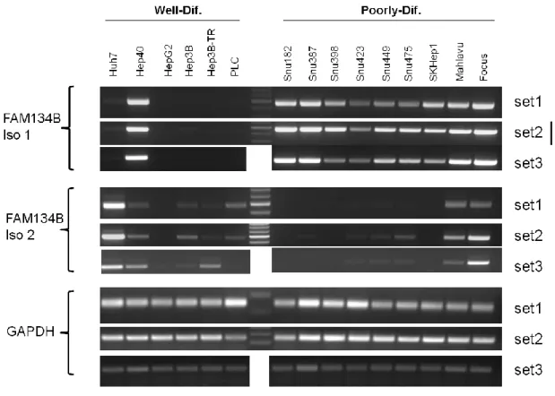
4.3 FAM134B expression in HCC cell lines and tissues ... 44
4.3.1 Detection of FAM134B mRNA levels in HCC cell lines ... 44
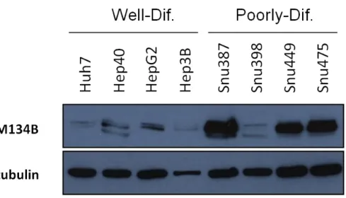
4.3.2 Detection of FAM134B protein levels in HCC cell lines ... 46
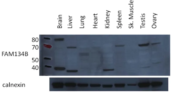
4.3.3 Detection of FAM134B protein levels in tissues ... 47
4.4 FAM134B expression is down-regulated in cancer ... 48
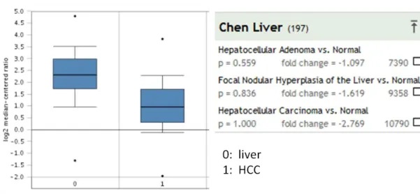
4.4.1 FAM134B in Chen liver data ... 48
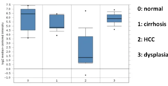
4.4.2 FAM134B in Wurmbach liver data ... 49
xiii
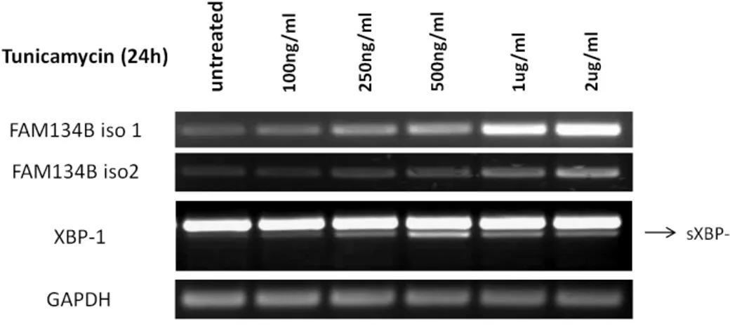
4.5.1 Tunicamycin treatment increases FAM134B expression in Huh7 cells ..
... 50
4.5.2 Dithiothreitol (DTT) treatment increases FAM134B expression in Huh7 cells ... 51
4.5.3 Thapsigargin treatment increases FAM134B expression in Huh7 cells .. ... 52
4.5.4 Detection of changes at protein levels of FAM134B in response to ER stress ... 53
4.5.5 Change in FAM134B expression at G2-M and G1 stages of cell cycle .. ... 54
4.6 Differential expression of FAM134B in well- and poorly-differentiated cell lines might be linked to their ER stress status... 55
4.7 FAM134B overexpression in Huh7 cells ... 56
4.7.1 Testing FAMB134 overexpressing stable Huh7 clones ... 57
4.7.2 FAM134B overexpression does not induce ER stress ... 57
4.7.3 FAM134B overexpression does not promote epithelial-to-mesenchymal transition in Huh7 cells ... 58
4.7.4 FAM134B overexpression does not induce autophagy in Huh7 cells . 60 4.8 Induction of ER stress impairs proliferation of Huh7 cells ... 61
4.9 FAM134B increases sensitivity to thapsigargin induced apoptosis in Huh7 cells ... 66
4.9.1 Western blot experiment ... 66
4.9.2 Cell cycle analysis with flow cytometry ... 67
4.10 FAM134B overexpression impairs cell proliferation during thapsigargin induced ER stress ... 69
4.11 FAM134B increases sensitivity to tunicamycin induced apoptosis in Huh7 cells ... 70
xiv
4.11.2 Cell cycle analysis with flow cytometry ... 71
4.12 FAM134B overexpression impairs cell proliferation during tunicamycin induced ER stress ... 72
4.13 FAM134B is likely to increase the severity of ER stress response ... 73
5. DISCUSSION AND CONCLUSION ... 75
5.1 FAM134B in the context of senescence ... 75
5.2 Differential expression of FAM134B in tissues ... 76
5.3 Characterization of FAM134B in the context of hepatocellular carcinoma 77 5.4 Describing FAM134B in the context of ER stress ... 78
5.5 FAM134B and morphology of Huh7 cells ... 80
5.6 FAM134B and autophagic response ... 80
5.7 FAM134B as a mediator of ER stress induced apoptosis ... 81
5.8 FAM134B in the pathogenesis of HCC ... 83
6. FUTURE PERSPECTIVES ... 84
xv
LIST OF TABLES
Table 3.1 : Primer list, sequences and Tm values ... 23 Table 3.2: Antibody list, catalog numbers and working dilutions ... 24
xvi
LIST OF FIGURES
Figure 4.1: Genomic region and transcripts of FAM134B ... 39
Figure 4.2: Genomic context of FAM134B gene ... 39
Figure 4.3: Domain representations of two isoforms of FAM134B ... 40
Figure 4.4: Adriamycin induces senescence in Huh7 cells, detected by SABG ... 41
Figure 4.5: Adriamycin treatment does not change FAM134B expression in Huh7 cells. ... 42
Figure 4.6: TGF-β treatment decreases FAM134B expression in Huh7 cells ... 43
Figure 4.7: TGF-β treatment causes a decrease in protein levels of FAM134B in HCC cells ... 43
Figure 4.8: FAM134B mRNA levels in HCC cell lines ... 45
Figure 4.9: FAM134B protein levels in HCC cell lines ... 46
Figure 4.10: Comparison of FAM134 protein levels in well- and poorly-differentiated HCC cell lines ... 46
Figure 4.11: FAM134B protein levels in rat tissues ... 47
Figure 4.12: FAM134B expression in Chen liver data ... 48
Figure 4.13: FAM134B expression in Wurmbach liver data ... 49
Figure 4.14: Tunicamyin treatment causes an increase in FAM134B expression ... 50
Figure 4.15: DTT treatment causes an increase in FAM134B expression ... 51
Figure 4.16: Thapsigargin treatment increases FAM134B expression in Huh7 cells 52 Figure 4.17: Tunicamycin increases FAM134B protein levels in Snu387 cells ... 53
Figure 4.18: Tunicamycin treatment does not affect FAM134B protein levels in Huh7 and Hela cells ... 53
Figure 4.19: FAM134B expression in synchronized Huh7 cells, detected by RT-PCR ... 54
Figure 4.20: ER stress status of HCC cells, detected by sXBP-1 RT-PCR ... 55
Figure 4.21: ER stress status of HCC cell lines, detected by western blot ... 56
Figure 4.22: Testing FAM134B overexpressing clones, detected by western blot ... 57
Figure 4.23: Testing ER stress in Huh7-FAM134B clones, detected by RT-PCR .... 58
xvii
Figure 4.25: Neither tunicamycin treatment, nor FAM134B expression induces EMT in Huh7 ... 59 Figure 4.26: Tunicamycin induces autophagy in Huh7 cells ... 60 Figure 4.27: FAM134B overexpression does not induce autophagy in Huh7 cells ... 61 Figure 4.28: Tunicamycin inhibits proliferation of Huh7 cells, detected by BrdU IF64 Figure 4.29: Tunicamycin induces pRb activation and cell cycle arrest in Huh7 cells ... 65 Figure 4.30: FAM134B increases sensitivity to thapsigargin induced apoptosis in Huh7 cells, detected by western blot ... 66 Figure 4.31: FAM134B increases sensitivity to thapsigargin induced apoptosis in Huh7 cells, detected by propidium iodide based cell cycle analysis ... 68 Figure 4.32: FAM134B overexpression impairs proliferation of Huh7 cells during thapsigargin induced ER stress ... 69 Figure 4.33: FAM134B increases sensitivity to tunicamycin induced apoptosis in Huh7 cells, detected by western blot ... 70 Figure 4.34: FAM134B increases sensitivity to thapsigargin induced apoptosis in Huh7 cells, detected by propidium iodide based cell cycle analysis ... 71 Figure 4.35: FAM134B overexpression impairs proliferation of Huh7 cells during thapsigargin induced ER stress ... 72 Figure 4.36: FAM134B increases the intensity of UPR following thapsigargin treatment ... 73
1
1. INTRODUCTION
1.1 Hepatocellular carcinoma
1.1.1 Epidemiology of hepatocellular carcinoma
Hepatocellular carcinoma (HCC) is the most common liver cancer type with a percentage between 80-90% among all primary liver cancer types. Furthermore, if all cancers are taken into consideration, HCC is classified in a substantial percentage, around 6% of all cancers (Parkin et al., 2005). HCC affects many populations in the world, taking a place among the most lethal cancers (Farazi and DePinho, 2006). After lung and stomach cancers, HCC is the third leading cause of cancer related deaths. It causes 600,000 deaths every year with a new occurrence of 500,000 – 600,000 cases per year (Faloppi et al., 2011). Occurrence of HCC is directly related to age, having the highest frequency in the population of over age 65 (Gomaa et al., 2008). Apart from the relationship between age and HCC, there is a significant sex ratio of 2,4:1, more common in men (Parkin et al., 2005).
1.1.2 Aetiologies and risk factors of hepatocellular carcinoma
Hepatocellular carcinoma is a cancer type with high complexity in terms of the aetiological factors leading to the occurrence of the hepatocarcinogenesis. Among these risk factors, the most major are viral factors such as chronic Hepatitis B and Hepatitis C, metabolic factors such as non-alcoholic fatty liver disease and diabetes, toxic factors such as aflatoxins and alcohol, immune related factors such as autoimmune hepatitis, and additional factors such as cirrhosis and hereditary haemochromatosis (Badvie, 2000; Farazi and DePinho, 2006; Parikh and Hyman, 2007). Although all of these risk factors are associated with the incidence of HCC, effectiveness and the prevalence of these factors are highly dependent on the geographic condition over the world. For instance, the difference in the geographical
2
distribution of HCC is mainly due to the variable distribution of Hepatitis B virus (HBV) and Hepatitis C virus (HCV) infections (Liu and Kao, 2007). However, even though HBV and HCV infections are important factors, they are not that much prominent in the US. Instead, alcoholic cirrhosis and dietary related non-alcoholic fatty liver diseases are the major factors leading hepatocarcinogenesis in most western countries (Stickel and Hellerbrand, 2010).
1.1.2.1 Role of aflatoxin in hepatocarcinogenesis
Aflatoxin B1 (AFB1) is a toxic factor for the liver produced by Aspergillus, which contaminate some foods, such as corn, peanut, and grain (Groopman et al., 1996). Therefore, digestion of such contaminated foods causes aflatoxin exposure in the liver and trigger hepatocarcinogenesis. The affect of aflatoxin on hepatocarcinogenesis is associated with its role in causing specific codon 249 mutation on tumor suppressor p53 gene (Bressac et al., 1991; Ozturk, 1991). Interestingly, this mutation has also been detected in patients with a history of HBV infection (Yu et al., 2005). Hence, aflatoxin exposure is likely to trigger hepatocyte carcinogenesis by inducing a common mutation that is highly responsible for the lack of tumor suppression activity of p53.
1.1.2.2 Role of alcohol in hepatocarcinogenesis
Alcohol is metabolized mainly by activity of alcohol dehydrogenase enzyme in cytoplasm of hepatocytes, as well as by some other mechanisms in endoplasmic reticulum and peroxisomes (McKillop and Schrum, 2005). Ethanol metabolism results in products of free radicals and acetaldehyde. Release of these free radicals and acetaldehyde affects the cellular homeostasis by inducing oxidative stress and also preventing some cellular activities, such as DNA repair, by directly binding to the cellular components (Sun et al., 2001). On the other hand, ethanol directly affects the activities of some other key regulators of proliferation signaling and cell cycle, such as protein kinase C, adenylate cyclase, STAT, JNK, NF-ĸB, and MAPK (McKillop and Schrum, 2005) as well as increasing the activity of G protein inhibitor
3
which is associated with HCC through triggering p42/p44 MAPK signaling (Diehl, 2005)
1.1.2.3 Hepatitis B virus induced hepatocarcinogenesis
Three decades ago, it has been shown that hepatitis B carriers have an increased annual incidence of HCC with an average of 0,5%, and 1% for population with age over 70 (Beasley et al., 1981). Further studies pointed out that HBV associated hepatocarcinogenesis is directly related to the necro-inflammation and fibrosis effects of this viral infection. These effects further pave the way for hepatocyte regeneration (Waris and Siddiqui, 2003). In addition, HBV infection can contribute to hepatocellular carcinogenesis through host genome integration, triggering some oncogenic pathways and even endoplasmic reticulum interaction and upregulation of oxidative stress (Farazi and DePinho, 2006). In terms of HBV and cirrhosis relationship, in East Asian countries, the incidence rate of HCC in HBV-related cirrhosis was shown to be 2,7% (Michielsen et al., 2005). This suggests that underlying liver diseases are undoubtedly key factors of acquiring HCC. In fact, HBV infection may also result in HCC independent of cirrhotic contribution, showing that not merely HBV and cirrhosis co-contribution but co-contribution of HBV with some other factors lead to hepatocarcinogenesis (Waris and Siddiqui, 2003).
1.1.2.4 Hepatitis C virus induced hepatocarcinogenesis
Hepatitis C virus infection is associated with hepatocyte transformation in different ways. Firstly, studies showed that HCV infection is most likely to turn into chronic infection, because about 80% of the infected patients cannot be totally disinfected in the acute phase. Resulting chronic infection results in prevailing of the effects of viral infection, such as inflammation and regeneration in hepatocytes. These in advance lead to chronic hepatitis and cirrhosis, which finally brings out hepatocyte carcinogenesis with the accumulation of chromosomal abnormalities (Ahn and Flamm, 2004; Suruki et al., 2006). The percentage of cirrhosis development and hepatocellular carcinogenesis in the chronic infection cases are 20% and 2,5%,
4
respectively (Bowen and Walker, 2005). In addition, HCV induced hepatocarcinogenesis is found to involve some key viral proteins in promotion of carcinogenesis. Among these, NS5A is known to control transcription and mitogenic signal transduction as well as the regulation of cell cycle (Tellinghuisen and Rice, 2002). Besides, important cell regulatory mechanisms, such as proliferation, apoptosis, transcription and key signaling factors of these regulations, such as NF-ĸB and MAP kinase are modulated by HCV core protein (Block et al., 2003).
1.1.2.5 Other factors inducing hepatocarcinogenesis
Apart from the major risk factors of hepatocellular carcinogenesis, there are some other aspects contributing to the initiation or development of HCC.
Diabetes is considered as one of the key additional factors for the onset of HCC. Studies of Davila et al. showed that the ratio of HCC patients with diabetes is substantially greater than the patients with diabetes within the control group without cancer. Furthermore, independent of the other foremost risk factors, diabetes is regarded as a self-determining risk factor with an effect of 2-3 fold increase in occurrence of HCC (Davila et al., 2005). The link between diabetes and the hepatocellular carcinogenesis is possibly due to the accumulation of free fatty acids in liver and the acquisition of insulin resistance in the liver of diabetic patient (Maclaren et al., 2007). These results cause development of fibrosis through dysfunctional effects on hepatocyte homeostasis, such as hepatocyte injury and hepatocyte apoptosis (Farrell and Larter, 2006).
Apart from the effect of diabetes on HCC, other disorders affecting liver are also involved in the increased risk of hepatocarcinogenesis. Among these, non-alcoholic steatohepatitis and non-alcoholic fatty liver disease are likely to promote HCC development in a way that they contribute to advance of liver fibrosis and cirrhosis (Adams and Angulo, 2005). Furthermore, some other genetic disorders, such as hereditary haemochromatosis and alpha1-antitrypsin deficiency are associated with the higher risk of developing HCC (Badvie, 2000). Hereditary haemochromatosis is a disease related to the excess iron taking into hepatocytes. Such an iron overload in hepatocytes brings about a hepatocellular damage, finally leading to
5
hepatocarcinogenesis (Pietrangelo, 2009). Another inherited disease alpha1-antitrypsin deficiency comes up with the emergence of alpha1-antitrypsin polymers in the liver cells. These antitrypsin polymers trigger hepatocyte fatality, and thereby, cirrhosis (Parfrey et al., 2003).
1.1.3 Molecular pathogenesis of hepatocellular carcinoma
It has been discussed that the molecular pathogenesis of hepatocellular carcinoma is a rather complex mechanism (Farazi and DePinho, 2006). Through the progress of HCC, different risk factors and modulations, such as mutation, altered pathways, genetic changes, epidgenetic changes, and chromosomal aberrations are involved. Accumulation of these changes through hepatocarcinogenesis paves the way for the development of neoplastic state in normal livers, non-cirrhotic livers and cirrhotic livers (Bruix and Sherman, 2005; Llovet et al., 2003). The basis of HCC originates initially from damaged hepatocytes which later on start proliferation and regeneration cycles. Increased regenerating activity of hepatocytes leads to the occurrence of cirrhosis and then dysplasia, finally to hepatocellular carcinoma with the help of addition effects and risk factors (Schlaeger et al., 2008). Nevertheless, details of the molecular events resulting in hepatocellular carcinoma are still not well known (Farazi and DePinho, 2006).
Normal liver to hepatocarcinogenic liver transition is a multistep mechanism mostly triggered by genetic changes resulting in firstly cirrhotic liver, and then cancer. This progress involves liver stem cells as well as hepatocytes (Llovet and Bruix, 2008). At the initial steps, HBV and HCV infections, amplified transforming growth factor alpha and insulin like growth factor 2 activities speeds up the early process of hepatocyte proliferation (Thorgeirsson and Grisham, 2002). During these stages, especially infection of HBV brings about oncogene activation, instable chromosomes and DNA rearrangements via integration of the viral genome (Ferber et al., 2003). On the other hand, DNA damage is further induced by occurrence of oxidative stress and chronic inflammation in hepatocytes (Hussain et al., 2007). At the preneoplastic stage of carcinogenesis, the process involves both genetic an epigenetic aberrations. In the mean time, dysplastic nodules are produced in a way that particular cell
6
populations with these aberrations are selected and grow up to form these dysplastic structures. This transformation is further supported by activation of survival and proliferation pathways, in addition to uncontrolled telomerase activity in order to serve for unlimited proliferative capacity of these cell populations (Llovet and Bruix, 2008). Dysplastic nodules are pre-malignant structures possessing abnormalities at the cellular level and are likely to transform into hepatocellular carcinoma thanks to the accumulation of genomic instability and loss of p53 function (Farazi and DePinho, 2006).
1.1.4 Genetics of hepatocellular carcinoma
As other cancer types, hepatocellular carcinoma is also highly associated with the genetic aberrations in the original cells. These aberrations involve changes in gene expression, somatic mutations, amplification or deletion of specific sites and epigenetic changes directly affecting the expression of key genes (Llovet and Bruix, 2008). Among these changes, genome wide alterations consisting of amplification or deletion of specific chromosomal sites have been detected in HCC cases. The most common amplification sites are 1q, 6p, 8q, 17q, and 20q, whereas deletion sites are 4q, 8p, 13q, 16q, and 17p (Chiang et al., 2008a; Thorgeirsson and Grisham, 2002). Besides, other critical amplification sites were detected at regions where cyclin D1 and VEGFA genes are located, at 11q13 and 6p21 sites, respectively (Chiang et al., 2008a). There are a few critical somatic mutations already found to be associated with hepatocarcinogenesis. One of the major tumor suppressors, TP53 gene, was found to be mutated with a frequency of 30% in HCC patients worldwide. And, this mutation at the codon 249 of TP53 gene was reported to be related to aflatoxin B1 exposure (Bressac et al., 1991). Besides, another cell cycle control gene Rb which is located at 13q site, which is a common loss of heterozygosity site, and inactivating mutations of Rb gene was reported to be at least 15% of HCC cases (Ozturk, 1999). In the aspect of genomic instability, the major alteration in the HCC cells is that hapatocarcinogenic cells have highly increased telomerase activity, approximately in 90% of HCC cases. Increased telomerase activity is most likely due to HBV genome integration into TERT locus, increased expression of telomerase RNA component
7
TERC, and loss of a chromosomal region involving telomerase repressor (Farazi et al., 2003). In addition to genetic alterations, epigenetic changes are also factors in promoting hepatocarcinogenesis. Expressions of some critical genes having tumor suppressor roles, such as p16INK4a, E-cadherin, BRCA1, and IGFR-II, are suppressed by hypermethylation of the promoter regions of these genes (Farazi and DePinho, 2006; Thorgeirsson and Grisham, 2002).
1.2 Senescence
1.2.1 Cellular senescence
Cellular senescence was first explained as a loss of proliferating ability of the cells in culture, resulting in the stop of population growth after an approximate number of passaging cells in vitro (Sherwood et al., 1988). Besides, cells with dividing and renewal ability can quit the cell cycle permanently due to various stress factors, such as DNA damage, mitogenic signals, change in the chromatin structure and dysfunctional telomeres (Campisi, 2005). Cellular senescence is a potential anti-cancer mechanism mainly regulated through p53 and RB proteins. Senescence response acts as a barrier for cancer in a way that damaged cells are prevented from aberrant proliferation which is a key factor of carcinogenesis (Campisi, 2005). However, senescent cells undergo significant changes at the cellular level, such as chromosomal aberrations, increased size and size heterogeneity (Sherwood et al., 1988). And, these changes at the cellular level may contribute to aging and some age related diseases (Campisi, 2005).
1.2.2 Replicative senescence
Replicative senescence refers to the induction of senescence response as a result of telomere shortening. Telomeres are repetitive hexamer units located at the ends of eukaryotic chromosomes. These structures act as caps for the linear end, having a length of a few to several kbs. Presence of telomeres at the end of chromosomes
8
provides protection against chromosomal degradation and chromosome end fusion (Reaper et al., 2004).
Telomere dependent senescence is associated with the telomere shortening. In the lack of adequate telomerase activity, a part of telomeres are lost during the replication process. Loss of telomeres in each replication results in drastically reduced telomere length, which is sensed as DNA damage signal (Ozturk et al., 2009). As a result of this DNA damage signal, key cell cycle regulators, such as p53, retinoblastoma protein, and p16INK4a are activated and cell cycle arrest occurs (Campisi, 2005).
1.2.3 Senescence in liver
Like other somatic cells, hepatocytes do not possess telomerase activity to compensate the telomeric loss at chromosome ends. Hence, during hepatocyte proliferation, shortening of telomeres is inevitable. Excess proliferation of hepatocytes results in progressive telomere shortening, eventually leading up to chronic liver diseases. Therefore, hepatocyte telomere shortening and resulting senescence phenotype are key features of liver cirrhosis (Wiemann et al., 2002). Cirrhosis is an important pathological factor for the development of HCC, and characterized by excessive collagen deposition. Collagen deposition in the liver causes the formation of nodules in the liver and acts as a key intermediate step for fibrosis (Farazi and DePinho, 2006).
Senescence response is a pathological condition in the liver, but development of hepatocarcinogenesis requires further factors at this step. In fact, senescence of hepatocytes is a barrier for carcinogenesis through which cells require additional aberrations to pass this barrier, regain proliferative ability, and trigger HCC (Paradis et al., 2001). The main aberrations required to overcome the senescence barrier are inactivation of pRb and p53 pathways (Ozturk et al., 2009). The most common of these are the inactivation mutation of p53 gene and silencing of p16INK4a at the epigenetic level (Ozturk et al., 2009; Soussi, 2007).
9
1.2.4 Spontaneous replicative senescence
Cancer cells have immortal characteristics. However, whether this characteristic is reversible or irreversible is still being questioned. In 2006, studies of Ozturk N. et al. showed that hepatocellular carcinoma cell line Huh7 derived cells can be reprogrammed into replicative senescence (Ozturk et al., 2006). In this study, Ozturk N. et al. made a long term culture of Huh7 cell line. After a long term culture, they obtained two sets of clones, namely C1/C3-Early/C3-Late and G11/G12-Early/G12-Late. The C3 clone stopped proliferation at 80 population doublings (PD), while C1 clone replicated over 150 PD. C3 clone was detected to be totally senescence-associated β-galactosidase (SABG) positive, and BrdU negative. C1 clone and C3-Early clones were similar. They showed normal Huh7 morphology, positive BrdU, and low percentage of SABG positive cells (Ozturk et al., 2006).
On the other hand, Ozturk N. et al. showed that while immortal clone of Huh7 had hTERT activity, the spontaneous senescent C3 clone did not possess hTERT activity. Besides, SIP1 gene (ZFHX1B, Zinc finger homeobox 1B) expression was detected to be inversely correlated with hTERT expression in C3 senescent clones. In order to further show the effect of SIP1 on hTERT activity, shRNA targeting of SIP1 was done. Results indicated that knockdown of SIP1 released hTERT expression and rescued spontaneous senescent C3 cells from senescence arrest (Ozturk et al., 2006). This study was continued further in detailed with the expression profiling of the immortal and senescent Huh7 clones. Gene expression profiling was done using HU133Plus2 Affymetrix Chips and data was analyzed using R software. According to the results, the list of genes with statistically different expression between immortal and senescent clones involves 3073 genes. There is also a number of significant genes with differential expressions in other two-group comparisons, 2149 genes between immortal (C1) and early senescent (C3-Early), and 2023 genes between early senescent (C3-Early) and senescent (C3) (Ozturk M. et al., unpublished data).
10
1.2.5 FAM134B as a senescence-associated gene
Data analysis of gene expression profiling in senescent, pre-senescent and immortal clones of Huh7 resulted in detection of a number of differentially expressed genes in within three groups. Among these genes, there are some genes that have been and being studied extensively, while some are totally novel genes that were identified in whole genome sequencing but have not been studied and characterized yet. FAM134B (Family with sequence similarity 13, member B) was detected as a significant gene associated with senescence phenotype in this microarray study. FAM134B gene was represented with two different probes in the HU133Plus2 Affymetrix chip. Probe codes are 218532_s_at and 218510_x_at. According to the probe 218532_s_at, FAM134B gene was detected to be 6.5 fold up-regulated in senescent clones in comparison to immortal clones, and 2.6 fold up-regulated in senescent clones compared to pre-senescent clones. On the other hand, microarray results according to other probe 218510_x_at, 4.9 fold up-regulation was detected in senescent clones with respect to immortal clones, while 2.8 fold up-regulation detected in senescent clones with respect to pre-senescent clones. Consequently, FAM134B was identified as one of the most significant genes showing differential expression between senescent and immortal clones with a p-value of 1.097 E-06 (Ozturk M., unpublished data). According to these results, FAM134B is a senescence associated gene, and supposedly has a role in obtaining senescent phenotype or acquisition of senescence phenotype brings about increased expression of this gene. Therefore, the relationship between FAM134B gene and senescence is worth investigating in detail, especially because of the fact that it is a novel gene possibly having important functions in the cell context.
1.3 Endoplasmic reticulum stress
Endoplasmic reticulum (ER) is a key organelle in the cell functioning in the regulation and secretion of all proteins. After translation of linear proteins, these peptides enter into the ER and become mature in this organelle. The ER ensures proper folding and post-translational modifications of entered proteins before these proteins are transported to Golgi.
11
The lumen of ER is an environment with very high calcium ion concentration. Calcium ions are continuously transported into the ER by active transport through calcium ATPases. The lumen with high calcium ion concentration is a suitable environment for formation of disulfide bonds of proteins. Thanks to such oxidative environment and presence of calcium-dependent chaperone proteins in the ER, protein folding is done within the ER and folded proteins are transported out (Xu et al., 2005).
ER works in a dynamic fashion. The flux into the ER is not always at the same level, changing according to the cells’ programs. Physiological state of the cell and environmental conditions are the main factors affecting the dynamic situation of the ER. ER always tries to keep protein mechanism well maintained. Therefore, protein folding capacity is adjusted in order to retain high fidelity. To maintain this homeostasis, ER requires sensors to determine the physiological conditions in the ER and signaling to regulate and maintain the homeostasis. However, homeostasis cannot be always well-maintained, resulting in an imbalance between the unfolded protein load in the ER and the effectiveness of ER machinery working on handling the situation. This imbalance is called endoplasmic reticulum stress (ER stress). The pathway reconciling the stress response and ER homeostasis is called unfolded protein response (UPR) (Ron and Walter, 2007). Presence of such a sensing mechanism and regulation was detected in a study showing that increasing the unfolded protein load of ER results in increased expression of ER lumen chaperones (Kozutsumi et al., 1988).
1.3.1 Handling mechanisms for endoplasmic reticulum stress
When an imbalance between accumulated unfolded proteins and the ER machinery occurs, the cell tries to overcome this situation by acquiring some adaptations. This intrinsic protection mechanism of cells is primarily focused on maintaining homeostasis at both cellular and tissue levels. Therefore, these adaptations may aim to relieve the stress or to remove the stressed cell from the tissue.
12
The first response against stressed ER is the reduction of unfolded protein load in the ER. This is a kind of transient solution for alleviating ER stress. The amount of accumulated unfolded proteins is reduced by some regulations, either aiming to decrease the amount of newly synthesized proteins or preventing entering of peptides into ER. Both of these regulations seek a solution for stressed ER by reducing the amount of proteins being processed in the ER. Secondly, apart from reducing the amount of proteins in the ER, cells acquire a further protection by increasing the working capacity of the ER machinery. This adaption is a long term response involving UPR target genes. Long term adaptation aims lessening ER stress by activating key UPR target genes encoding ER chaperone proteins. In this case, the more chaperones are produced and work in the ER, the faster ER stress is alleviated thanks to increased activity of these chaperones in proper folding of proteins. If both of these mechanisms do not result in alleviation of ER stress, cells activate a third mechanism, which is programmed cell death. This takes place in situations where ER homeostasis is no longer possible to be set up again. Hence, keeping homeostasis at organism level is preferred and stressed cell is programmed into controlled cell death (Ron and Walter, 2007).
1.3.2 Components of unfolded protein response
Unfolded protein response is the signal transduction response activated as a result of stress in the ER. So far, three different ER stress signal transducers have been identified in the ER. Each of these three initiates a signal transduction through a particular branch of UPR. These three elements are Inositol requiring protein 1 (IRE1), activating transcription factor 6 (ATF6), and PKR-like ER kinase (PERK) (Ron and Walter, 2007).
Inositol requiring protein 1 is the highly conserved arm of UPR. This element was firstly identified in yeast and found to be encoded by IRE1 gene. This gene encodes a ER transmembrane protein with a luminal and a cytoplasmic domain (Cox et al., 1993). Cytoplasmic domain of IRE1 consists of a kinase activity, which is triggered by activation of ER stress signal coming from the luminal part. Following the activation signal, IRE1 activates itself by oligomerization and
trans-13
autophosphorylation of the kinase domains (Credle et al., 2005; Zhou et al., 2006). Activation of the kinase domain of IRE1 also activates its other functional response, endonucleolytic cleavage. The substrate of this endonucleolytic activity is a transcription factor Hac1 in yeast (Cox and Walter, 1996; Mori et al., 1996), and X-box binding protein 1 (XBP1) in metazoans (Calfon et al., 2002). IRE1 dependent transcriptional regulation works through its endonucleolytic activity to cleave and remove an intron from XBP1 mRNA. This cleavage makes XBP1 transcription factor active by leading spliced XBP1 mRNA to translation (Lee et al., 2002; Yoshida et al., 2001). Apart from activation of XBP1 by endonucleolytic cleavage, XBP1 expression is also controlled at the transcription level during UPR. XBP1 mRNA levels are increased as UPR is induced (Yoshida et al., 2006). Functional XBP1 protein works as a transcription factor at the promoter sites of some genes having role in endoplasmic reticulum associated protein degradation and transport of unfolded proteins out of ER (Rao and Bredesen, 2004). Furthermore, with accompany of NF-Y, XBP1 binds to two different types of cis-acting elements, which are ER stress enhancer and UPR element (Yoshida et al., 2001). On the other hand, IRE1 is responsible for activation of kinases having role in inflammation and cell death machinery in association with TRAF2. Downstream of IRE1, TRAF2 activates kinase Ask1 and stress induced Jun N-terminal kinase (JNK) (Urano et al., 2000), as well as caspase-12 of apoptotic pathway (Yoneda et al., 2001).
Other arm of the UPR signals through activating transcription factor 6. ATF6 was firstly identified as a new class of UPR signal transducer specifically in metazoans (Haze et al., 1999). ATF6 is found tethered to ER membrane as an inactive precursor. Upon activation of UPR, ATF6 is transported from ER membrane to Golgi apparatus. In Golgi, ATF6 undergoes two protease cleavages, which are processed by site 1 protease (S1P) and by site 2 protease (S2P), respectively. After cleavage, remaining active ATF6 fragment translocates into the nucleus to activate expression of target genes (Haze et al., 1999; Ye et al., 2000). ATF6 activates expression of a set of UPR genes including XBP1 (Yoshida et al., 2001). When UPR signal is activated, IRE1 and ATF6 arms of UPR act cooperatively in a way that while ATF6 increases the transcription of XBP1, IRE1 undertakes endonucleolytic cleavage and activating role of XBP1 to trigger expression of target alarm genes.
14
The third arm of UPR involves PKR-like ER kinase. PERK is activated similar to IRE1 by oligomerization of PERK and trans-autophosphorylation of its cytoplasmic domain (Bertolotti et al., 2000). In addition to its autophosphorlyation function, PERK also phosphorylates eukaryotic translation initiation factor-2-alpha (eIF2α) at Ser51 site. Ser51 phosphorylation of eIF2α results in inhibition of eIF2α re-cycling by guanine nucleotide exchange factor eIF2B. Lack of eIF2B functioning on eIF2α decreases the amount of active GTP-bound form of eIF2α. As a result of less active eIF2α, general translation initiation is halted, thereby reducing the amount of newly synthesized proteins (Harding et al., 1999). Activation of the PERK arm not only signals through reduction of translation, but also regulates activation of some downstream targets. For instance, activating transcription factor-4 (ATF4) and nuclear factor ĸB (NF-ĸB) are activated at translational and post-translational levels via PERK signaling (Ron and Walter, 2007). ATF4 activated another important transcription factor C/EBP homologous protein (CHOP). Signaling cascade continues with the targets of CHOP, which are growth arrest and DNA
damage-inducible protein-34 (GADD34) and ER oxidase-1 (ERO1) (Marciniak et al., 2004).
1.3.3 Sensing the stress
Activation of UPR is dependent on sensing stressed ER. Because ER is stressed due to the accumulation of unfolded proteins, heavy unfolded protein load must be sensed in order to start a signal transduction through three main arms of UPR. The most common chaperone protein BiP is the key sensor for ER stress (Rutkowski and Kaufman, 2004). One of the three main elements of UPR, ATF6, is held in the ER via protein-protein interaction with BiP (Shen et al., 2002). Other UPR element, IRE1 is also retained associated with BiP via its lumenal domain (Liu et al., 2003). And, the last arm of UPR, PERK, is also located on ER membrane and bound to BiP through hydrophobic regions on its lumenal domain (Ma et al., 2002).
Sensing stress via BiP relies on the ability of BiP to bind peptide binding regions of proteins. BiP binds to both these three UPR elements and unfolded proteins thanks to this region. In the unstressed ER, the most abundant chaperone in the ER, BiP, binds to lumenal domains of PERK and IRE1, preventing dimerization of these UPR
15
elements to become active. In the stressed ER, due to excess unfolded proteins, BiP starts binding to unfolded proteins, decreasing its affinity to PERK, IRE1 and ATF6. Dissociation of BiP from PERK and IRE1 allows homodimerization and autophosphorylation of these proteins, which activates these two arms of UPR. On the other hand, dissociation of BiP from ATF6 releases ATF6 to transport into Golgi and become activated by proteolytic cleavage (Rutkowski and Kaufman, 2004).
1.3.4 Deciding survival or death during ER stress
Accumulation of excess unfolded proteins in the ER triggers UPR to somehow overcome this unfavorable situation. Three main elements of UPR control activities of some key regulators functioning in the cell death response. While the decision of cells’ survival or death relies on the activities of these key UPR elements, actual factor behind this scenario is dependent on how severe the cell experiences a stress in the ER and how persistent the stress is.
ER associated cell death is likely to be controlled by the level of calcium ions in the ER. Calcium ions might take a role in activation of proteases in cytoplasm to induce cell death (Nakagawa and Yuan, 2000). However, there is no detected relationship between the ER stress and release of calcium from the ER so far. ER stress might contribute to apoptosis through some key apoptotic regulator, such as BAX and BAK. These death factors normally reside in the ER, but translocating onto mitochondria membrane, where they can function upon ER stress (Scorrano et al., 2003; Wei et al., 2001). ER stress mediated cell death might also signal through IRE1 branch of UPR. In this case, caspase-12 activation with the help of TRAF2 triggers death in mice (Nakagawa et al., 2000). Paralog of mice caspase-12 in human, caspase-4 is also associated with the ER functioning, possibly performing a similar role of its paralog in mice (Hitomi et al., 2004). Furthermore, IRE1 mediated Ask1 activation leads to JNK activation, resulting in the phosphorylation of JNK targets. Among these targets, phosphorylation of Bcl-2 inhibits its anti-apoptotic activity (Yamamoto et al., 1999), whereas phosphorylation of Bim results in activation of its pro-apoptotic function (Lei and Davis, 2003). At the transcription factor level, ER stress induced apoptosis is regulated by CHOP (GADD153). CHOP can repress the
16
expression of anti-apoptotic Bcl-2, resulting in the promotion of cell death (McCullough et al., 2001). In brief, at least two of the three branches of UPR have already been detected as factors in ER stress induced cell death. Therefore, cell death response is likely to be resulted from the cooperative role of different UPR signaling elements. Even though the primary goal of UPR is to alleviate the stress level in the ER, if the improper folding process in the ER is excessive or persistent, UPR
response aims directing cells to death, typically apoptosis (Xu et al., 2005).
1.4 Liver and ER stress
The major cell type in the liver, hepatocytes, is a type of cell specialized in metabolic activities. Hepatocytes are responsible for high amount of protein synthesis and secretion in the body. Since the endoplasmic reticulum is the site where post translation control of synthesized proteins is done, ER in hepatocytes has to work at maximum capacity. Under normal circumstances, hepatocytes can handle this massive work of protein synthesis, folding and port-translational modification, and secretion in a routine way thanks to presence of abundant endoplasmic reticulum. However, hepatocyte ER might be stressed upon emergence of some internal and external factors. Viral infections to hepatocytes, alcohol and drug usage, metabolic disorders, and mutations in ER protein encoding genes are the main causes of stressed ER in the liver (Ji and Kaplowitz, 2006).
Stressed ER in hepatocytes can be overcome by common adaptive stress response, UPR. When ER stress is induced by the abovementioned factors, UPR is activated and tries to overcome the stress by inducing ER resident proteins having role in protein folding, triggering degradation of unfolded proteins, and decreasing the total protein synthesis in ribosomes. In a case that ER stress cannot be assuaged by these adaptations, continual stress brings about pathological outcomes in the liver. Hepatocyte death, inflammation and accumulation of hepatic fat are among these pathological consequences or uncontrolled ER stress. These conditions may lead to liver diseases and contribute to the occurrence of liver injury and exacerbate the prevailing liver disease of other aetiological factors, such as viral infection or diabetes associated liver disease (Ji and Kaplowitz, 2006).
17
1.5 FAM134B in the literature
Family with sequence similarity 134, member B (FAM134B) is a novel gene belonging to the same family with FAM134A and FAM134C. Even though there is a few publications related to this gene, its function is still not known. All the information that has been identified so far is summarized below.
In 2007, the studies of Tang et al. were published in an article titled ‘Oncogenic properties of a novel gene JK-1 located in chromosome 5p and its overexpression in human esophageal squamous cell carcinoma.’ This research showed that novel gene JK-1, which is the name of FAM134B isoform number 2, is associated with esophageal squamous cell carcinoma (ESCC) and has a transforming capacity in normal cells. Firstly, their results indicated that JK-1 is overexpressed in 69% (9/13) of ESCC cell lines and 30% (9/30) of ESCC patient samples. Furthermore, when they overexpressed JK-1 in normal cell types, NIH3T3 and HEK293, these cells possessed anchorage dependent and anchorage independent growth. And also, Tang et al. reported that subcutaneous injection of JK-1 overexpressing NIH3T3 cells created subcutaneous sarcomas in all (3/3) three mice. In brief, this study provided the first evidence for JK-1’s transforming capacity and JK-1 may be associated with the pathogenesis of ESCC (Tang et al., 2007).
In 2009, Kurth et al. published an important article, titled ‘Mutations in FAM134B, encoding a newly identified Golgi protein, cause severe sensory and autonomic neuropathy’, indicating the first evidence that FAM134B mutation is associated with a neurodegenerative disease. In their study, genome-wide homozyosity mapping in the sample family having hereditary sensory and autonomic neuropathy type II (HSANII) disease resulted in focusing on a candidate region of 5p15.1. Further analysis pointed out a homozygous nonsense mutation in the FAM134B gene. In order to detect the specific tissues expressing this gene, they performed an in situ hybridization assay. The results showed that FAM134B is predominantly expressed in sensory and autonomic ganglia, while other family members FAM134A and FAM134C are strongly expressed in central nervous system as wells as organs like liver, lung and kidney. Their localization studies resulted in the detection of FAM134B protein co-localized with cis-Golgi marker giantin in N2a cells,
18
autonomic ganglion neurons related tumor cell line. In order to test the role of FAM134B in the structure of Golgi, they knocked down FAM134B with a lentiviral approach in N2a cells. Knock down of FAM13B4 resulted in a significant reduction in the cis-Golgi size, with a reduction rate of 38% and 40% for two different RNAi. On the other hand, they noticed induction of apoptosis in cultured dorsal root ganglia (DRG) neurons when they are infected with lentiviral approach to knock down FAM134B. However, unlike DRG neurons, when mouse hippocampal pyramidal neurons were infected with the same RNAi approach, these cells were not affected. This might point out that nociception neurons are specifically sensitive to FAM134B depletion. In brief, this study indicated that FAM13B4 has an important role in survival of sensory and autonomic ganglia neurons. Because other members of the same family, family with sequence similarity 134, were also detected to be predominantly expressed in the nervous system, it is likely to state that this family of proteins might have a general role in the maintenance of neurons (Kurth et al., 2009). Previous studies in our group detected FAM134B as a spontaneous replicative senescence-associated gene (Ozturk et al., unpublished data). Therefore, our group aimed to find out the relationship between FAM134B and senescence. To do that, firstly, its association with senescence, which was previously detected in the microarray study, was tested with semi-quantitative RT-PCR. Results showed that FAM134B is indeed over expressed in C3 senescent clone of Huh7 cells in comparison with parental Huh7 and C1 immortal Huh7 clone. Furthermore, localization immunofluorescence studies resulted in the detection of FAM134B colocalization with endoplasmic reticulum protein calnexin. On the other hand, because Huh7 HCC cells have low level of FAM134B, FAM134B overexpressing stable Huh7 cells were created. FAM134B is associated with replicative senescence. However, it was not known whether this is the cause or the result of senescence. To test this, senescence-associated β-galactosidase experiments were performed. Results showed that FAM134B overexpression does not induce senescence in Huh7 cells. Besides, further BrdU incorporation studies indicated that FAM134B does not induce or repress Huh7 cell proliferation (Tasdemir et al, unpublished data).
19
Studies on FAM134B so far come up briefly with the following information: its possible relationship with ESCC, its role in survival of specific neurons and neurodegenerative disease, its localization to endoplasmic reticulum or cis-Golgi, and its structural function in cis-Golgi. However, we still do not know much about the function of this protein in the contexts of general cell biology and hepatocellular carcinoma.
20
2. OBJECTIVES AND RATIONALE
Being one of the most common and lethal cancer types in the human population worldwide, hepatocellular carcinoma is a significant cancer type that awaits a cure. However, the molecular pathogenesis of hepatocellular carcinoma is a rather complex mechanism (Farazi and DePinho, 2006). Indeed, through the progress of HCC, different risk factors and modulations, such as mutation, altered pathways, genetic changes, epigenetic changes, and chromosomal aberrations are involved (Bruix and Sherman, 2005).
Studies of Ozturk et al. showed that immortal HCC cell line Huh7 can be reprogrammed into replicative senescence, resulting in the loss of tumorigenic capacity of parental immortal cells (Ozturk et al., 2006). Acquisition of such a non-proliferative capacity of immortal cells came up with the idea that directing immortal cells into senescence might be a potential therapeutic approach to tumorigenesis. Designing promising therapies relies on the characterization of the features of immortal and replicative senescent cells in detail. As being one of the most significantly upregulated gene in senescent clones, FAM134B might undertake a critical function in the context of HCC.
On the other hand, Studies of Kurth et al. indicated that mutation in FAM134B is associated with HSANII neurodegenerative disease. Knock down of FAM134B in neural cells results in decreased cis-Golgi size, and induction of apoptosis in some neurons (Kurth et al., 2009). Therefore, FAM134B gene might have a significant role in the survival of neurons. Because senescence is also a survival but non-proliferative response, increased FAM134B expression in senescent cells is also correlated with its survival function in neuron.
Lastly, FAM134B seems to have a structural function in the cis-Golgi and/or ER. Apart from its structural function, it might possess other critical functions related to the survival of the cells, cellular homeostasis and pathogenesis of diseases.
21
3. MATERIALS AND METHODS
3.1 MATERIALS
3.1.1 General Laboratory Reagents
The reagents used in this research were bought from major biochemical companies such as Sigma-Aldrich (St. Louis, MO, USA) and Merck (Darmstadt, Germany). X-gal was purchased from MBI Fermentas GmbH (Germany). Ethanol, methanol, haematoxylene and Bradford reagents were from Sigma-Aldrich (St. Louis, MO, USA). DMSO and Ponceau S were purchased from Applied Biochemia (Darmstadt, Germany). Plasmid maxi-prep kit used for plasmid extraction was purchased from Quiagen (Chatsworth, CA, USA). Nucleospin RNA II total RNA isolation kit was from Macherey-Nagel (Duren, Germany). Agarose was purchased from Sigma Biosciences (St. Louis, MO, USA). ECL+ blot detection kit and western blot membranes were purchased from Amersham Pharmacia Biotech Company. Yeast extract, agar and tryptone were from Gibco (Carlsbad, CA, USA) and BRL Life Technology Inc. (Gaithersburgs, MD, USA). TGF-β1 was purchased from R&D Systems (Minneapolis, USA).
3.1.2 Tissue culture materials and reagents
All plastic materials used in cell culture, such as petri dishes, plates, flask were purchased from Corning Life Sciences Inc. (USA). Dulbecco’s modified Eagle’s medium (DMEM) and Roswell Park Memorial Institute (RPMI) cell culture mediums were bought from GIBCO (Invitrogen, Carlsbad, CA, USA). Optimem transfection medium and lipofectamine 2000 transfection reagents were from GIBCO and Invitrogen, respectively. Furthermore, other cell culture reagents such as trypsin
22
EDTA, fetal bovise serum (FCS), penicillin/strepromycin antibiotics, and L-glutamine were also purchased from GIBCO.
3.1.3 Bacterial Strains
The bacteria strain used in this research was E. coli DH5α strain.
3.1.4 cDNA synthesis
Fermentas RevertAid First strand cDNA synthesis kit (MBI Fermentas, Germany) was used for preparation of cDNAs for expression analysis.
3.1.5 Polymerase chain reaction
Semi quantitative polymerase chain reaction (PCR) reagents, 10X Taq DNA polymerase Buffer (+(NH4)2SO4 – MgCl2), Taq DNA polymerase, 2 mM dNTPs, 25 mM MgCl2 were bought from MBI Fermentas. Quantitative RT-PCR reaction reagents DyNAmo HS SYBR Green qPCR Kit F-410 was purchased from Finnzymes.
3.1.6 Nucleic acids
DNA molecular weight markers were purchased from MBI Fermentas (Germany). pCMV10-FLAG and pCMV14-FLAG plasmids were from Sigma-Aldrich (St.Louis, MO, USA). pGIPZ and pGIPZ-shFAM134B plasmids were purchased from Open Biosystems.
3.1.7 Oligonucleotides
Primer Sequence Tm
hFAM134B isoform 1 For. CAAGAGGTGCACAGTTGTGGAGAA 58
23
hFAM134B isoform 2 For. CTCGAGAAGCTTATGCCTGAAGGTGAAGACTT 58
hFAM134B isoform 2 Rev. GCAACCGTGAGGCTAATCTTAGGA 58
hXBP-1 For. TTACGAGAGAAAACTCATGGCC 58
hXBP-1 Rev. GGGTCCAAGTTGTCCAGAATGC 62
hGAPDH For. GGCTGAGAACGGGAAGCTTGTCAT 60
hGAPDH Rev. CAGCCTTCTCCATGGTGGTGAAGA 60
hVimentin For. CGTCACCTTCGTGAATACCA 60
hVimentin Rev. CCAGAGGGAGTGAATCCAGA 60
hFAM134B isoform 1 For. TTGGGCGTGTTATTATGCAA 53
hFAM134B isoform 1 Rev. GCCAGGGCTCTGCTGTTTA 58
hFAM134B isoform 2 For. CACATTAGCCGTGGTTAGCA 57
hFAM134B isoform 2 Rev. TTCTGCAATACAGTGGCTGAG 58
hFAM134B common For. TGGGACCTTCAACCTTTCAG 57
hFAM134B common Rev. ATTGCGTCTCTTTGCTTGGT 55
Table 3.1 : Primer list, sequences and Tm values
3.1.8 Electrophoresis, photography and spectrophotometry
Agarose used for gel electrophoresis was purchased from Sigma Biosciences Chemical Company (St. Louis, MO, USA). Electrophoresis apparatus was from Thermo Electron Corporation. Power supplies PAC-200 and PAC-300 were bought from Bio Rad Laboratories (CA, USA). Nucleic acid concentration were measured by using NanoDrop from Thermo Scientific (Wilmington, USA). Bradford based protein concentration measurements were done using spectrophotometer Beckman Du640 from Beckman Instruments Inc. (CA, USA).
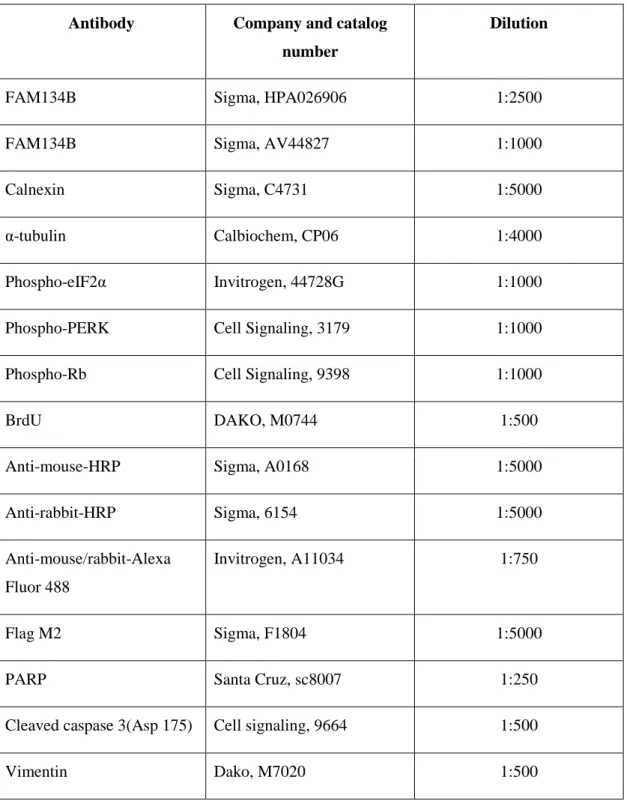
3.1.9 Antibodies
Antibodies used in this study, their catalog numbers, and working dilutions are given below.
24
Antibody Company and catalog number
Dilution
FAM134B Sigma, HPA026906 1:2500
FAM134B Sigma, AV44827 1:1000
Calnexin Sigma, C4731 1:5000
α-tubulin Calbiochem, CP06 1:4000
Phospho-eIF2α Invitrogen, 44728G 1:1000
Phospho-PERK Cell Signaling, 3179 1:1000
Phospho-Rb Cell Signaling, 9398 1:1000
BrdU DAKO, M0744 1:500
Anti-mouse-HRP Sigma, A0168 1:5000
Anti-rabbit-HRP Sigma, 6154 1:5000
Anti-mouse/rabbit-Alexa Fluor 488
Invitrogen, A11034 1:750
Flag M2 Sigma, F1804 1:5000
PARP Santa Cruz, sc8007 1:250
Cleaved caspase 3(Asp 175) Cell signaling, 9664 1:500
Vimentin Dako, M7020 1:500
25
3.2 SOLUTIONS AND MEDIA
3.2.1 General solutions
50X Tris Acetate EDTA (TAE) 242 g Tris base, 57.1 ml glacial acetic acid,
18.6 EDTA were dissolved in 1 liter ddH20 Working dilution is 1X
10X Phosphate Buffered Saline (PBS) 80 g NaCL, 2 g KCl, 14.4 g Na2HPO4, 2.4 g KH2PO4 in 1 litre ddH2O
Working dilution is 1X
Ethidium bromide 10 mg/ml dissolved in ddH2O (stock)
Working concentration is 30 µg/ml
3.2.2 Bacteria solutions
Luria-Bertani medium (LB medium) 10g bacto-tryptone, 5 g bacto-yeast extract and 10g NaCl for 1 litre. Additionally, 15 g/L bacto agar for LB agar plates
Glycerol stock solution Final concentration of glycerol is 25% in LB
Ampicillin 100 mg/ml stock solution in ddH2O
Working solution is 100 µg/ml
3.2.3 Tissue culture solutions
DMEM/RPMI media Complete medium contains 10% Fetal
bovine serum, 1% penicillin/streptomycin, 1% non-essential amino acids, stored at 4 ̊C