https://dergipark.org.tr/tr/pub/bursauludagziraat http://www.uludag.edu.tr/ziraatdergi
Aralık/2020, 34(2), s. 303-315
ARAŞTIRMA MAKALESİ RESEARCH ARTICLE
Geliş Tarihi (Received): 26.02.2020 Kabul Tarihi (Accepted): 26.08.2020
The Evulation of Stress Related Gene Expression Level and Relationship to
Cellular H
2O
2in Chickpea (Cicer arietinum L.) Under Copper Stress
A
Musa KAR
1*Nuriye ÖZTÜRK
2Abstract: The aim of this study is to determine the changes in expression levels of stress genes in chickpea (Cicer arietinum) plant leaves exposed to copper (Cu) at different times and concentrations. Also; the relationship between the changes in gene expression and cellular H2O2 was investigated. In this context, the
amount of malondialdehyde (MDA) and reactive oxygen species (ROS) hydrogen peroxide (H2O2) levels were
determined. Furthermore, the changes in gene expressions of Metallothionein (MT), Catalase (CAT) and superoxide dismutase (Cu / Zn-SOD) enzymes were determined based on the actin expression level that selected as a housekeeping gene. It was determined that MDA content increased significantly due to time and concentration, In all duration and concentrations, the expression of stress-related genes significantly differed from the control group. Hleowever, a decrease has been determined by all gene expressions after the highest expression. This phenomenon is associated with cellular H2O2, which shows a steady increase in stress. At the
end of the study, it was concluded that the elevating duration and concentration of Cu induced oxidative stress and caused the expression of stress-related genes. Furthermore, cellular H2O2 might be acting as a signal
molecule that, up-regulate gene expressions until a certain concentration and down-regulate until a certain concentration. Thanks to the results of this study; Plants in agricultural areas can be exposed to a certain concentration of H2O2 to provide an earlier response to oxidative stress against biotic or abiotic stresses. In this
way, the use of chemical pesticides can be minimized due to obtaining more durable products.
Keywords: Cicer arietinum, Cu, H2O2, Oxidative signaling, Stress-related genes.
A Bu çalışma yüksek lisans tezinden üretilmiştir.
*
Sorumlu yazar/Corresponding Author: 1 Musa KAR Nevşehir Hacı Bektaş Veli Üniversitesi Fen-Edebiyat Fakültesi,
Moleküler Biyoloji ve Genetik Bölümü, Nevşehir, Türkiye, [email protected], OrcID 0000-0001-7983-4814 2 Nuriye ÖZTÜRK Nevşehir Hacı Bektaş Veli Üniversitesi, Fen Bilimleri Enstitüsü, Biyoloji Bölümü, Nevşehir, Türkiye, [email protected], OrcID 0000-0003-4857-013
Atıf/Citation: Kar, M. ve Öztürk, N. 2020. Determination of the Expression Level of Stress-Related Genes in chickpea
(Cicer arietinum) under Short-Term Heavy Metal Stress and the Relationship to Cellular H2O2 concentrations. Bursa
Bakır Stresi altında Nohut Bitkisinde (Cicer arietinum L.) Stresle İlişkili Gen
Ekspresyon Seviyelerinde Meydana Gelen Değişimlerin Belirlenmesi ve
Hücresel H
2O
2 ile İlişkisiÖz: Bu çalışmanın amacı, nohut (Cicer arietinum) bitkisinde farklı zaman ve konsantrasyonlarda bakıra maruz kalmış bitki yapraklarındaki stres genlerinin ekspresyon seviyelerindeki değişimlerin belirlenmesidir. Ayrıca; gen ekspresyonlarındaki değişim ile hücresel H2O2arasındaki ilişki tespit edilmeye çalışılmıştır. Bu bağlamda
hücrede oksidatif stresin göstergesi olan melondialdehit (MDA) miktarı ve reaktif oksijen türlerinden (ROS) H2O2 miktarları tespit edilmiştir. Ayrıca antioksidan savunma elemanlarından Metallothionein (MT), Catalase
(CAT) ve superoksit dismutaz (Cu/Zn-SOD) enzimlerinin gen ekspresyonlarındaki değişim house-keeping gen olarak seçilmiş aktinin ekspresyon düzeyi baz alınarak tespit edilmiştir. Çalışma sonucunda MDA içeriğinin süre ve konsantrasyona bağlı olarak belirgin bir şekilde arttığı, stres alakalı gen ekpresyonlarının bütün konsantrasyonlarda kontrolden yüksek düzeyde eksprese edildiği ancak en yüksek ekspresyon gerçekleştikten sonra süre ve konsantrasyondan arttıkca ekpsreyon düzeylerinde bir azalma olduğu belirlenmiştir. Bu durum stres anındaki kararlı bir artış gösteren hücresel H2O2 ile ilişkilendirilmiştir. Çalışma sonucunda Cu
maruziyetinin oskidatif strese neden olarak stres alakalı genlerin ekpresyonlarını indüklediği tespit edilmiştir. Ayırca hücresel H2O2’nin belirli konsantrasyona kadar gen ekpresyonunu up-regule ederken belirli
konsantrasyondan sonra down regule etmiş olduğu düşünülmektedir. bu calısmanın sonucları sayesinde; tarım alanlarındaki bitkiler belirli konsantrasyonda H2O2 ye maruz bırakılarak biyotik ve ya abiyotik streslere karsı
oksidadatif stres cevabını daha erken evrede vermesi sağlanabilir. Bu sayede, daha dayanıklı ürünler elde edilmesine bağlı olarak kimyasal zirai ilac kullanımı asgari düzeye indirilebilir.
Anahtar Kelimeler: Cicer aretinum, Cu, H2O2, Oksidatif Sinyazlizasyon, Stres alakalı genler.
Introduction
The rapid increase in the world population, industrial development, and global warming have caused a dramatic decrease in the usable agricultural lands in recent years. It is estimated that abiotic stress conditions are one of the most important causes of future arable land loss because 19% of world soil is hyper arid and 56% is at risk of desertification (FAO, 2004)
For these reasons, it is necessary to increase the production of high-quality foods while decrease costs and losses are reduced. In this regard; the physiological responses of the plants to the biotic and abiotic stress is the most common research topic among the scientist. Due to their sessile life plants are exposed to various abiotic stresses such as drought, temperature, salinity, and heavy metal exposure (Sewelam et al., 2016). Heavy metal exposure is one of the most common stress factors, especially in farmlands. Heavy metal exposure can cause quite different cellular effects in plants. In general, the deterioration of ion balance in plants, cell membrane permeability and DNA damage because of very destructive effects. (Leblebici and Aksoy, 2011).
Cu is an essential element that plays an important role in many metabolic events required for growth and development in all plants. It has a vital role in many physiological processes such as photosynthesis, respiration, reactions to oxidant stress, cell wall metabolism and hormone perception (Hernández-Hernández et al., 2018). On the other hand, excess Cu concentrations have several side effects. These are an imbalance in photosynthesis and pigment synthesis causes oxidative stress and other metabolic disorders. (Yruela, 2005).
Abiotic conditions lead to excessive production of reactive oxygen species that are highly toxic to plants and cause cellular damage. Cellular ROS reaches excessive concentrations, it can also cause significant damage by reacting with lipids, proteins, DNA, and several cellular components. This may result in a structural change and/or inhibition of the function. Because of these toxic effects of ROS, it is very important to keep its production and toxins under strict control. Plants have a very complex antioxidant system to avoid potential damages caused by ROS to cellular compartments. The main enzymatic components of the defense system are superoxide dismutase (SOD), catalase (CAT), ascorbate peroxidase (APX), glutathione reductase (GR) (Ahmad, 2014; Sahin et al., 2017).
Plants can react to biotic and abiotic difficulties with rapid changes in stress-related gene expression and protein synthesis. The determination of roles of functional genes or proteins against metal toxicity is very important in explaining the molecular mechanisms of metal tolerance. In recent years, scientists have suggested that H2O2 acts as a protective or signaling factor, except oxidative damage (Mittler et al., 2004). Despite our
knowledge of the mechanisms of detoxification of antioxidant enzymes in metal exposure, we have inadequate information about signal transduction for the regulation and activation of these enzymes. Because H2O2
production is an immediate response to increased Cu-exposure, it has been emphasized by many researchers that it can be a key molecule that can trigger signal transduction events in plants after exposure to the metal (Farnese et al., 2016; Gupta et al., 2016). Vanderauwera et al. emphasized that reactive oxygen species are not as dangerous as they are considered and that H2O2 signaling effect is required in certain concentrations in order to
activate the antioxidant defense system (Vanderauwera et al., 2009)
In this study, MDA and H2O2 concentrations were determined in leaf cells of chickpea plants exposed to Cu
heavy metal at different times and concentrations. In addition, changes in mRNA transcription levels of stress-related genes were investigated and their relations with reactive oxygen species, H2O2, were explained.
Material and Method
Plant Material and Growing Conditions
Cicer arrinetium seeds were purchased from Nevşehir Directorate of Provincial Agriculture and Forestry one commercal genotype (Kocbası) used in this study. The seeds were subjected to germination in 250C of moist
perlite until the roots and the first leaves sprouted. The plants were taken to beakers with Hoagland solution to elongate their roots. After the root length was 10-15 cm (14 days), CuSO4 was added to the beakers to be 50, 100 and 200 µM, respectively. They were exposed to Cu for 1, 3 and 5 days. The light / dark photoperiod is set to 16: 8 in the growth chamber. All applications are made as three replicates.
MDA Determination
The 500 mg leaf sample was homogenized with 3 mL of a solution containing 20% TCA and 0.5% TBA ((w/v). For lipid peroxidation, the homogenate was allowed to stand for 30 minutes at 95 ° C, then 10 minutes ice was used to stop the reaction. The samples were centrifuged at 12,000 g for 15 minutes. and the absorbance of the supernatant was recorded at 532 and 600 nm. The amount of malondialdehyde (MDA) (extinction coefficient of 155 mM−1 cm−1) was calculated by subtracting the non-specific absorbance at 600 nm from the absorbance at 532 nm.
H2O2 determination
Root tissues (500 mg) were homogenized at + 4 ° C with 5 ml trichloracetic acid (TCA) in 0.1% (w / v) and centrifuged at 12,000 rpm for 15 minutes. Afterward, 0.5 ml of the supernatant, 0.5 ml of 10 mM potassium phosphate buffer (pH 7.0) and 1 M potassium iodide (1 ml) was added to a separate tube. The solution incubated at room temperature. Absorbance was measured 280 nm and H2O2 concentration in the cell was determined
according to the standard curve calculated using gradual H2O2 concentrations (Junglee et al., 2014).
RNA isolation and cDNA synthesis
Thermo Gene JET Plant RNA Purification Kit used for total RNA isolation. Yielded RNA quality was determined by measuring the absorbance values in 260/280 and 230/260 nanometers by using Donovix micro-volume spectrophotometer and used in cDNA synthesis. For the first-strand cDNA synthesis, Thermo RevertAid First Strand cDNA Synthesis Kit was used.
The quantitative real-time PCR analysis
For quantitative real-time analysis accuPower®, DualStar ™ qPCR PreMix was used. For the preparation of this premix, 1 µM forward primer, 1 µM reverse primer, 5 µM cDNA and 13 µM DEPC-water were mixed and the final volume was adjusted to 20 µM. Actin gene was selected as a house-keeping gene. The primer sequences are given in Table 1.
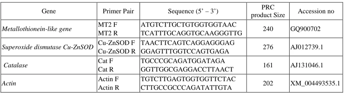
Table 1. Primer sequences for PCR amplification and product sizes
Gene Primer Pair Sequence (5’ – 3’) PRC
product Size Accession no
Metallothionein-like gene MT2 F
MT2 R
ATGTCTTGCTGTGGTGGTAAC
TCATTTGCAGGTGCAAGGGTTG 240 GQ900702
Superoxide dismutase Cu-ZnSOD Cu-ZnSOD F
Cu-ZnSOD R TAACTTCAGTCAGGAGGGAG GGAGTTTGGTCCAGTGAGA 276 AJ012739.1 Catalase Cat F Cat R TGCCCGCAGATGGATAGA GGTTGGCGAGGACCTTAACT 161 AJ131046.1 Actin Actin F Actin R TGTCTTGAGTGGTGGTTCTAC CTTGCCGCCCAGATATTGTA 202 XM_004493535.1
Statistical Analysis
2∆ct calculation method by Livak and Schmittgen (2001) was used to determining changes in stress-related gene expression levels.
Analysis of variance (ANOVA) was performed to confirm significant differences among treatments. Post-hoc Duncan’s test used to determine differences among groups (p<0.05). All statistical analyses were performed with the SPSS 22.0 software package.
Result and Discussion
The amount of MDA was calculated to determine the damage caused by the cell membrane and to determine the oxidative stress status of the cell.
MDA concentration increased as Cu duration and concentration increased. Statistically the highest MDA concentration on day 5, 200-µM concentration. (Fig 1 A) (P<0.05).
Fig.1. The differential of MDA content (A) and cellular H2O2 concentration (B) in Cu exposed Chickpea leaves.
Different letters show the significance between groups (p<0.05). The bar shows standard errors. (x axis: Different Cu concentrations)
The change in the amount of cellular H2O2 gives an idea about the oxidative state of the cell. Cellular H2O2
concentration determined in chickpea leaves exposed to Cu is showing a significant increase depending on duration and concentration. Statistically, the highest H2O2 concentration was found in the 5th day at a
concentration of 200 µM (Fig. 2-B) According to the post hoc analysis, it was determined that all groups were different from each other (p <0.05).
Relative changes of stress-related antioxidant genes expression levels were calculated based on changes of actin gene expression which selected as a house-keeping gene.
0 0,2 0,4 0,6 0,8 1 1,2 1,4 1,6 1,8 2 0 50 100 200 M DA C O N CE N TR AT ıO N
1.Day 3.day 5.day
ab
a a a
bc
c
b
ab
b
bc
b
c
A
0 2 4 6 8 10 12 14 16 18 20 0 50 100 200 H2O 2 CO N CN ET RA Tı O N1.day 3.day 5.day
a a
b
c
c
d
ef
f
ef
f
g
B
a
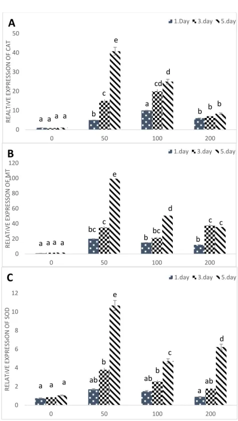
When the CAT expression was examined, the highest exposure level was observed on day 5 at a concentration of 50 µM. In prolonged duration and concentrations, CAT enzyme expression was statistically different from the control group. However, a statistically significant decrease was found with respect to the concentration at which the highest expression was observed. (Fig 3-A) (p <0.05).
In the 50 µM concentration 5th day application, MT gene expression was found to be approximately 100-fold higher than actin gene expression. Afterward, it was found that there were a decrease in expression levels at 100 and 200 µM concentrations according to the concentration level of 50 µM. In addition, no statistically significant difference was found between Day 3 and Day 5 200 µM concentrations. (Fig 3-B) (p<0.05).
When Cu/Zn- SOD gene expression is examined; similar to the other stress-related gene expressions, the highest expression level was found at 5th day 50 µM concentration. On the other hand; the expression levels are reduced moreover, there was no statistically significant difference between the expression level at 1st day 200 µM and the control group (Fig. 2-C) (p<0.05).
In this study, the product of lipid peroxidation and an indicator of oxidative stress MDA concentration and a by-product of oxidative stress ROS/H2O2 concentrations investigated. In addition, changes in expression levels of
MT, Cu-Zn SOD and CAT antioxidant stress-related genes were determined based on housekeeping gene actin expression. The linkage between cellular H2O2 concentration and changes in the levels of expression was
explained.
Free radicals that cause oxidative stress can react with all the cellular structures, but the most sensitive component to these interactions are lipids. Malondialdehyde (MDA) is a highly reactive metabolic product that occurs as a result of a series of reactions during lipid peroxidation, which is caused by the effects of free oxygen radicals on tissues (Gaweł et al., 2004). In this study, a steady increase in all concentrations was observed depending on the Cu exposure. In previous similar studies, they emphasized that the primary indicator of stress caused by heavy metal exposure is lipid peroxidation and MDA content (Duman et al., 2010).
MTs are low molecular mass proteins that are thought to play a key role in response to metal toxicity and oxidative stress through Cys thiol groups. (Gu et al., 2014; Guo et al., 2008; Mir et al., 2004). Kumar et al. reported that; MT gene expression is elevated as a result of the different concentrations of Cu exposure to Neurospora crassada (Kumar et al., 2005). In the study of Tamas et al., where they investigated the effect of Cd on barley plant roots, it was found that as Cd concentration increases in plant roots MT expression level also increased (Tamás et al., 2008). Jain et al., in the study they conducted with sugar cane plants exposed to selenium (Se), found that the MT gene was expressed at a higher level than all concentrations. However, they found that MT expression level increased up to a certain Se concentration and decreased after a certain Se concentration (Jain et al., 2015). In the study conducted by Tombuloğlu et al. on the effect of boron element on the tomato plant, they found that MT gene expression level increased up to a certain concentration in root and shoot parts of the plant and but decreases after a certain concentration(Tombuloglu et al., 2012). In this study we found that; MT gene expression increased all durations and concentrations according to the control group. Liu et al. conducted a study with rice exposed to Cu at a different time and concentration they put out that; there was a decrease in MT expressions as the prolonged duration and concentration. As a result, they stated that; the OsMT expression increase is not related to Cu concentration it is directly regulated by H2O2 (Liu et al., 2015). Our
Fig. 2. Relative expressions of stress-related genes; Catalase (A) Metallothionein (B) and Cu/Zn-SOD (C) in Cu
exposed Cicer arietinum leaves. Different letters show the significance between groups (p<0.05). The bar shows standard errors. ( x axis: Different Cu concentrations)
0 10 20 30 40 50 0 50 100 200 REA LT ıV E EX PR ES Sı ON OF C AT
1.Day 3.day 5.day
a a a a
b
c
a
b b
b
cd
d
e
A
0 20 40 60 80 100 120 0 50 100 200 REL AT ıV E EX PR ES SON OF M T1.day 3.day 5.day
a a a a
bc
c
b
bc
b
c c
d
e
B
0 2 4 6 8 10 12 0 50 100 200 REL AT ıV E EX PR ES Sı ON OF S OD1.day 3.day 5.day
a a a
ab
ab
a
b
ab
b
c
d
e
C
The most important function of the catalase is to detoxify H2O2 as H2O and O2. Rosattao et al. reported that
CAT expression levels are increased under stress factors (Rossatto et al., 2017). Kim et al. stated that; as a result of salt stress in rice CAT gene expression induced positively (Kim et al., 2007). In our study in parallel with these studies, there was a significant increase in CAT expression levels as a result of stress. It is notable that; a decrease of CAT gene expression, after the highest expressions observed, is not distinct as other studied gene expressions. Rossatto et al. emphasized that; the signaling molecule effect of H2O2 on CAT expressions is lower
than that of other antioxidant genes. In their studies, similar results reported from Kar and Sougir et al. (Kar, 2018; Souguir et al., 2013). Our findings are in parallel with the literature.
In addition, in the study where Rossatto et al. applied salt stress to rice plants, CAT expression was found to be higher at all application periods compared to control and it was stated that the expression level increased as the application period was prolonged (Rossatto et al., 2017). In the study of Souguir et al., in the rice seedlings to which Cd was applied, it was found that CAT expression level increased depending on the increasing concentration and the prolonged period (Souguir et al., 2013). Luna et al. concluded that CAT regulation serves to limit excessive H2O2 accumulation while allowing essential signaling functions to occur (Luna et al., 2005).
Contrary to these findings, in our study, CAT expression did not depend on the elevation of H2O2 concentration
and continued to express in chickpea roots.
SOD plays a decisive role in protecting against the toxic effects of oxidative stress by scavenging superoxide radicals and ensuring their conversion to O2 and H2O2 (Verma and Dubey, 2003). Many studies have reported
that ROS associated with oxidative stress under drought and salt stress in plants induces SOD enzyme activity (Baloǧlu et al., 2012; Ishibashi et al., 2011). Soydam et al. determined that; tomato plants subjected to salt and drought stress and there was an increase of SOD enzyme expressions. (Aydin et al., 2014). Similarly, in this study, an increase in SOD enzyme expression is observed in response to oxidative stress triggered by Cu exposure.
Also; Rossatto et al. found that SOD expression increased in rice plants up to 15-days exposure but decreased after 20-day salt stress exposure (Rossatto et al., 2017). In the current study, also found a significant decrease in SOD expression due to increased time and concentration. Dai et al. applied different concentrations of salt stress to the canola plant. As a result of the study, they found that the SOD expression level increased up to 200 mmol L-1 concentration and that it decreased in 250 and 300 mmol L-1 concentrations (Dai et al., 2009). Rossatto et al., who worked with rice plants under salt stress found that SOD expression increased compared to control up until 15 days of exposure but decreased after 20 days of exposure (Rossatto et al., 2017). In our study, which is similar to the studies in the literature, SOD expression level increased up to 50 µM Cd application and reached the highest level in 12 hours 50 µM Cd application, but as the concentration amount increased, SOD expression level decreased in all application periods. Although the expression levels detected as a result of prolonged exposure were higher than the control levels, there was no statistical difference between the expression levels. This shows that there is a gradual increase in expression with the introduction of the defense mechanism in the early times when the plant encounters stress and a decrease in expression when the plant is unable to tolerate stress.
In recent years; scientists have been approached a different view of cellular H2O2 function and emphasized
that; reactive oxygen species are less harmful than formerly known. (Farnese et al., 2016; Gupta et al., 2016; Del Río, 2015). Sharma et al. reported that; H2O2 and other ROS species can serve as a signal molecule because it is
useful in low concentrations and toxic when excess a certain concentration (Sharma et al., 2012). Rosatta et al. emphasized that; after salt stress in rice plant H2O2 concentration was low and do not cause any damage to cells,
on the contrary, they reported that low H2O2 expression of stress-related genes. (Rossatto et al., 2017). Gill and
Tuteja have stated that because of the long life of H2O2 and the permeability of the membranes, ROS is
considered as a secondary messenger and acts as a key regulator in a wide range of physiological processes. (Gill and Tuteja, 2010). Choudhury et al. stated that ROS affects the expression of some genes and that ROS acts as a biological signal in the regulation of stresses. They emphasized that O2
•
and H2O2 considered as the primary
ROS in plants serves as secondary messengers regulating various functions in the growth and development of the plant (Choudhury et al., 2017). Apel and Hirt maintained that activate signal transmission could affect gene expression in 3 different ways by (1) activating the ROS sensors, (2) oxidizing the signal path components directly by ROS and (3) by changing the activity of transcription factors of ROS (Apel and Hirt, 2004). In the study they conducted, Gill and Tuteja maintained that since H2O2 is long-lived and the permeability between
membranes is high, regarding the signals produced through ROS, ROS was started to be accepted as a secondary precursor. They also stated that ROS acts as a key regulator in a wide range of physiological processes (Gill and Tuteja, 2010). Laloi et al. emphasized that the increases in H2O2 concentration were usually based on different
mechanisms and that H2O2 served as a signal (Laloi et al., 2004). Kar, exposed chickpea the plant roots to Cd
and reported that; antioxidant gene expression is associated with H2O2 concentration. In a certain level of H2O2
concentration stress-related gene expressions up-regulated and a significant decrease observed in gene expressions after a certain H2O2 concentration. (Kar, 2018). In addition, Del-rio emphasized the cellular
importance of H2O2 to secondary messenger tasks and emphasized that it is appropriate to use the term
“oxidative signaling” for H2O2 activities in the cell. (Del Río, 2015). Our findings support these findings.
Conclusions
As a result of the study, Cu exposure to chickpea at different duration and concentrations increases cellular MDA content and H2O2 accumulation. In order to prevent oxidative damage of Cu exposure, stress-related genes
were highly expressed than the control group at all application concentrations. Thanks to the results of this study; Plants in agricultural areas can be exposed to a certain concentration of H2O2 to provide an earlier response to
oxidative stress against biotic or abiotic stresses. In this way, the use of chemical pesticides can be minimized due to obtaining more durable products.
Additionally; expressions of stress-related genes were highest when H2O2 concentration was at a certain level but decreased expression levels due to increased H2O2 concentration. The findings may contribute further oxidative signaling studies.
Acknowledgment
All authors participated in the conception of the topic. M.K and N.Ö conducted %50-%50 laboratory studies. M.K and N.Ö have written %75-%25 of the manuscript. All authors read and approved the final manuscript after critically revising it for important content. The authors declare that there is no conflict of interest regarding the publication of this article.
References
Ahmad, P. 2014. Oxidative Damage to Plants: Antioxidant Networks and Signaling (Elsevier GmbH.).
Apel, K. and Hirt, H. 2004. REACTIVE OXYGEN SPECIES: Metabolism, Oxidative Stress, and Signal Transduction. Annual Review of Plant Biology 55(1): 373–399.
Aydin, S. Büyük, I. and Aras, E.S. 2014. Expression of SOD gene and evaluating its role in stress tolerance in NaCl and PEG stressed Lycopersicum esculentum. Turkish Journal of Botany 38(1): 89–98.
Baloǧlu, M.C. Kavas, M. Aydin, G. Öktem, H.A. and Yücel, A.M. 2012. Antioxidative and physiological responses of two sunflower (Helianthus annuus) cultivrs under PEG-mediated drought stress. Turkish Journal of Botany 36707–714.
Choudhury, F.K. Rivero, R.M. Blumwald, E. and Mittler, R. 2017. Reactive oxygen species, abiotic stress and stress combination. Plant Journal 90(5): 856–867.
Dai, Q. lin Chen, C. Feng, B. Liu, T. ting Tian, X. Gong, Y. ya Sun, Y. kun Wang, J. and Du, S. zhang 2009. Effects of different NaCL concentration on the antioxidant enzymes in oiLseed rape (Brassica napus L.) seedlings. Plant Growth Regulation 59273–278.
Duman, F. Urey, E. Temizgul, R. and Bozok, F. 2010. Biological responses of a non-target aquatic plant (Nasturtium officinale) to the herbicide, tribenuron-methyl. Weed Biology and Management 10(2): 81–90. FAO 2004. FAO. 2004. Data sets, indicators, and methods to assess land degradation in drylands. World Soil
Resources Report No. 100. Rome.
Farnese, F.S. Menezes-Silva, P.E. Gusman, G.S. and Oliveira, J.A. 2016. When Bad Guys Become Good Ones: The Key Role of Reactive Oxygen Species and Nitric Oxide in the Plant Responses to Abiotic Stress. Frontiers in Plant Science 7(April): 471.
Gaweł, S. Wardas, M. Niedworok, E. and Wardas, P. 2004. Malondialdehyde (MDA) as a lipid peroxidation marker. Wiadomości Lekarskie (Warsaw, Poland : 1960) 57(9–10): 453–455.
Gill, S.S. and Tuteja, N. 2010. Reactive oxygen species and antioxidant machinery in abiotic stress tolerance in crop plants. Plant Physiology and Biochemistry 48(12): 909–930.
Gu, C.S. Liu, L. qin Zhao, Y.H. Deng, Y. ming Zhu, X. dong and Huang, S.Z. 2014. Overexpression of Iris. lactea var. chinensis metallothionein llMT2a enhances cadmium tolerance in Arabidopsis thaliana. Ecotoxicology and Environmental Safety 10522–28.
Guo, W.-J. Meetam, M. and Goldsbrough, P.B. 2008. Examining the Specific Contributions of Individual Arabidopsis Metallothioneins to Copper Distribution and Metal Tolerance. Plant Physiology 146(4): 1697– 1706.
Gupta, K. Sengupta, A. Chakraborty, M. and Gupta, B. 2016. Hydrogen Peroxide and Polyamines Act as Double Edged Swords in Plant Abiotic Stress Responses. Frontiers in Plant Science 7(September): 1343. Hernández-Hernández, H. Juárez-Maldonado, A. Benavides-Mendoza, A. Ortega-Ortiz, H. Cadenas-Pliego, G.
Sánchez-Aspeytia, D. and González-Morales, S. 2018. Chitosan-PVA and Copper Nanoparticles Improve Growth and Overexpress the SOD and JA Genes in Tomato Plants under Salt Stress. Agronomy 8(9): 175. Ishibashi, Y. Yamaguchi, H. Yuasa, T. Iwaya-Inoue, M. Arima, S. and Zheng, S.H. 2011. Hydrogen peroxide
spraying alleviates drought stress in soybean plants. Journal of Plant Physiology 168(13): 1562–1567. Jain, R. Verma, R. Singh, A. Chandra, A. and Solomon, S. 2015. Influence of selenium on metallothionein gene
expression and physiological characteristics of sugarcane plants. Plant Growth Regulation.
Junglee, S. Urban, L. Sallanon, H. and Lopez-lauri, F. 2014. Optimized Assay for Hydrogen Peroxide Determination in Plant Tissue Using Potassium Iodide. American Journal of Analytical Chemistry 5(August): 730–736.
Kar, M. 2018. Determination of the expression level of stress-related genes in Cicer arietinum root cell under Cd stress and the relationship to H2O2 concentrations. Ecotoxicology 27(8): 1087–1094.
Kim, D.-W. Shibato, J. Agrawal, G.K. Fujihara, S. Iwahashi, H. Kim, D.H. Shim, I.-S. and Rakwal, R. 2007. Gene transcription in the leaves of rice undergoing salt-induced morphological changes (Oryza sativa L.). Molecules and Cells 24(1): 45–59.
Kumar, K.S. Dayananda, S. and Subramanyam, C. 2005. Copper alone, but not oxidative stress, induces copper-metallothionein gene in Neurospora crassa. FEMS Microbiology Letters 242(1): 45–50.
Laloi, C. Apel, K. and Danon, A. 2004. Reactive oxygen signalling: The latest news. Current Opinion in Plant Biology.
Leblebici, Z. and Aksoy, A. 2011. Growth and Lead Accumulation Capacity of Lemna minor and Spirodela polyrhiza (Lemnaceae): Interactions with Nutrient Enrichment. Water, Air, & Soil Pollution 214(1–4): 175– 184.
Liu, J. Shi, X. Qian, M. Zheng, L. Lian, C. Xia, Y. and Shen, Z. 2015. Copper-induced hydrogen peroxide upregulation of a metallothionein gene, OsMT2c, from Oryza sativa L. confers copper tolerance in Arabidopsis thaliana. Journal of Hazardous Materials 29499–108.
Livak, K.J. and Schmittgen, T.D. 2001. Analysis of relative gene expression data using real-time quantitative PCR and the 2(-Delta Delta C(T)) Method. Methods (San Diego, Calif.) 25(4): 402–408.
Luna, C.M. Pastori, G.M. Driscoll, S. Groten, K. Bernard, S. and Foyer, C.H. 2005. Drought controls on H2O2 accumulation, catalase (CAT) activity and CAT gene expression in wheat. Journal of Experimental Botany 56(411): 417–423.
Mir, G. Domènech, J. Huguet, G. Guo, W.J. Goldsbrough, P. Atrian, S. and Molinas, M. 2004. A plant type 2 metallothionein (MT) from cork tissue responds to oxidative stress. Journal of Experimental Botany 55(408): 2483–2493.
Del Río, L.A. 2015. ROS and RNS in plant physiology: An overview. Journal of Experimental Botany 66(10): 2827–2837.
Rossatto, T. do Amaral, M.N. Benitez, L.C. Vighi, I.L. Braga, E.J.B. de Magalhaes Junior, A.M. Maia, M.A.C. and da Silva Pinto, L. 2017. Gene expression and activity of antioxidant enzymes in rice plants, cv. BRS AG, under saline stress. Physiology and Molecular Biology of Plants : An International Journal of Functional Plant Biology 23(4): 865–875.
Sahin, O. Taskin, M.B. Kaya, E.C. and Taskin, H. 2017. Poultry Manure Biochar Reduces Arsenic Induced Oxidative Stress and Arsenic Levels in Rice Plants Tavuk Gübresi Biyokömürünün Çeltik Bitkisi Arsenik Al ı m ı ve Arsenik Düzeyleri Üzerine Etkisi ve Oksidatif Stres İ le İ li ş kisi. Bursa Uludag Üniv. Ziraat Fak. Derg. 113103–113.
Sewelam, N. Kazan, K. and Schenk, P.M. 2016. Global Plant Stress Signaling: Reactive Oxygen Species at the Cross-Road. Frontiers in Plant Science 7(February): 187.
Sharma, P. Jha, A.B. Dubey, R.S. and Pessarakli, M. 2012. Reactive Oxygen Species, Oxidative Damage, and Antioxidative Defense Mechanism in Plants under Stressful Conditions. Journal of Botany 2012(1): 1,26. Souguir, D. El Ferjani, E. Ledoigt, G. and Goupil, P. 2013. Transcript accumulation of stress-related genes in
Vicia faba roots under a short exposure to cadmium. Plant Biosystems - An International Journal Dealing with All Aspects of Plant Biology 3504(August): 1–9.
Tamás, L. Dudíková, J. Ďurčeková, K. Halušková, L. Huttová, J. Mistrík, I. and Ollé, M. 2008. Alterations of the gene expression, lipid peroxidation, proline and thiol content along the barley root exposed to cadmium. Journal of Plant Physiology 165(11): 1193–1203.
Tombuloglu, H. Semizoglu, N. Sakcali, S. and Kekec, G. 2012. Boron induced expression of some stress-related genes in tomato. Chemosphere 86(5): 433–438.
Verma, S. and Dubey, R.S. 2003. Lead toxicity induces lipid peroxidation and alters the activities of antioxidant enzymes in growing rice plants. Plant Science 164(4): 645–655.