MJCCA9 – 782 ISSN 1857-5552 e-ISSN 1857-5625
Received: March 1, 2018 DOI: 10.20450/mjcce.2019.1403
Accepted: November 26, 2019 Original scientific paper
A DETAILED STUDY ON THE OPTICAL PROPERTIES OF 3-BENZOYL-7-HYDROXY
COUMARIN COMPOUND IN DIFFERENT SOLVENTS AND CONCENTRATIONS
Adnan Kurt1,*, Bayram Gündüz2, Murat Koca3
1Department of Chemistry, Faculty of Science and Arts, Adiyaman University, Adiyaman, Turkey 2Department of Engineering Basic Sciences, Faculty of Engineering and Natural Sciences, Malatya Turgut
Özal University, Malatya, Turkey
3Department of Pharm. Chemistry, Pharmacy Faculty, Adiyaman University, Adiyaman, Turkey [email protected]
A coumarin-derived compound, 3-benzoyl-7-hydroxy coumarin (BHYC), is synthesized to deter-mine its optoelectronic properties, including absorbance band edge, optical band gap, transmittance, re-fractive index, electrical susceptibility, volume-surface energy loss functions and optical/electrical con-ductance parameters. The absorbance spectra of BHYC in dimethylformamide (DMF) and dimethyl-sulfoxide (DMSO) solvents exhibit maximum peaks at 350 and 353 nm, respectively, in the near-ultraviolet region. The absorbance band edge values of BHYC in DMF and DMSO are 2.526 and 2.500 eV, respectively. The optical band gap of BHYC varies from 2.560 to 2.408 eV with increasing molarity. In contrast, the refractive index of BHYC increases from 2.47 to 2.95 with changing molarity. The ob-tained results show that 3-benzoyl-7-hydroxy coumarin exhibits a semiconductor behavior and it may be an important candidate for many optoelectronic devices, such as diodes, photodiodes and sensors. Keywords: coumarin derivative; synthesis and characterization; optoelectronic parameters; dispersion parameters; solvent and concentration effects
ЕДНО ПОДРОБНО ИСПИТУВАЊЕ НА ОПТИЧКИТЕ СВОЈСТВА НА 3-БЕНЗОИЛ-7-ХИДРОКСИ КУМАРИН ВО РАЗЛИЧНИ РАСТВОРУВАЧИ И КОНЦЕНТРАЦИИ Извршена е синтеза на дериват на кумарин, 3-бензоил-7-хидрокси кумарин (BHYC), со цел да се определат оптичкоелектронските совјства, вклучувајќи ги границата на апсорпционата лента, оптичкиот процеп на лентата, трансмитанцата, рефрактивниот индекс, електричната чувствителност, функциите на губењето на енергијата на волуменска површина и оптичките/електричните параметри на кондукција. Апсорпционите спектри на BHYC во диметилформамид (DMF) и во диметилсулфоксид (DMSO) манифестираат максимуми, соодветно, на 350 и 353 nm, во блиската ултравиолетова област. Вредностите на граничните апсорпциски ленти на BHYC во DMF и во DMSO, соодветно, се 2,526 и 2,500 eV. Оптичкиот процеп на лентата варира од 2,560 до 2,408 eV со зголемување на моларноста. Наспроти тоа, рефрактивниот индекс со промена на моларноста расте од 2,47 до 2,95. Добиените резултати покажуваат дека 3-бензоил-7-хидрокси кумарин манифестира полуспроводнички својства и може да биде важен кандидат за оптоелектронски инструменти, како што се диоди, фотодиоди и сензори. Клучни зборови: дериват на кумарин; синтеза и карактеризација; оптоелектронски параметри; параметри на дисперзија; влијание на растворувачи и на концентрација
1. INTRODUCTION
Natural and synthetic coumarins are im-portant classes of oxygenated heterocyclic com-pounds [1]. These heterocyclic molecules of both natural and synthetic origin have attracted consid-erable attention from organic and medicinal chem-ists for many years due to their unique features. The applications of these molecules vary in a wide range depending on their biological, physical and chemical properties. These compounds show sig-nificant biological or pharmacological activities, including antibiotic, antibacterial, antitumor, viral, anticoagulants, antipsoriatic, HIV, anti-inflammatory and enzyme inhibitor properties [1– 6]. In addition, these kinds of molecules have in-tensively π-conjugated bond systems like other aromatic heterocycles. This feature makes them important in terms of their photophysical and spec-troscopical properties [7]. Based on these com-pounds, some applications, such as organic light-emitting diodes, electroluminescence and fluores-cence materials, nonlinear optical materials, pho-toalignment of liquid crystalline molecules, charge-transfer agents, two photon absorption ma-terials, organic-inorganic hybrid materials and la-ser dyes, have been realized in various industrial and technological platforms [8–16].
The π-electron system present in the struc-ture of these molecules also causes them to be ac-tive in the UV-visible absorption range [7]. Some studies have determined the optical properties of coumarins based on their UV sensitivities [17–22]. However, sufficient studies on the optical proper-ties of coumarins, such as absorbance band edge, optical band gap, transmittance, refractive index, electrical susceptibility, volume-surface energy loss functions and optical/electrical conductance parameters, in various solvents and concentrations are not yet available in the literature according our current knowledge. In light of this literature defi-ciency, we synthesize a coumarin-derived com-pound, 3-benzoyl-7-hydroxy coumarin (BHYC), to determine its optoelectronic properties. While there are a few papers on the synthesis of 3-benzoyl-7-hydroxy coumarin, none of them are related to its optical properties. For example, in one study re-ported on this compound, Raju and co-workers synthesized a series of 3,4- and 3,6-disubstituted chromenones (coumarins), including 3-benzoyl-7-hydroxy coumarin, and reported their α-glucosidase inhibitory antihyperglycemic activities [1]. In another study, Secci et al. synthesized
sev-eral coumarin derivatives substituted with carbon-yl, acyl and carboxyhydrazido groups at the 3-position of molecules (including 3-benzoyl-7-hydroxy coumarin), which were tested in vitro for their human monoamine oxidase A and B (hMAO-A and hM(hMAO-AO-B) inhibitory activity [23]. In anoth-er study, Hanoth-eravi and co-workanoth-ers developed a new strategy for the synthesis of 3-acyl coumarins with various functional groups and substitutions, includ-ing also 3-benzoyl-7-hydroxy coumarin usinclud-ing a mesoporous molecular sieve, MCM-41, as a novel and efficient catalyst [24]. However, they only re-ported the synthesis of coumarins and did not in-vestigate any of their properties.
As discussed above, there are a number of studies involving the synthesis, characterization and investigation of various properties of many coumarin derivatives with different functional groups, with the exception of 3-benzoyl-7-hydroxy coumarin. However, the majority of these studies focused on fluorescence emissions or biological activities. Therefore, we aim to synthesize a cou-marin-derived compound, BHYC, with intensive conjugated π-electrons to investigate its optoelec-tronic parameters in various solvents and concen-tration systems. The obtained results show that 3-benzoyl-7-hydroxy coumarin exhibits a semicon-ductor behavior and it may be an important candate for many optoelectronic devices, such as di-odes, photodiodes and sensors.
2. EXPERIMENTAL 2.1. Instrumental techniques
A Bruker 300 MHz Ultrashield TM instru-ment was used to characterize BHYC by nuclear magnetic resonance (1H-NMR) spectroscopy at room temperature using a deuterated DMSO sol-vent and tetramethylsilane (TMS) as an internal standard. The infrared characterization was ob-tained with a Perkin Elmer Spectrum 100 FTIR spectrometer with an ATR accessory. The UV measurements of the BHYC solutions were record-ed with a Shimadzu UV-1800 spectrophotometer.
2.2. Materials
As analytical reagents, 2,4-dihydroxy-benzaldehyde, ethyl benzoylacetate and piperidine were purchased from Sigma-Aldrich. The used solvents, N,N-dimethylformamide (DMF),
dime-thyl sulfoxide (DMSO), acetone, methanol and ethanol were also obtained from Sigma-Aldrich.
2.3. Synthesis of 3-benzoyl-7-hydroxy coumarin
(BHYC)
In general, 3-substituted carbonyl coumarins may be synthesized by the Knoevenagel condensa-tion of salicylaldehydes and β-ketoesters with cata-lytic amounts of piperidine in the presence of vari-ous solvents, such as acetonitrile, ethanol and so on. We herein synthesized the 3-benzoyl-7-hydroxy coumarin compound according to the
pathway reported in previous studies [1, 23]. Brief-ly, BHYC was synthesized as follows: 2,4-dihydroxybenzaldehyde (2.762 g), ethyl ben-zoylacetate (3.844 g), three drops of piperidine and 50 ml of acetone were dissolved in a three-necked reaction balloon. Then, the mixture was refluxed and stirred on a magnetic stirrer for 2 h. After this, the organic raw mixture was precipitated in excess methanol and the rose-colored product (BHYC) was separated and dried. Finally, it was purified by recrystallization twice in ethanol. The synthesis of BHYC is shown in Scheme 1.
Scheme 1. Synthesis of BHYC
2.4. Preparation of BHYC solutions
To determine the effect of concentration on the optical parameters of BHYC, a series of BHYC/DMSO solutions at different concentrations of 10, 15, 58, 186 and 270 µM was prepared. In addition, to determine the solvent effect on the op-tical parameters, the BHYC solutions in DMSO and DMF solvents were kept at 12 µM and the op-tical measurements for both solvent systems were performed. The required amounts of BHYC for all concentrations were obtained with an AND-GR-200 Series analytical balance.
3. RESULTS AND DISCUSSION
The FTIR spectrum of BHYC is illustrated in Figure 1, where the vibration at 3171 cm–1 is characteristic for –OH stretching. The absorption bands at 3062–2930 and 2900–2825 cm–1 were attributed to aromatic and aliphatic C-H stretching, respectively. Stretching for benzoyl and lactone carbonyls was observed at frequencies of 1710and 1682 cm–1, respectively. The absorptions at 1650 and 1609 cm–1 were recorded for the lactone C=C stretching in the coumarin group and the aromatic C=C stretching vibrations, respectively.
Fig. 1. FTIR spectrum of BHYC
The spectral characterization ofBHYC was also performed by 1H-NMR spectroscopy. The 1 H-NMR spectrum of BHYC is shown in Fig. 2. The singlet signal observed at 10.98 ppm is attributed to the O-H proton in the coumarin ring. The signal at 8.35 ppm is attributed to the coumarin lactone =CH- proton. The chemical shifts observed be-tween 7.88 and 6.79 ppm are characteristic for ar-omatic =CH- protons in benzoyl and coumarin rings, respectively. Another two resonances at 3.3 and 2.5 ppm are also reasoned from the DMSO solvent.
Fig. 2. 1H-NMR spectrum of BHYC
3.1. UV spectral characteristics and absorbance
band edges of BHYC solutions
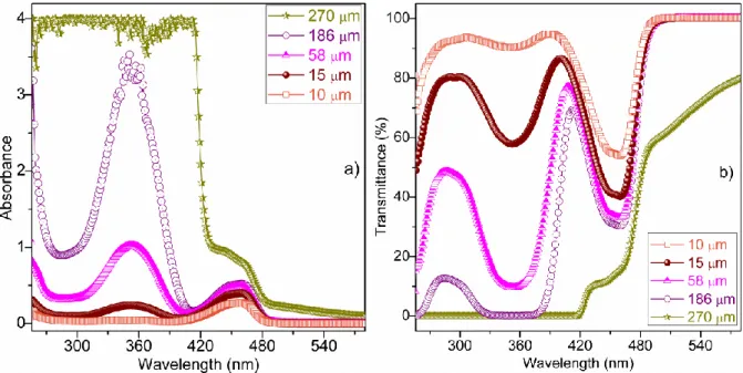
Generally, an absorbance spectrum gives the absorption properties of a material and is signifi-cant to obtain its crucial optical parameters. We obtained the absorbance and transmittance spectra of the BHYC solutions for various concentrations of 10, 15, 58, 186 and 270 µM in DMSO solvent and also for various solvents, such as DMF and DMSO, at a fixed concentration of 12 µM.
Figures 3(a,b) indicate the absorbance and transmittance spectra of the BHYC solutions for 10, 15, 58, 186 and 270 µM molarities, respective-ly. As seen in Figs. 3(a,b), the transmittance values of the BHYC solutions decrease with increasing molarity, while the absorbance values of the BHYC solutions increase with increasing molarity. The absorbance and transmittance spectra of the BHYC are not observed at higher molarities than ~186 µM. The obtained results show that the UV spectra of the BHYC solutions change significantly with molarity.
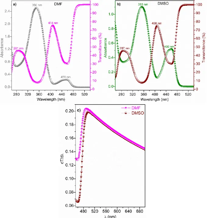
We then investigated the UV spectra of BHYC for the DMF and DMSO solvents. Figures 4(a,b) show the absorbance and transmittance spectra of BHYC for the DMF and DMSO sol-vents, respectively. As seen in Fig. 4a, the absorb-ance spectra of the BHYC for DMF exhibit the maximum peak at 350 nm, which is in the near-ultraviolet (NUV) region and exhibits a small peak at 456 nm, which is in the visible (V) region. In contrast, the transmittance spectra of BHYC for DMF exhibit a medium peak at 281 nm, which is in the NUV region and exhibits a high peak at 414 nm, which is in the V region. As seen in Figure 4b,
the absorbance spectra of BHYC for DMSO exhib-it a maximum peak in the NUV region (at 353 nm) and a medium peak in the V region (at 460 nm). However, the transmittance spectra of BHYC for DMSO exhibit a medium peak in the NUV region (at 287 nm) and a high peak in the V region (at 408 nm). These results suggest that the curves of the UV spectra for DMF and DMSO are similar, whereas their peak positions, i.e., wavelengths, are different from each other. The DMF and DMSO solvents have dramatic effects on the UV spectra of BHYC.
Fig. 4. Absorbance and transmittance spectra of BHYC for a) DMF and b) DMSO and c) their dT/dλ curves vs. wavelength (λ)
The absorbance band edge (EAbs-be) gives use-ful information regarding the absorption properties and estimates the optical band structure of a material. For this, we have obtained the EAbs-be values of BHYC for the DMF and DMSO solvents using the maximum peak values (491 and 496 nm, respective-ly) of Figure 4c, which gives the dT/dλ curves vs. wavelength (λ). The (EAbs-be) values of BHYC for DMF and DMSO are found to be 2.526 and 2.500 eV, respectively. These results show that the absorb-ance band edge of BHYC for DMF is higher than the absorbance band edge of BHYC for DMSO.
3.2. Optical band gaps and refractive indices
of BHYC solutions
The optical band gap (Eg) and refractive in-dex (n) play a role in investigating the optoelec-tronic properties of the optoelecoptoelec-tronic materials and devices. The Eg values of the BHYC for 10,
15, 58, 186 and 270 µM, and for DMF and DMSO, are obtained from the (𝛼𝐸)2 curves vs. photon
en-ergy (E) [25] (α is absorption coefficient) using the Tauc model [26], as seen in Figures 5(a,b) for dif-ferent molarities and solvents, respectively.
The optical band gap values of BHYC are calculated to be 2.560, 2.549, 2.543, 2.539 and 2.408 eV at concentrations of 10, 15, 58, 186 and 270 µM, respectively. As seen by these values, the
Eg value for 270 µM is the lowest (2.408 eV) while the Eg for 10 µM is the highest value (2.560 eV). According to these results, we can say that the Eg values of BHYC may be controlled with molarity and the lowest optical band gaps may also be ob-tained with a higher molarity. Furthermore, the Eg value calculated for the DMSO solvent (2.552 eV) is lower than the Eg value for the DMF solvent (2.591 eV). The optical band gaps are consistent with the absorbance band edges of BHYC.
Fig. 5. (𝛼𝐸)2 curves vs. photon energy (E) of BHYC for different molarities (a) and solvents (b)
Depending on the optical band gaps of BHYC, we calculated the refractive indices for the Kumar-Singh, Herve-Vandamme, Reddy, Ravindra and Moss relations [27–30]. The n value of BHYC varies from 2.47 to 2.95 by changing the molarity. The n curves of BHYC for different molarities and solvents are shown in Figures 6(a,b), respectively.
As seen in Figure 6a, the n values obtained from the Reddy relation are the highest, while the n val-ues obtained from the Moss and Herve-Vandamme relations are the lowest. A similar result is obtained for DMF and DMSO, as seen in Figure 6b. The refractive indices of BHYC for DMF are lower than the refractive indices of BHYC for DMSO.
Fig. 6. Refractive index (n) curves of BHYC for different molarities (a) and solvents (b)
3.3. Electrical susceptibility, volume-surface
energy loss functions and optical/electrical conductance parameters of BHYC
for different solvents
Electrical susceptibility (χc) depends on the carrier transitions and is determined from the opti-cal constants by the following equation [25, 31]:
𝜒𝑐 = 1 4𝜋(𝑛
2− 𝑘2− 𝜀
0) (1)
where k = αλ/4π and ε0 is the dielectric constant in the absence of any contribution from free carriers. The χc values of BHYC for DMF and DMSO are calculated from Eq. (1). Figure 7 indicates the χc curves vs. λ of BHYC. As seen in Figure 7, the χc exhibits the maximum peaks at ~305–385 nm and the χc values of BHYC for DMF are much higher than the χc values of BHYC for DMSO.
Fig. 7. Electrical susceptibility (χc) curves vs. λ of BHYC
for DMF and DMSO solvents
Volume and surface energy loss functions (VELF and SELF), which are dispersion parame-ters, depend on the real (ε1 = 𝑛2− 𝑘2) and imagi-nary (ε2 = 2𝑛𝑘) parts of the dielectric constant. The VELF and SELF can be obtained by [32, 33]:
VELF = 𝜀22
(𝜀12−𝜀22) (2)
and
SELF = 𝜀22
((𝜀1+1)2+𝜀22) (3)
The VELF and SELF values of BHYC for DMF and DMSO are calculated from Eqs. (2) and (3), respectively. The VELF and SELF curves vs. E of BHYC for DMF and DMSO are shown in Figure 8. The scatter ones belong to VELF, while the dot ones belong to SELF. As seen in Figure 8, the VELF values of the BHYC are close to SELF values of the BHYC, and the VELF values of the BHYC for DMSO are higher than the VELF values of the BHYC for DMF solvent.
Fig. 8. VELF and SELF curves vs. E of BHYC for DMF and DMSO solvents
The conductance of a material is important for the efficiencies of electronic and optoelectronic devices. For this, we investigated the optical con-ductance (σopt) and electrical conductance (σelect) values of the BHYC for DMF and DMSO using the following equations [34]:
𝜎𝑜𝑝𝑡= 𝛼𝑛𝑐 4𝜋 (4) and 𝜎𝑒𝑙𝑒𝑐𝑡= 2𝜆𝜎𝑜𝑝𝑡 𝛼 (5)
where c is the velocity of light. The σopt and σelect
values obtained from Eqs. (4) and (5) are empha-sized in Figures 9 (a, b), which show the σopt and
σelect curves vs. λ of BHYC, respectively. As seen
in Figures 9 (a, b), the σopt and σelect exhibit the
maximum peaks at ~305–385 nm, and the σopt and
σelect values of the BHYC for DMSO are much
lower than the σopt and σelect values of BHYC for
DMF.
Fig. 9. (a) Optical conductance (σopt) and (b) electrical conductance (σelect) curves vs. λ of BHYC for DMF and DMSO solvents
4. CONCLUSIONS
The absorbance spectra of BHYC in DMF and DMSO solvents exhibited the maximum peaks at 350 and 353 nm in the near-ultraviolet region, respective-ly. The absorbance band edge values of BHYC for DMF and DMSO were found to be 2.526 and 2.500 eV, respectively. The optical band gap of BHYC var-ied from 2.560 to 2.408 eV with increasing molarity. In contrast, the refractive index of BHYC shifted from 2.47 to 2.95 with increasing molarity. We ob-tained and compared the electrical susceptibility, vol-ume-surface energy loss functions and opti-cal/electrical conductance parameters of BHYC for DMF and DMSO solvents. The obtained results show that 3-benzoyl-7-hydroxy coumarin exhibits a semiconductor behavior and it may be an important candidate for many optoelectronic devices, such as diodes, photodiodes and sensors.
REFERENCES
[1] B. C. Raju, A. K. Tiwari, J. A. Kumar, A. Z. Ali, S. B. Agawane, G. Saidachary, K. Madhusudana,
α-Glucosidase inhibitory antihyperglycemic activity of substituted chromenone derivatives, Bioorg. Med.
Chem. 18, 358–365 (2010).
DOI: 10.1016/j.bmc.2009.10.047.
[2] D. Srikrishna, C. Godugu, P. K. Dubey, A Review on pharmacological properties of coumarins, Mini Rev.
Med. Chem. 18, 113–141 (2018).
DOI: 10.2174/1389557516666160801094919.
[3] M. J. Matos, D. Vina, C. Picciau, F. Orallo, L. Santana, E. Uriarte, Synthesis and evaluation of 6-methyl-3-phenylcoumarins as potent and selective MAO-B inhibitors, Bioorg. Med. Chem. Lett. 19, 5053–5055 (2009). DOI: 10.1016/j.bmcl.2009.07.039.
[4] P. Manojkumar, T. K. Ravi, G. Subbuchettiar, Synthesis of coumarin heterocyclic derivatives with antioxidant activity and in vitro cytotoxic activity against tumour cells, Acta Pharm. 59, 159–170 (2009).
DOI: 10.2478/v10007-009-0018-7.
[5] I. Kostova, Coumarins as-inhibitors of HIV reverse transcriptase, Curr. HIV Res. 4, 347–363 (2006). DOI: 10.2174/157016206777709393.
[6] G. Melagraki, A. Afantitis, O. Igglessi-Markopoulou, A. Detsi, M. Koufaki, C. Kontogiorgis, D. J. Hadjipavlou-Litina, Synthesis and evaluation of the antioxidant and anti-inflammatory activity of novel
coumarin-3-aminoamides and their alpha-lipoic acid adducts, Eur. J.
Med. Chem., 44, 3020–3026 (2009).
DOI: 10.1016/j.ejmech.2008.12.027.
[7] M. Tasior, D. Kim, S. Singha, M. Krzeszewski, K. H. Ahn, D. T. Gryko. π-Expanded coumarins: synthesis, optical properties and applications, J. Mater. Chem. C, 3, 1421–1446 (2015). DOI: 10.1039/c4tc02665a. [8] S. A. Swanson, G. M. Wallraff, J. P. Chen, W. J. Zhang,
L. D. Bozano, K. R. Carter, J. R. Salem, R. Villa, J. C. Scott, Stable and efficient fluorescent red and green dyes for external and internal conversion of blue OLED emission, Chem. Mater. 15, 2305–2312 (2003). DOI: 10.1021/cm021056q.
[9] H. Zhang, T. Yu, Y. Zhao, D. Fan, L. Qian, C. Yang, K. Zhang, Syntheses, characterization and fluorescent properties of two triethylene-glycol dicoumarin-3-carboxylates, Spectrochim. Acta A Mol. Biomol.
Spectrosc. 68, 725–727 (2007).
DOI: 10.1016/j.saa.2006.12.052.
[10] G. Jones, M. A. Rahman, Fluorescence properties of coumarin laser dyes in aqueous polymer media. Chromophore isolation in poly(methacrylic acid) hypercoils, J. Phys. Chem. 98, 13028–13037 (1994). DOI: 10.1021/j100100a035.
[11] D. Gindre, K. Iliopoulos, O. Krupka, M. Evrard, E. Champigny, M. Sallé, Coumarin-containing polymers for high density non-linear optical data storage,
Molecules 21, article no:147 (2016).
DOI: 10.3390/molecules21020147.
[12] C. Kim, A. Trajkovska, J. U. Wallace, S. H. Chen, New insight into photoalignment of liquid crystals on coumarin-containing polymer films, Macromolecules, 39, 3817–3823 (2006). DOI: 10.1021/ma060269o. [13] J. Donovalova, M. Cigan, H. Stankovicova, J. Gaspar,
M. Danko, A. Gaplovsky, P. Hrdlovic, Spectral properties of substituted coumarins in solution and polymer matrices, Molecules, 17 3259–3276 (2012). DOI: 10.3390/molecules17033259.
[14] D. Gindre, K. Iliopoulos, O. Krupka, E. Champigny, Y. Morille, M. Sallé, Image storage in coumarin-based copolymer thin films by photoinduced dimerization.
Optics Letters, 38, 4636–4639 (2013).
DOI: 10.1364/OL.38.004636.
[15] W. Chen, U. S. Tong, T. Zeng, C. Streb, Y. F. Song, Reversible photodimerization of coumarin-modified Wells–Dawson anions, J. Mater. Chem. C 3, 4388–4393 (2015). DOI: 10.1039/c5tc00379b.
[16] G. Bakhtiari, S. Moradi, S. Soltanali. A novel method for the synthesis of coumarin laser dyes derived from 3-(1H-benzoimidazol-2-yl) coumarin-2-one under
micro-wave irradiation, Arab. J. Chem. 7, 972–975 (2014). DOI: 10.1016/j.arabjc.2010.12.012.
[17] S. Sinha, A. P. Kumaran, D. Mishra, P. Paira, Synthesis and cytotoxicity study of novel 3-(triazolyl)coumarins based fluorescent scaffolds, Bioorg. Med. Chem. Lett. 26, 5557–5561 (2016).
DOI: 10.1016/j.bmcl.2016.09.078.
[18] S. Pajk, Synthesis and fluorescence properties of environment-sensitive 7-(diethylamino)coumarin deriva-tives, Tetrahedron Lett. 55, 6044–6047 (2014). DOI: 10.1016/j.tetlet.2014.09.019.
[19] E. S. Aazam, A. F. El Husseiny, H. M. Al-Amri, Synthesis and photoluminescent properties of a Schiff-base ligand and its mononuclear Zn(II), Cd(II), Cu(II), Ni(II) and Pd(II) metal complexes, Arab. J. Chem. 5, 45–53 (2012). DOI: 10.1016/j.arabjc.2010.07.022. [20] A. Rabahi, M. Makhloufi-Chebli, S. M. Hamdi, A. M. S.
Silva, D. Kheffache, B. B. Kheddis, M. Hamdi, Synthesis and optical properties of coumarins and iminocoumarins: Estimation of ground- and excited-state dipole moments from a solvatochromic shift and theoretical methods. J. Mol. Liq. 195, 240–247 (2014). DOI: 10.1016/j.molliq.2014.02.029.
[21] X. Liu, Z. Xu, J. M. Cole, Molecular design of UV−vis absorption and emission properties in organic fluorophores: toward larger bathochromic shifts, enhanced molar extinction coefficients, and greater stokes shifts, J. Phys. Chem. C 117, 16584−16595 (2013). DOI: 10.1021/jp404170w.
[22] Y. Bai, J. Du, X. Weng, Synthesis, characterization, optical properties and theoretical calculations of 6-fluoro coumarin, Spectrochim. Acta A Mol. Biomol. Spectrosc. 126, 14–20 (2014). DOI: 10.1016/j.saa.2014.01.123. [23] D. Secci. S. Carradori, A. Bolasco, P. Chimenti, M.
Yáñez, F. Ortuso, S. Alcaro. Synthesis and selective human monoamine oxidase inhibition of carbonyl, 3-acyl, and 3-carboxyhydrazido coumarin derivatives, Eur.
J. Med. Chem. 46, 4846–4852 (2011).
DOI: 10.1016/j.ejmech.2011.07.017.
[24] M. M. Heravi, N. Poormohammad, Y. S. Beheshtiha, B. Baghernejad, R. Malakooti, A New strategy for the synthesis of 3-acyl-coumarin sing mesoporous molecular sieve MCM-41 as a novel and efficient catalyst, Chin. J. Chem. 27, 968–970 (2009).
DOI: 10.1002/cjoc.200990164.
[25] B. Gündüz, Effects of molarity and solvents on the optical properties of the solutions of tris[4-(5-dicyanomethylidenemethyl-2-thienyl)phenyl]amine (TDCV-TPA) and structural properties of its film, Opt.
Mater. 36, 425–436 (2013).
DOI: 10.1016/j.optmat.2013.10.005.
[26] J. Tauc, A. Menth, States in the gap, J. Non-Cryst.
Solids 8–10, 569–585 (1972).
DOI: 10.1016/0022-3093(72)90194-9.
[27] S. K. Tripathy, Refractive indices of semiconductors from energy gaps, Opt. Mater. 46, 240–246 (2015). DOI: 10.1016/j.optmat.2015.04.026.
[28] M. Cabuk, B. Gündüz, Controlling the optical properties of polyaniline doped by boric acid particles by changing their doping agent and initiator concentration, Appl.
Surf. Sci. 424, 345–351 (2017).
DOI: 10.1016/j.apsusc.2017.03.010.
[29] M. Kurban, B. Gündüz, Physical and optical properties of DCJTB dye for OLED display applications: experimental and theoretical investigation, J. Mol.
Struct. 1137, 403–411 (2017).
DOI: 10.1016/j.molstruc.2017.02.064.
[30] C. Orek, B. Gündüz, O. Kaygili, N. Bulut, Electronic, optical, and spectroscopic analysis of TBADN organic semiconductor: Experiment and theory, Chem. Phys.
Lett. 678, 130–138 (2017).
[31] F. Abeles, Optical Properties of Solids, North-Holland Publishing Company, Amsterdam, London, 1972. [32] J. I. Pankove, Optical Processes in Semiconductors.
Dover Publication Institute, New York, 1971.
[33] B. Gündüz, Investigation of the spectral, optical and surface morphology properties of the
N,N′-Dipentyl-3,4,9,10-perylenedicarboximide small molecule for optoelectronic applications, Polym. Adv. Technol. 27, 144–155 (2016). DOI: 10.1002/pat.3607.
[34] J. O. Akinlami, I. O. Olateju, Reflection coefficient and optical conductivity of gallium nitride GaN, Quant.
Electron. Optoelectron. 15, 281–284 (2012).