Kabul (Accepted) :06/03/2018
Application of Taqman RTi-PCR Assay in Specific Detection of Klebsiella
pneumoniae from Surface Waters
Esen Tutar1*, Kübra Sueda Akıncı2, İsmail Akyol3
1 Kahramanmaraş Sütçü İmam University, Science and Letters Faculty,
KAHRAMANMARAŞ
2 Health Sciences University, Health Sciences Faculty, Department of Nutrition and
Dietetics, İSTANBUL
3 Kahramanmaraş Sütçü İmam University, Agriculture Faculty, Agricultural Biotechnology
Department, KAHRAMANMARAŞ *e-mail: [email protected]
Abstract: Klebsiella pneumoniae is an opportunistic pathogen causing nosocomial infections. The normal habitat of K. pneumoniae is the human intestines and the bacterium causes no infection in normal flora. Although, many Klebsiella infections are hospital-acquired infections, K. pneumoniae may also be transferred from environmental sources due to its widely distribution in nature. The environmental isolates of K.
pneumoniae additionally pose a risk to humans as clinical isolates. The present study aims to investigate the
potential of K. pneumoniae in surface waters by using PCR and RTi-PCR assays. We have optimized PCR and RTi-PCR assays with high sensitivity and specificity for K. pneumoniae. Surface waters samples were collected from different regions and analyzed by using PCR and RTi-PCR assays. The results indicated that all tested water samples are contaminated with K. pneumoniae at different levels. The RTi-PCR findings were confirmed by conventional PCR.
Keywords: K. pneumoniae, Taqman assay, RTi-PCR, surface waters
Yüzey Sularında Bulunan Klebsiella pneumoniae'nin Özgül Olarak BelirlenmesindeTaqman RTi-PCR Yönteminin Uygulanması
Öz: Klebsiella pneumoniae, nozokomiyal enfeksiyonlara neden olan fırsatçı bir patojendir. Klebsiella pneumoniae'nin normal habitatı insan bağırsaklarıdır ve bakteri normal florasında hiçbir enfeksiyona neden
olmaz. Çoğu Klebsiella enfeksiyonu hastane kaynaklıdır ancak K. pneumoniae doğada yaygın olarak bulunmasından dolayı çevresel kaynaklardan da transfer edilebilir. Ayrıca, K. pneumoniae'nin çevresel izolatları, klinik izolatlar gibi insanlar için bir risk oluşturmaktadır. Bu çalışma ile PCR ve RTi-PCR analizlerini kullanarak yüzey sularında K. pneumoniae'nin potansiyelini araştırmak amaçlanmıştır. Klebsiella pneumoniae için yüksek hassasiyet ve özgüllük ile PCR ve RTi-PCR analizleri optimize edilmiştir. Yüzey suları örnekleri farklı bölgelerden toplanmış ve PCR ve RTi-PCR deneyleri kullanılarak analiz edilmiştir. Sonuçlar, test edilen su örneklerinin K. pneumoniae ile farklı seviyelerde kontamine olduğunu göstermiştir. RTi-PCR bulguları geleneksel PCR ile teyit edilmiştir.
Anahtar kelimeler: K. pneumoniae, Taqman yöntemi, RTi-PCR, Yüzey suları
1. Introduction
Klebsiella pneumoniae is an important opportunistic pathogen causing severe morbidity and mortality in humans,
especially the newborn, the elderly and immunocompromised individuals (Kurupati et al., 2004). K. pneumoniae known as one of the major nosocomial pathogen generally
colonizes the gastrointestinal tract, skin and nasopharynx and may lead to serious infections such as necrotizing pneumoniae, pyogenic liver abscesses and endogenous endophthalmitis (Pitout et al., 2015; Vuotto et al., 2014). Biofilm formation and multidrug-resistance phenotypes of K. pneumoniae are thought to be significant factors in its pathogenesis. K. pneumoniae has the ability to form biofilm, which protect the bacterium against host defense mechanisms and antibiotics. In addition, antibiotics treatment is difficult in Klebsiella infection because of its multidrug-resistance phenotypes (Clegg and Murphy, 2016; Li et al., 2014). There are some virulence factors including capsular polysaccharides, type 1 and type 3 pili, factors involved in aggregative adhesions and siderophores and these virulence factors play a key role in Klebsiella infection (Vuotto et al., 2014).
K. pneumoniae is a widespread pathogen in nature and environmental sources such as plants, soil and surface waters. Although many studies have been conducted with clinical isolates of K. pneumoniae (Chen et al., 2014; Deleo et al., 2014; Gadsby et al., 2015), it is limited to investigations related to identification of the pathogen in environmental isolates (Shannon et al., 2007; Struve and Krogfelt, 2004). K. pneumoniae is a fecal coliform bacterium and the prevalence of the pathogen in nature is recognized as an
indicator of fecal contamination (Barati et al., 2016). However, environmental isolates is similar to clinical isolates in terms of virulence factors and it is considered that environmental isolates may be a serious threat to humans (Barati et al., 2016; Podschun et al., 2001; Struve and Krogfelt, 2004). Therefore, K. pneumoniae is needed to be well identified to control and prevent Klebsiella infection and its potential role in the pathogenesis should be resolved. Pathogen microorganisms have been widely identified by using PCR and by Real Time PCR (RTi-PCR) technology (Dong et al., 2015; Kong et al., 2002; Ramalingam et al., 2010; Xiao et al., 2014). Especially, RTi-PCR has convenience on identification of pathogenic microorganisms with superior features like sensitivity, a wide dynamic range, specificity, speed, closed system, application of quantitative analysis and detection at low limits.
The present study aimed to investigate the prevalence and characterization of K. pneumoniae by using PCR and RTi-PCR in surface waters. There is no research performed on PCR and RTi-PCR to detect K. pneumoniae in surface waters to our best knowledge. In this study, K. pneumoniae was identified by two molecular methods (PCR and RTi-PCR) for verification. The potential risk of K. pneumoniae pathogen causing diseases in water was qualitatively
and quantitatively determined by PCR and RTi-PCR assay.
2. Materials and Methods
2.1. Bacterial strain and culture condition
K. pneumoniae strains ATCC 29544 was supplied from Refik Saydam National Type Culture Collections, Ankara, Turkey. Reference strains were grown aerobically at 37 °C for 18-24 hours in Tryptic Soy medium (Merck, Germany).
2.2. DNA extraction and the total viable count of microorganism
Reference strains were grown in 10 mL medium at 37 °C for 18-24 hours and 1 mL of the bacterial cultures was used to DNA extraction. Genomic DNAs were isolated using GF-1 Nucleic Acid Extraction Kits (Vivantis, Malaysia) according to the manufacturer’s instruction. The quality and quantities of the isolated DNAs were measured using the NanoDrop 2000 spectrophotometer (Thermo Scientific, Waltham, MA, USA). Serial dilutions (100
to 10-7) of overnight bacterial cultures were prepared and 1 ml of aliquots was spread on agar media. Plates were incubated at 37 °C for 24 h and the colony-forming units estimated.
2.3. Conventional PCR and nucleotide sequencing
The primers and details used in conventional PCR are given in Table 1. PCR reactions were carries out in 40 μl of mixture containing 1 μl (20 pmol) forward primer, 1 μl (20 pmol) reverse primer, 1 μl dNTP (1 mM), 4 μl buffer (NH4)2SO4 (10X),
1,6 μl MgCl2, 1 μl DNA polymerase (5
U/μl) [25 mM Tris–HCl (pH 7.5), 0.1 mM EDTA, 1 mM DTT ve %50 (v/v) glycerol], 1 μl template DNA (*400 ng/ml) and 29.4 μl ddH2O. PCR amplifications were performed
in a DNA thermal cycler with the following thermal cycling program: initial cycle of 2 min at 95 oC; 35 cycles each consisting of
30 s at 95 oC, 45 s at 55 oC, 45 s at 72 oC
and a final cycle of 7 min at 72 oC. The PCR
products (5 μl plus 1 μl of 6X loading dye) were run on a 1% agarose gel.
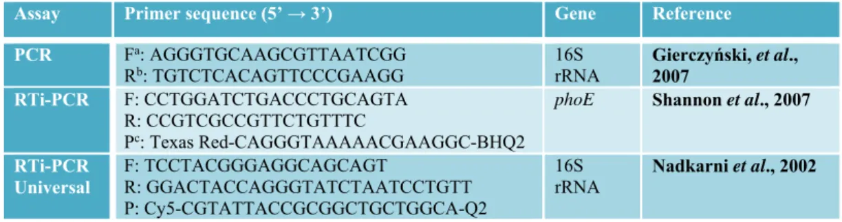
Table 1. Primers and probe utilized for K. pneumoniae
Assay Primer sequence (5’ → 3’) Gene Reference PCR Fa: AGGGTGCAAGCGTTAATCGG
Rb: TGTCTCACAGTTCCCGAAGG 16S rRNA Gierczyński, et al., 2007
RTi-PCR F: CCTGGATCTGACCCTGCAGTA R: CCGTCGCCGTTCTGTTTC
Pc: Texas Red-CAGGGTAAAAACGAAGGC-BHQ2
phoE Shannon et al., 2007
RTi-PCR Universal F: TCCTACGGGAGGCAGCAGT R: GGACTACCAGGGTATCTAATCCTGTT P: Cy5-CGTATTACCGCGGCTGCTGGCA-Q2 16S
rRNA Nadkarni et al., 2002
The nucleotide sequences of PCR fragments were confirmed with an Applied Biosystems (Foster City, CA, USA) DNA sequencer (model 3130xl). DNA sequencing reactions were conducted using the DNA sequencing kits (ABI BigDye®) supplied by Applied Biosystems. pJET1.2 forward and pJET1.2 reverse primers were used with the DNA sequencing kits. The results of sequencing were analyzed using Chormas Pro 2.6.4 (Technelysium Pty Ltd, South Brisbane, Australia) and Clone Manager 9 (Scientific & Educational Software, Cary, NC) programs. Consensus sequences were compared with the sequences from GenBank database and accuracy of species identification was verified.
2.4. RTi-PCR assay
Primers and probes used for detection of K. pneumoniae were listed in Table 1. RTi-PCR amplifications were monitored using the ABI Fast 7500 RTi-PCR platform and the experiments were run in triplicate. RTi-PCR amplifications were optimized to determine the presence of K. pneumoniae. In RTi-PCR assays, the target region was the phoE gene and size of amplified products were 69 bp. Positive control amplifications
were performed using primers and probe designed on the 16S rRNA sequence and DNA free amplifications were used as negative control. The RTi-PCR reaction mixtures contained 1X Reaction Buffer, 1μl dNTP (2mM), 1μl forward primer (10 μM), 1μl reverse primer (10 μM), 1μl prob (10 μM), 0.5 μl universal 16S rRNA forward primer (10 μM), 0.5 μl universal 16S rRNA reverse primer (10 μM), 0.5 μl universal 16S rRNA probe (10 μM) and 1μl Taq DNA polymerase (5 U/μl), and the mixtures were completed to 30 μl with nuclease free water. Amplification conditions were: one cycle of 5 min at 95 oC and following 40 cycles of 30
s at 94 oC and 1 min at 50 oC for RTi-PCR
assays.
2.5. Standard curve analysis
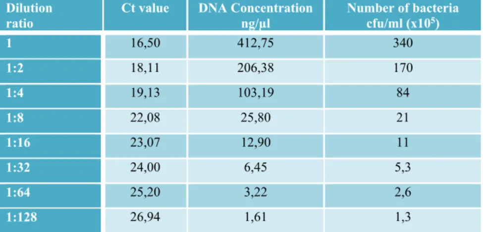
Isolated genomic DNAs (known total viable count of microorganisms) were diluted in deionized water eight times with 1:2 dilution factor (Table 2). Each dilution was used as template DNA and RTi-PCR amplifications were carried out in optimized conditions. Standard curves were generated with Ct values versus Log DNA concentrations and Log microorganism counts.
Table 2. Serial dilutions for standard curves in RTi-PCR to identify K. pneumoniae
2.6. Detection of K. pneumoniae in surface water samples
Water samples (n=20) were randomly collected from five different sites located in Kahramanmaraş city (Turkey) at the middle of the body of water 1 m below the surface. Samples were collected aseptically in pre-sterilized screw caped bottles and transported to the laboratory as soon as possible. Each 1 ml sample was inoculated into 90 ml tryptic soy broth. Following, 1 ml of samples incubated overnight at 37 °C was used for DNA isolation. Twenty water samples were examined by using PCR and RTi-PCR for the qualitative and quantitative analysis of pathogenic microorganism.
2.7. Data analysis
All quantitative analyses were applied only with cycle threshold (Ct) values <40, and all samples were analysed in three replicates. Microsoft Excel was used for all data analysis.
Standard curves of the multiplex RTi-PCR assay were obtained by plotting the mean Ct values vs log total viable count of
microorganisms (cfu/ml). The number of microorganisms in water samples was calculated by comparison with the standard curves.
3. Results and Discussion
Conventional PCR experiments were performed to identify K. pneumoniae using 16S rRNA gene. Nucleotide sequences of the 16S rRNA fragment obtained from the PCR amplification were used to confirm reference pathogen (data not shown). The sequences matched against the GenBank database and K. pneumoniae pathogen was identified accurately. The 16S rRNA region is one of the preferred gene regions to determine the relationship between taxa and to distinguish between genera and species (Rijpens and Herman, 2002). It was considered as the appropriate target region because 16S rRNA gene has the important properties such as the length of the 16S rRNA region (about 1500 bp), the number of multiple copies present in all bacteria and conserved and variable regions among the bacteria (Beneduce et al., 2007).
Dilution
ratio Ct value DNA Concentration ng/µl Number of bacteria cfu/ml (x105)
1 16,50 412,75 340 1:2 18,11 206,38 170 1:4 19,13 103,19 84 1:8 22,08 25,80 21 1:16 23,07 12,90 11 1:32 24,00 6,45 5,3 1:64 25,20 3,22 2,6 1:128 26,94 1,61 1,3
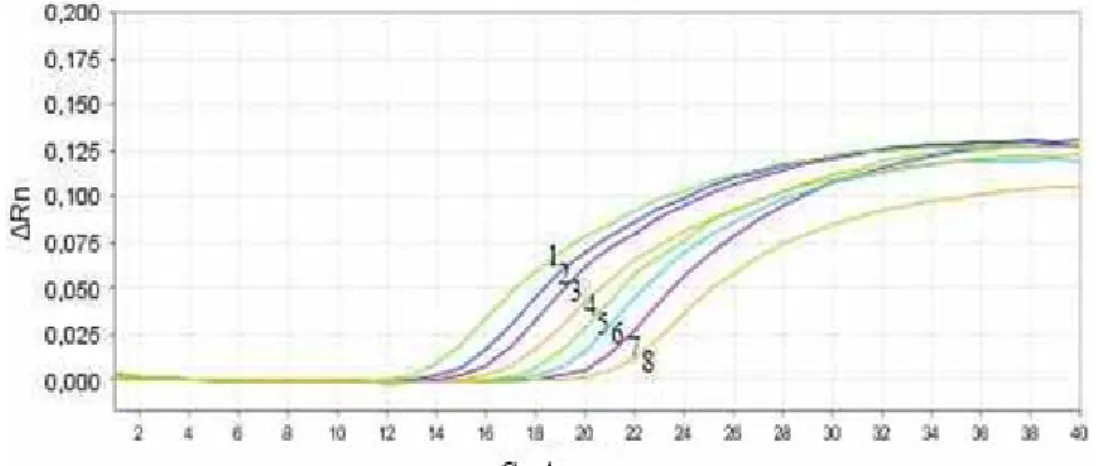
RTi-PCR amplifications were carried out in single format and the universal 16S rRNA probe and primers were included as a positive control for all reactions. RTi-PCR conditions were optimized to identify K. pneumoniae and the diluted DNAs of reference strains tested by RTi-PCR assays (Figure 1). In RTi-PCR assays, the coefficient of determination value (r2) was plotted between Ct values and pathogen numbers. Moreover, the coefficient of
determination was calculated between Ct values and DNA concentrations (data not shown). The data depending on the different dilution levels for Ct value, DNA concentration and number of bacteria are shown in Table 2. RTi-PCR amplified products were run on agarose gels for confirmation (data not shown). K. pneumoniae was correctly identified in all the RTi-PCR experiments and no cross reaction or false positive results were found.
Figure 1. Amplification plot of K. pneumoniae DNA dilutions. 1: 1X, 2: 1:2X, 3: 1:4X, 4: 1:8X, 5: 1:16X, 6: 1:32X, 7: 1:64X, 8: 1:128X.
Microbial pathogens may pose a significant threat for food safety and human health, therefore one of most effective ways to prevent and control infections and illnesses is the accurate detection of them, even at low presence. Conventional methods do not provide the desired accuracy, precision and speed, and sufficient information on quantitative detection of microorganisms compared to Real Time PCR technology (Nannapaneni et al., 2012). Real Time PCR assays have powerful and
convenient methods for pathogenic identification such as a wide dynamic range, specificity, application of quantitative analysis and detection at low limits. Thereby, it is eliminated many limitations of conventional methods with utilizing Real Time PCR technology on identification of foodborne pathogens (Velusamy et al., 2012; Aytaç et al., 2014).
RT-PCR technology has different strategies for identification and quantification in its application such as
SYBR Green, Taqman, FRET. SYBR Green and Taqman techniques are widely used for microbial identification. However, Taqman technique is more sensitive than SYBR Green technique (Delibato et al., 2011; Ding et al., 2017; Fukushima et al., 2010; He et al., 2016; Seo and Brackett, 2005). In present study, Taqman technique was performed to identify of K. pneumoniae because of its advantages.
In the RTi-PCR assay, Ct values were ranged from 16 to 26 and total viable count of reference microorganisms were varied from 1,3x10⁵ to 3,40x10⁷ for phoE gene. The correlation coefficients (r2 values) was 0.996 for K. pneumoniae. The results of regression analysis were indicated that standard curve has a good linearity to identify K. pneumoniae (Figure 2). The regression equation based on total viable count of microorganisms was used to characterize K. pneumoniae in water samples, as described below.
Figure 2. Linear regressions of RTi-PCR amplification (Ct value) versus log10 cfu of K.
pneumoniae
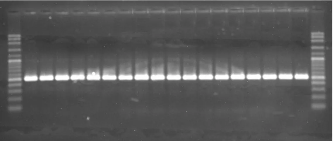
In total, 20 water samples were tested to detect desired pathogen by using PCR and RTi-PCR. According to PCR results, K. pneumoniae was found in all samples analyzed (Figure 3). Moreover, this pathogen was determined in all water samples at different contamination levels by using RTi-PCR. K. pneumoniae displays broad-range dynamic spectrum in microbial load with 1,0x10⁴ to 4,0x10⁹ cfu/mL (on
average; 5,9x10⁸ cfu/mL).
Figure 3 PCR gel image showing amplification of 16S rRNA in water samples r² = 0,996 10 20 30 40 0 1 2 3 Cy cle T hr eshold (Ct) log10cfu/mL
In a similar study, K. pneumoniae was identified using TaqMan primer and probe targeting phoE gene and detected at all different processes of wastewater, between the raw wastewater and final effluent stage (Shannon et al., 2007). Barati et al. (2016) investigated the occurrence of K. pneumoniae in terms of phenotype and genotype properties in water and sediment samples collected from the Matang mangrove estuary. All samples were tested by microbiological methods and confirmed by biochemical assays and PCR. Many samples were contaminated with K. pneumoniae. Additionally, K. pneumoniae isolates were determined to be potentially virulent to humans according to results of the phenotypic and genotypic analysis. In a different study, well water samples in Samaru (Nigeria) were analyzed using presumptive multiple tube fermentation and confirmatory tests for total and fecal coliforms. All the well water samples were contaminated with one or more bacterial pathogens, Escherichia coli 20%, Klebsiella pneumoniae 100% and Proteus mirabilis 40% (Aboh et al., 2015). Furthermore, the microbiological quality of different water sources were investigated and K. pneumoniae was found in most of the tested samples. It is considered that the contaminated water might not safe for human health (Aboh et al., 2015; Miah et al.,
2016; Samuel et al., 2016; Tabassum et al., 2015). Klebsiella spp. are usually abundant in water and has a low clinical significances (June et al., 2016). However, pathogenic features of K. pneumoniae isolated from environmental sources resemble those of clinical isolates of this pathogen (Barati et al., 2016; Struve and Krogfelt, 2004). Thereby, nonclinical isolates posed a potential risk should be investigated more detail in the way of pathogenic mechanism.
In conclusion, we have been determined the potential hazards of K. pneumoniae in surface waters in the present study. Both PCR and RTi-PCR assays were successfully performed and demonstrated a high sensitivity, specificity and accuracy in K. pneumoniae identification. The findings of this work indicated that all the tested water samples were contaminated with K. pneumoniae at different levels. In addition, the analysis results of two assays were shown to be compatible with each other. The present work may be contributed to better understanding of K. pneumoniae pathogenicity in environmental sources. Acknowledgement
This work was supported by the Scientific and Technological Research Council of Turkey (TÜBİTAK) with Project No: 115 O 099 and TUBITAK BIDEB 2211-C Domestic Doctoral Fellowship Program (Esen TUTAR, Ph. D.).
References
Aboh EA, Giwa FJ, Giwa A (2015). Microbiological assessment of well waters in Samaru, Zaria, Kaduna, State, Nigeria. Annals of African Medicine 14: 32‑38.
Aytaç SA, Taban BM (2014). Food-borne microbial diseases and control: food-borne ınfections and ıntoxications. In Malik A, Erginkaya Z, Ahmad S, Erten H (eds) Food Processing: Strategies for Quality Assessment. 1st ed. New York, NY: Springer, New York. p. 191–224.
Barati A, Ghaderpour A, Chew LL, Bong CW, Thong KL, Chong VC, Chai LC (2016). Isolation and characterization of aquatic-borne Klebsiella pneumoniae from tropical estuaries in Malaysia. International Journal of Environmental Research and Public Health 13.
Beneduce L, Fiocco D, Spano G (2007). Development of PCR-based molecular tools for the detection of emerging food and water-borne pathogenic bacteria. In Mendez-Vilas A, (ed) Communicating Current Research and Educational Topics and Trends in Applied Microbiology. 1st ed. Spain: Formatex, Spain. p. 569–576.
Chen L, Chavda KD, Findlay J, Peirano G, Hopkins K, Pitout JDD, Kreiswirth BN (2014). Multiplex PCR for identification of two capsular types in epidemic KPC-producing Klebsiella pneumoniae sequence type 258 strains. Antimicrobial Agents and Chemotherapy 58: 4196‑4199.
Clegg S, Murphy CN (2016). Epidemiology and virulence of Klebsiella pneumoniae. Microbiology spectrum 4: 1‑17.
Deleo FR, Chen L, Porcella SF, Martens CA, Kobayashi SD, Porter AR, Kreiswirth BN (2014). Molecular dissection of the evolution of carbapenem-resistant multilocus sequence type 258 Klebsiella pneumoniae. Proc Natl Acad Sci USA 111: 4988‑4993. Delibato E, Fiore A, Anniballi F, Auricchio B, Filetici E, Orefice L, De Medici D (2011).
Comparison between two standardized cultural methods and 24 hour duplex SYBR green real-time PCR assay for Salmonella detectionin meat samples. The New Microbiologica 34: 299‑306.
Ding T, Suo Y, Zhang Z, Liu D, Ye X, Chen S, Zhao Y (2017). A multiplex RT-PCR assay for S. aureus, L. monocytogenes, and Salmonella spp. detection in raw milk with pre-enrichment. Frontiers in Microbiology 8: 1‑11.
Dong D, Liu W, Li H, Wang Y, Li X, Zou D, Yuan J (2015). Survey and rapid detection of Klebsiella pneumoniae in clinical samples targeting the rcsA gene in Beijing, China. Frontiers in Microbiology 6: 1‑6.
Fukushima H, Kawase J, Etoh Y, Sugama K, Yashiro S, Iida N, Yamaguchi K (2010). Simultaneous screening of 24 target genes of foodborne pathogens in 35 foodborne outbreaks using multiplex Real-Time SYBR green PCR analysis. International Journal of Microbiology 1-18.
Gadsby NJ, McHugh MP, Russell CD, Mark H, Conway-Morris A, Laurenson IF, Templeton KE (2015). Development of two real-time multiplex PCR assays for the detection and quantification of eight key bacterial pathogens in lower respiratory tract infections. Clinical Microbiology and Infection 21: e788.
Gierczyński R, Jagielski M, Rastawicki W, Kałuzewski S (2007). Multiplex-PCR assay for identification of Klebsiella pneumoniae isolates carrying the cps loci for K1 and K2 capsule biosynthesis. Polish Journal of Microbiology 56: 153‑156.
He P, Zhu G, Luo J, Wang H, Yan Y, Chen L, Chen Z (2016). Development and application of a one-tube multiplex real-time PCR with melting curve analysis for simultaneous detection of five foodborne pathogens in food samples. Journal of Food Safety 1‑7. June M, Paul O, Kiplagat K (2016). Bacteriological quality of water from selected water
sources in Samburu South – Kenya. Imperial Journal of Interdisciplinary Research 9: 310‑316.
Kong RYC, Lee SKY, Law TWF, Law SHW, Wu RSS (2002). Rapid detection of six types of bacterial pathogens in marine waters by multiplex PCR. Water Research 36: 2802‑2812.
Kurupati P, Chow C, Kumarasinghe G, Poh CL (2004). Rapid detection of Klebsiella pneumoniae from blood culture bottles by Real-Time PCR rapid detection of Klebsiella pneumoniae from blood culture bottles by Real-Time PCR. Journal of Clinical Microbiology, 42: 8‑12.
Li B, Zhao Y, Liu C, Chen Z, Zhou D (2014). Molecular pathogenesis of Klebsiella pneumoniae. Future Microbiology 9: 1071‑1081.
Miah B, Majumder AK, Latifa GA (2016). Evaluation of microbial quality of the surface waters of Hatirjheel in Dhaka City. Stamford Journal of Microbiology 6: 30‑33. Nadkarni MA, Martin FE, Jacques NA, Hunter N (2002). Determination of bacterial load by
real-time PCR using a broad-range (universal) probe and primers set. Microbiology (Reading, England) 148: 257‑66.
Nannapaneni R (2012). Methods for ıdentification of bacterial foodborne pathogens. In Oyarzabal OA, Backert S (eds). Microbial Food Safety An Introduction. 1st ed. Springer-Verlag New York. p. 45–55
Pitout JDD, Nordmann P, Poirel L (2015). Carbapenemase-producing Klebsiella pneumoniae, a key pathogen set for global nosocomial dominance. Antimicrobial Agents and Chemotherapy 59: 5873‑5884.
Podschun R, Pietsch S, Höller C, Ullmann U (2001). Incidence of Klebsiella species in surface waters and their expression of virulence factors. Appl Environ Microbiol 67(7): 3325–3327.
Ramalingam N, Rui Z, Liu HB, Dai CC, Kaushik R, Ratnaharika B, Gong HQ (2010). Real-time PCR-based microfluidic array chip for simultaneous detection of multiple waterborne pathogens. Sensors and Actuators, B: Chemical 145: 543‑552.
Rijpens NP, Herman LMF (2002). Molecular methods for identification and detection of bacterial food pathogens. Journal of AOAC International 85(4): 984–995.
Samuel O, Florence N, Ifeanyi O (2016). Microbial quality assessment of commercial bottled water brands in major markets in Awka, Nigeria. Universal Journal of Microbiology Research 4: 1‑5.
Seo KH, Brackett RE (2005). Rapid, specific detection of Enterobacter sakazakii in infant formula using a real-time PCR assay. J Food Prot 68: 59‑63.
Shannon KE, Lee DY, Trevors JT, Beaudette LA (2007). Application of real-time quantitative PCR for the detection of selected bacterial pathogens during municipal wastewater treatment. Science of The Total Environment 382: 121‑129.
Struve C, Krogfelt KA (2004). Pathogenic potential of environmental Klebsiella pneumoniae isolates. Environmental Microbiology 6: 584‑590.
Tabassum A, Saha ML, Islam MN (2015). Prevalence of multi-drug resistant bacteria in selected street food and water samples. Bangladesh J Bot 44: 621‑627.
Velusamy V, Arshak K, Korostynka O, Vaseashta A, Adley C (2012). Real Time detection of foodborne pathogens applications in food quality monitoring and biosecurity. In Vaseashta A, Braman E, Susmann P, (eds). Technological Innovations in Sensing and Detection of Chemical, Biological, Radiological, Nuclear Threats and Ecological Terrorism. Springer, Netherlands. p. 149–58.
Vuotto C, Longo F, Balice M, Donelli G, Varaldo P (2014). Antibiotic resistance related to biofilm formation in Klebsiella pneumoniae. Pathogens 3: 743‑758.
Xiao X, Zhang L, Wu H, Yu Y, Tang Y, Liu D, Li X (2014). Simultaneous detection of Salmonella, Listeria monocytogenes, and Staphylococcus aureus by multiplex Real-Time PCR assays using high-resolution melting. Food Analytical Methods 7: 1960‑1972.