Super-mini percutaneous nephrolithotomy (SMP)
vs retrograde intrarenal surgery for the treatment
of 1
–2 cm lower-pole renal calculi: an
international multicentre randomised controlled
trial
Guohua Zeng* , Tao Zhang* , Madhu Agrawal
†, Xiang He
‡, Wei Zhang
§,
Kefeng Xiao
¶, Hulin Li**, Xuedong Li
††, Changbao Xu
‡‡, Sixing Yang
§§, Jean J. de la
Rosette
¶¶***, Junhong Fan*, Wei Zhu* and Kemal Sarica
†††Department of Urology *Minimally Invasive Surgery Center, The First Affiliated Hospital of Guangzhou Medical University, **Guangdong Key Laboratory of Urology, ZhuJiang Hospital of Southern Medical University, Guangzhou, ‡Zhejiang Provincial People’s Hospital, Zhejiang,§The First Affiliated Hospital With Nanjing Medical University, Nanjing, ¶Shenzhen People’s Hospital, Shenzhen,††The Second Affiliated Hospital of Harbin Medical University, Harbin,‡‡The Second Affiliated Hospital of Zhengzhou University, Zhengzhou,§§Renmin Hospital of Wuhan University, Wuhan, China, †Centre for Minimally-Invasive Endourology, Global Rainbow Healthcare, Agra, India,¶¶Istanbul Medipol University, Istanbul, Turkey, ***AMC University Hospital, Amsterdam, The Netherlands, and†††Dr. Lutfi Kirdar Kartal Research and Training Hospital, Istanbul, Turkey
Objectives
To compare the safety and effectiveness of super-mini-percutaneous nephrolithotomy (SMP) and retrograde intrarenal surgery (RIRS) for the treatment of 1–2 cm lower-pole renal calculi (LPC).
Patients and Methods
An international multicentre, prospective, randomised, unblinded controlled study was conducted at 10 academic medical centres in China, India, and Turkey, between August 2015 and June 2017. In all, 160 consecutive
patients with 1–2 cm LPC were randomised to receive SMP or RIRS. The primary endpoint was stone-free rate (SFR). Stone-free status was defined as no residual fragments of ≥0.3 cm on plain abdominal radiograph of the kidneys, ureters and bladder, and ultrasonography at 1-day and on computed tomography at 3-months after operation. Secondary endpoints included blood loss, operating time, postoperative pain scores, auxiliary procedures,
complications, and hospital stay. Postoperative follow-up was scheduled at 3 months. Analysis was by intention-to-treat. The trial was registered at http://clinicaltrials.gov/ (NCT02519634).
Results
The two groups had similar baseline characteristics. The mean (SD) stone diameters were comparable between the
groups, at 1.50 (0.29) cm for the SMP group vs 1.43 (0.34) cm for the RIRS group (P= 0.214). SMP achieved a
significantly better 1-day and 3-month SFR than RIRS (1-day SFR 91.2% vs 71.2%, P= 0.001; 3-months SFR 93.8% vs 82.5%, P= 0.028). The auxiliary procedure rate was lower in the SMP group. RIRS was found to be superior with lower haemoglobin drop and less postoperative pain. Blood transfusion was not required in either group. There was no significant difference in operating time, hospital stay, and complication rates, between the groups.
Conclusions
SMP was more effective than RIRS for treating 1–2 cm LPC in terms of a better SFR and lesser auxiliary procedure rate. The complications and hospital stay were comparable. RIRS has the advantage of less postoperative pain.
Keywords
super-mini percutaneous nephrolithotomy, retrograde intrarenal surgery, lower-pole renal calculi
Introduction
The prevalence of stone disease is increasing and the optimal treatment of symptomatic lower-pole renal calculi (LPC) remains quite challenging [1]. Hence, current treatment modalities for 1–2 cm LPC continue to be contested [2–4]. According to the 2017 European Association of Urology (EAU) guidelines, shockwave lithotripsy (SWL), percutaneous nephrolithotomy (PNL), and retrograde intrarenal surgery (RIRS) are recommended treatment options for 1–2 cm LPC [3]. Although the non-invasive nature and high patient acceptance rates are the main advantages of SWL, low stone-free and higher retreatment rates constitute the major drawbacks of this approach in the management of LPC [4–6]. RIRS is increasingly performed and has a good safety profile for the surgical management of LPC. Whereas, PNL has a higher stone-free rate (SFR), but it is more invasive and has higher complication rates compared to RIRS [3].
Taking the higher risk of complications associated with standard PNL into account, endourologists aimed to decrease the rate of severe complications (mainly bleeding) by using reduced tract sizes to limit the trauma induced in the renal parenchyma [7]. With this concept of reducing the risk of PNL-related complications, we developed the super-mini-PNL (SMP), which refers to an access sheath size of 10–14 F. It provides a safe and effective treatment method for renal calculi of≤2.5 cm [8]. It also has been suggested as an alternative treatment technique in the management of patients with LPC not amenable to RIRS [8]. However, Level-1 evidence comparing the two modalities for treating Level-1–2 cm LPC is lacking. To investigate this, we carried out a
multicentre, prospective, randomised controlled trial to compare the safety and effectiveness of SMP and RIRS for the treatment of patients with 1–2 cm LPC.
Patients and Methods
Patient Population
A prospective, randomised, unblinded controlled study was conducted at eight centres in China, one centre in India, and one centre in Turkey, from August 2015 to July 2017. Ethics Committee approval was obtained at each site (ClinicalTrials.gov NCT02519634). Table 1 presents the details of the inclusion criteria. The primary endpoint was the SFR at 3-months after surgery. Stone-free status was defined as no residual fragments of ≥0.3 cm on plain abdominal radiograph of the kidneys, ureters and bladder (KUB) and ultrasonography at 1-day and CT at 3-months after operation [2,9]. Secondary endpoints included haemoglobin decrease, transfusion rate, operating time, postoperative pain, auxiliary procedures, hospital stay, and complications (using the Clavien–Dindo grading system [10]).
The sample size was determined based on historical data of the SFR at 3-months after SMP and RIRS for LPC (98% and 85%, respectively). According to non-continuous sample size calculation formula, the minimum sample size for each group was estimated to be 72 (power>0.80) with a type-I error rate <0.05. To account for patients lost to follow-up and study withdrawals, this number was increased to 80.
Parallel randomisation was conducted at a ratio of 1:1, stratified by site, and was carried out using computer-generated random numbers. The coordinating centre revealed randomisation to the operating surgeon at the time of surgical scheduling. The number of patients enrolled in the study at each participating institution was equally distributed. The surgeons and patients were not blinded to group
assignment. Investigators who had not been involved and who were blinded to the surgical procedures performed the postoperative clinical assessment. In each centre, surgeries were performed only by one surgeon experienced in both procedures (≥50 SMPs and RIRSs per year).
Endoscopic Procedure
Preoperative assessment was the same for the entire study group. IVU and non-contrast CT were done in all patients to assess stone characteristics and renal anatomy. All procedures were carried out under general anaesthesia. Prophylactic antibiotics were given to all patients according to local antimicrobial guidelines.
Super-Mini-Percutaneous Nephrolithotomy
Super-mini-percutaneous nephrolithotomy surgical procedure was performed as previously described [8]. In summary, under general anaesthesia, in lithotomy position, a 5-F open-end ureteric catheter was placed under ureteroscopic guidance Table 1Patient selection criteria.
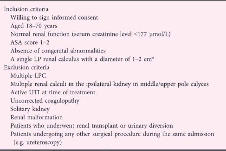
Inclusion criteria
Willing to sign informed consent Aged 18–70 years
Normal renal function (serum creatinine level<177 lmol/L) ASA score 1–2
Absence of congenital abnormalities
A single LP renal calculus with a diameter of 1–2 cm* Exclusion criteria
Multiple LPC
Multiple renal calculi in the ipsilateral kidney in middle/upper pole calyces Active UTI at time of treatment
Uncorrected coagulopathy Solitary kidney Renal malformation
Patients who underwent renal transplant or urinary diversion
Patients undergoing any other surgical procedure during the same admission (e.g. ureteroscopy)
ASA, American Society of Anesthesiology; LP, lower pole; LPC, lower-pole renal calculi.*The stone size was defined as the maximum diameter as determined by non-contrast CT.
into the renal pelvis. The patient was then turned to prone position. Percutaneous access was achieved using either ultrasonographic orfluoroscopic guidance. Tract dilatation was performed with fascial dilators up to 14 F. Next a 14-F irrigation-suction sheath was introduced into the pelvicalyceal system. Lithotripsy was performed using a 200-lm holmium-laserfibre at an energy level of 8–20 W. At the end of procedure,fluoroscopic images were taken to assess stone clearance. A 6-F JJ stent or 5-F ureteric catheter was inserted only in the presence of pyelocalyceal blood clots and/or pelvic perforation. Indications for 12-F nephrostomy tube placement included significant residual stone fragments requiring a second-look procedure and significant pyelocalyceal blood clots or bleeding. The operating time for SMP was recorded from the time of thefirst percutaneous renal puncture to wound closure.
Retrograde Intrarenal Surgery
The patient was placed in lithotomy position, and a 0.089 cm guidewire wasfirst placed into the renal pelvis. A 12/14-F ureteric access sheath was then advanced into the proximal ureter over the guidewire. An Olympus-P5 or Storz-X2 flexible ureteroscope was passed through the ureteric access sheath. Lithotripsy was performed using a 200-lm holmium-laserfibre at an energy level of 8–20 W. We attempted to mobilise the LPC to the upper or middle calyx before fragmentation. If this was not successful, the calculus was fragmented in the lower calyx. Large stone fragments were removed with a nitinol stone basket and sent for stone analysis. Stone fragments of<2 mm were left in situ for spontaneous passage. A 6-F JJ stent was routinely placed at the end of the procedure. The operating time was recorded from insertion of the endoscope into the urethra to the completion of stent placement.
Postoperative Evaluation and Follow-Up
KUB and ultrasonography were performed on postoperative day 1 to assess the one-session stone-free status. Low-dose CT, with a 2-mm section thickness, was obtained for all patients at the 3-month follow-up to evaluate the stone-free status. This interval was chosen to allow time for patients in the RIRS group to pass remaining stone fragments
spontaneously.
Postoperative pain severity was assessed at 6, 24 and 48 h postoperatively using a visual analogue scale (VAS; range: 1– 10) [6]. Hospital stay was rounded to the nearest whole day and calculated from the day of surgery to the day of discharge. Second-look SMP, SWL and external physical vibration lithecbole (EPVL) were auxiliary procedures used to treat patients with significant residual stones postoperatively. The JJ stents were removed 2 weeks after the procedures.
Statistical Analysis
Statistical analysis was performed using the Statistical Package for the Social Sciences (SPSSâ), version 20.0 (SPSS Inc., IBM Corp., Armonk, NY, USA). Continuous variables were analysed using Student’s t-test to compare the two means. Categorical variables between groups were analysed using the chi-squared or Fisher’s exact tests. The analysis was done according to the intention-to-treat principle. We also did a per-protocol analysis of the primary endpoints, in which we compared patients who underwent the assigned RIRS (i.e., those who did not have a conversion to SMP and those who did not have purulent urine in the kidney) with those who had been randomly assigned to and underwent SMP. A P< 0.05 was considered statistically significant.
Results
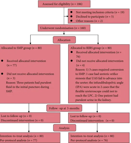
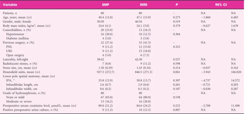
Theflow chart for trial inclusion is shown in Fig. 1. Of the 186 eligible patients, 160 patients were randomised into the two groups. Three patients in the SMP group were found to have purulentfluid in the collecting system during SMP puncture. Thus, a nephrostomy tube was placed and SMP postponed avoiding septic complications for these patients. In the RIRS group, three patients required conversion to SMP. One had an unfavourable ureteric orifice for RIRS access; two had steep infundibulopelvic angles (IPAs) preventing access of the LPC byflexible ureterorenoscopy. Additionally, one patient was found to have purulent urine in the kidney. A JJ stent was inserted for this patient and the procedure was postponed to avoid septic complications. The baseline characteristics of the two groups were comparable (Table 2). The SFR after a single treatment was significantly higher in the SMP group than in the RIRS group (91.2% vs 71.2%, P= 0.001). Clinically significant residual fragments (CSRF) were detected in four patients in the SMP group after a single treatment session. Of these cases, whilst one patient required a second-look SMP, one patient needed subsequent SWL to achieve stone clearance, and two patients required EPVL to remove residual fragments. For the RIRS group, CSRF were detected in 19 patients after a single treatment session. Nine of the 19 patients achieved complete stone clearance with subsequent SWL, whilst 10 patients required EPVL. At the 3-month postoperative follow-up, patients in the SMP group continued to have a significantly higher SFR than those in the RIRS group (93.8% vs 82.5%, P= 0.028; Table 3).
The mean operating times were similar between the groups (58.6 vs 52.3 min, P= 0.081). Intraoperatively, one patient had a minor renal pelvic perforation in the SMP group and required prolonged JJ stenting for 2 weeks (Clavien–Dindo Grade II). One patient in the RIRS group was found to have an iatrogenic false passage at the distal ureter requiring prolonged JJ stenting for 4 weeks (Clavien–Dindo Grade II)
after abandoning the procedure. The mean pain VAS score at 6, 24, and 48 h was significantly lower in the RIRS group, at 4.1 vs 3.2 (P= 0.001), 2.7 vs 2.0 (P = 0.004), and 1.7 vs 1.3 (P = 0.043), respectively.
The haemoglobin drop was significantly lower in the RIRS group (4.3 vs 10.2 g/L, P< 0.001). Blood transfusion was not required in either group. Three patients in the SMP group and one patient in the RIRS group had mild haematuria, all lasting for <6 h and which settled spontaneously.
Postoperative fever occurred in four patients in the SMP group and six patients in the RIRS group (P = 0.499). Two patients in each group required additional i.v. antibiotic treatment (Clavien–Dindo Grade II). None of the patients developed urosepsis postoperatively. All the patients had stone analysis and there was no significant difference in stone composition between the groups (P = 0.695). There was no significant difference in hospital stay and postoperative creatinine levels between the groups.
The per-protocol analysis incorporated the 77 patients who underwent a completed SMP and the 76 patients who were
assigned to and received RIRS. SMP in the per-protocol analysis achieved significantly better 1-day and 3-month SFRs than RIRS (1-day SFR 94.8% and 75.0%, P = 0.001; 3-month SFR 97.4% and 86.8%, P= 0.015).
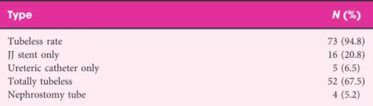
In the 77 patients who were randomly assigned to and received SMP, 52 patients (67.5%) did not require any upper tract drainage catheter (total tubeless). Amongst the patients who did require catheters, 16 (20.8%) had a JJ stent for 2 weeks and five (6.5%) had ureteric catheters for 1 day after the procedure. Four patients (5.2%) required a nephrostomy tube (Table 4).
Discussion
The management of LPC is more demanding than treatment of kidney stones in other locations due to the inherent anatomical challenges. Techniques such as RIRS and PNL have their advantages and disadvantages, and the optimal primary treatment for patients with 1–2 cm LPC is yet to be determined [2–4]. PNL provides overall significantly higher Allocation
Not meeting inclusion criteria (n = 19) Declined to participate (n = 5) Other reasons (n = 2)
Allocated to RIRS group (n = 80) Received allocated intervention (n = 76)
Did not receive allocated intervention (n = 4)
Reason: 1) 3 cases required conversion to SMP: 1 case had ureteric orifice stenosis that UAS fail to advance into the ureter; the infundibulopelvic angle (IPA) were acute in 2 cases that the flexible ureteroscope could not to reach the LPC. 2) One patient had purulent urine in the kidney. Follow -up at 3-months
Lost to follow-up (n = 0) Discontinued intervention (n = 0)
Underwent randomisation (n = 160) Assessed for eligibility (n = 186)
Lost to follow-up (n = 0) Discontinued intervention (n = 0) Analysis Intention-to-treat analysis (n = 80) Per-protocol analysis (n = 77) Intention-to-treat analysis (n = 80) Per-protocol analysis (n = 76) Allocated to SMP group (n = 80)
Received allocated intervention (n = 77)
Did not receive allocated intervention (n = 3)
Reason: Three patients had purulent fluid in the initial puncture during SMP.
Table 2Patient’s demographics and preoperative clinical characteristics in the two study groups.
Variable SMP RIRS P 95% CI
Patients, n 80 80 – NA NA
Age, years, mean (SD) 49.4 (12.8) 47.1 (13.9) 0.275 1.860 6.485
Gender, male: female 50:30 46:34 0.519 NA NA
Body mass index, kg/m2, mean (
SD) 24.6 (4.1) 24.1 (3.0) 0.379 0.627 1.639 Comorbidities, n (%) 20 (25.0) 13 (16.3) – NA NA Hypertension 16 (20.0) 10 (12.5) 0.384 Diabetes mellitus 4 (5.0) 3 (3.8) Previous surgery, n (%) 22 (27.4) 33 (41.3) – NA NA PNL 9 (11.2) 12 (15.0) 0.323 URS 9 (11.2) 15 (18.8) Open surgery 4 (5.0) 6 (7.5) Laterality, left:right 38:42 42:38 0.527 NA NA Radiolucent stones, n (%) 7 (8.8) 9 (11.2) 0.598 NA NA Stone size, cm, mean (SD) 1.50 (0.29) 1.43 (0.34) 0.214 0.037 0.162 Hounsfield units, mean (SD) 927.5 (272.7) 846.5 (271.5) 0.061 3.943 166.020
Lower-pole spatial anatomy, mean (SD)
IPA,° 55.8 (12.9) 50.8 (13.7) 0.307 4.737 14.572 Infundibular length, cm 2.6 (0.7) 2.9 (0.6) 0.261 0.721 0.203 Infundibular width, cm 0.6 (0.2) 0.5 (0.2) 0.107 0.030 0.287 Grade of hydronephrosis, n (%) 80 80 – NA NA None or mild 67 (83.8) 64 (80.0) 0.538 Moderate or severe 13 (16.2) 16 (20.0)
Preoperative serum creatinine level,lmol/L, mean (SD) 89.0 (21.2) 84.6 (24.2) 0.223 2.708 11.498 Positive preoperative urine culture, n (%) 9 (11.2) 10 (12.5) 0.807 NA NA
NA, not applicable; RIRS, retrograde intrarenal surgery; SMP, super-mini-percutaneous nephrolithotomy; URS, ureteroscopy.
Table 3Comparison of intraoperative and postoperative variables in the SMP and RIRS groups.
Variable SMP RIRS P 95% CI
Operating time, min, mean (SD) 58.6 (21.6) 52.3 (22.4) 0.081 0.771 13.282
Intraoperative complications, n (%) 1 1 – NA NA
Minor pelvic perforation (Clavien–Dindo Grade II) 1 (1.3) 0 0.319 False passage (Clavien–Dindo Grade II) 0 1 (1.3) 0.313 Pain VAS score (range 1–10), mean (SD)
At 6 h 4.1 (1.6) 3.2 (1.6) 0.001 0.344 1.365 At 24 h 2.7 (1.7) 2.0 (1.5) 0.004 0.246 1.288 At 48 h 1.7 (1.4) 1.3 (1.2) 0.043 0.015 0.861 One-session SFR, n (%) Intention-to-treat analysis 73 (91.2) 57 (71.2) 0.001 NA NA Per-protocol analysis 73 (94.8) 57 (75.0) 0.001 NA NA Haemoglobin drop, g/L, mean (SD) 10.2 (8.9) 4.3 (8.8) <0.001 3.148 8.820
Blood transfusion rate 0 0 – – –
Hospital stay, day, mean (SD) 2.5 (1.1) 2.2 (1.1) 0.156 0.099 0.612
Postoperative complications, n (%) 7 7 – NA NA
Mild haematuria (Clavien–Dindo Grade I) 3 (3.9) 1 (1.3) 0.317 Fever (>38.5 °C) 4 (5.2) 6 (7.9) 0.499 Clavien–Dindo Grade I 2 (2.6) 4 (5.3) 0.396 Clavien–Dindo Grade II 2 (2.6) 2 (2.6) 0.989
Postoperative serum creatinine level,lmol/L, mean (SD) 90.1 (23.6) 85.3 (21.7) 0.193 2.453 12.056
Auxiliary procedures, n (%) 4 (5.2) 19 (25.0) 0.001 NA NA
Second-look SMP 1 (1.3) – –
SWL 1 (1.3) 9 (11.8) 0.008
EPVL 2 (2.6) 10 (13.2) 0.015
Final SFR at 3-month postoperatively, n (%)
Intention-to-treat analysis 75 (93.8) 66 (82.5) 0.028 NA NA Per-protocol analysis 75 (97.4) 66 (86.8) 0.015 Stone composition, n (%) 80 80 – NA NA Calcium oxalate 58 (72.5) 52 (65.1) 0.695 Uric acid 8 (10.0) 9 (11.2) Struvite 6 (7.5) 10 (12.5) Carbonate apatite 8 (10.0) 9 (11.2) NA, not applicable; RIRS, retrograde intrarenal surgery; SMP, super-mini-percutaneous nephrolithotomy.
SFRs than RIRS, at the expense of higher complication rates, blood loss, and longer hospital stay [3].
To decrease PNL-related morbidity rates, possibly caused by its large tract size, we developed and have previously described the use of SMP [8]. It is a modified mini-PNL technique using a miniaturised scope through a smaller, 10– 14 F nephrostomy tract. With the newly designed irrigation-suction sheath, the critical limitations of new miniaturised PNLs, i.e., poor irrigation and challenging stone extraction, were completely addressed [11]. Several studies have
suggested that SMP is highly effective and safe for moderate-sized stones [8,11].
In our present study, SMP had a significantly higher SFR for treating 1–2 cm LPC when compared to RIRS. This was probably due to the favourable location and angulation of the lower calyx of the kidney for SMP puncture, meaning that percutaneous access could be achieved directly in line with the target stones in the lower pole. This‘direct-attack’ approach enhanced the economy of movement of stone extraction, resulting into the effective removal of all stone fragments in a single treatment session. Another advantage of SMP procedure was the use of the negative pressure
aspiration component of the device, which actively removed the relatively smaller stone fragments by safely controlled suction [8].
Maximal ureterorenoscope deflection is commonly required with RIRS to access renal stones located in the lower pole. Prolonged laser lithotripsy at an acute IPA, in particular for those of <30°, could make complete stone clearance
challenging [12]. Another anatomical restriction for access to the lower pole in RIRS is the presence of a long lower pole calyx infundibulum [13]. As such, in the present study there was a significantly higher auxiliary procedure rate in the RIRS group when compared with the SMP group (25.0% vs 5.2%). It is noteworthy that three patients in the RIRS group had their procedure abandoned, and required conversion to SMP. One was due to ureteric orifice stenosis, and two were due to acute the IPA hindering access to the LPC by the flexible ureterorenoscope.
Renal haemorrhage is one of the most worrisome and common complications of a percutaneous procedure. In
contrast, RIRS has the advantage of not violating the renal parenchyma, which might be an important consideration for patients with 1–2 cm LPC. In the present study, the mean drop in haemoglobin level in SMP group was significantly greater than for the RIRS group. However, none of the patients required a blood transfusion. Significant bleeding in patients who underwent SMP was infrequent and most probably due to the reduced access-tract size. Therefore, we found no evidence to support the claim that SMP would require more transfusions in patients with 1–2 cm LPC. In the present study, other complications, including mild haematuria and fever, were comparable between the groups. Patients in the RIRS group had lower pain scores than those in the SMP group at initial points postoperatively. However, the difference becomes less prominent at 48 h. In our present study, a high proportion of patients underwent tubeless SMP, which seems to have favourable characteristics in terms of pain intensity.
A study conducted by Ozayar et al. [14] suggested that PNL requires a longer operating time as compared to RIRS for LPC, as the stones were usually fragmented and basket extraction was not required routinely during RIRS.
However, in our present study, we did not identify any significant differences in operating time between the groups. The reason might be that SMP utilised active suction to remove the stone fragments. In SMP, the shattered stones tend to aggregate at the opening of the sheath instead of scattering, resulting in a more comfortable and quicker lithotripsy.
With regards to the hospital stay, a recently published meta-analysis showed a significantly shorter hospital stay for patients who underwent RIRS when compared with patients who had PNL for the treatment of renal stones [15]. But, in patients who were treated ‘tubeless’ following PNL, the hospital stay was reduced significantly [16,17]. In our present study, 94.8% of the patients did not require a nephrostomy tube after SMP, and 67.5% did not require a nephrostomy tube or JJ stent following SMP. This may explain why the mean hospital stay was similar in the groups.
We acknowledge that there are limitations to the present study. Firstly, the mean hospital stay was longer than that reported in the Western literature. The reason is that most patients in China do not leave hospital until they can return to normal activities. Secondly, we did not evaluate the cost-effectiveness of the two procedures. Thirdly, surgeries were only performed by expert/sub-specialised endo-urologists, who perform >50 cases/year of both SMP and RIRS, and therefore the results may not be generalisable to lower-volume surgeons. Finally, most participants in the present study were Asian, therefore the results might not be applicable to a non-Asian population.
Table 4Tubeless rate of 77 patients who were randomly assigned to and received SMP.
Type N (%)
Tubeless rate 73 (94.8)
JJ stent only 16 (20.8)
Ureteric catheter only 5 (6.5) Totally tubeless 52 (67.5)
Nephrostomy tube 4 (5.2)
Conclusions
Our present study shows that both SMP and RIRS are safe and feasible surgical options in the treatment of 1–2 cm LPC. SMP was more effective than RIRS in terms of a better SFR and lesser auxiliary procedure rate. The operating time, complications, and hospital stay were comparable. RIRS has the advantage of less postoperative pain.
Acknowledgements
Kemal Sarica, Guohua Zeng and Tao Zhang designed the research; Guohua Zeng, Madhu Agrawal, Xiang He, Wei Zhang, Kefeng Xiao, Hulin Li, Xuedong Li, Changbao Xu, Sixing Yang and Kemal Sarica conducted the research; Guohua Zeng, Tao Zhang, Junhong Fan, Wei Zhu and de la Rosette JJ wrote the paper; Guohua Zeng and Kemal Sarica had primary responsibility for thefinal content. All authors read and approved thefinal manuscript.
Source of Funding
This study was supported in part by National Natural Science Foundation of China (No. 81670643, No. 81601273 and No. 81370804). Additional funding was provided by Science and Technology Planning Project of Guangdong Province (No. 2017B030314108) and Guangzhou Science Technology and Innovation Commission (No. 201604020001 and No. 201704020193).
Con
flicts of Interest
The authors have no conflicts of interest.
References
1 Donaldson JF, Lardas M, Scrimgeour D et al. Systematic review and meta-analysis of the clinical effectiveness of shock wave lithotripsy, retrograde intrarenal surgery, and percutaneous nephrolithotomy for lower-pole renal stones. Eur Urol 2015; 67: 612–6
2 Singh BP, Prakash J, Sankhwar SN et al. Retrograde intrarenal surgery vs extracorporeal shock wave lithotripsy for intermediate size inferior pole calculi: a prospective assessment of objective and subjective outcomes. Urology 2014; 83: 1016–22
3 T€urk C, Knoll T, Petrik A et al. Specific management of ureteral stones. In: Guideline on Urolithiasis. European Association of Urology, 2017: 24–5. Available at: http://uroweb.org/guideline/urolithiasis/. Accessed June 2018
4 Soliman T, Sherif H, Sebaey A, Mohey A, Elmohamady BN. Miniperc vs shockwave lithotripsy for average-sized, radiopaque lower pole calculi: a prospective randomized study. J Endourol 2016; 12: 69–87
5 Raman JD, Pearle MS. Management options for lower pole renal calculi. Curr Opin Urol 2008; 18: 214–9
6 Sabnis RB, Ganesamoni R, Doshi A, Ganpule AP, Jagtap J, Desai MR. Micropercutaneous nephrolithotomy (microperc) vs retrograde intrarenal surgery for the management of small renal calculi: a randomized controlled trial. BJU Int 2013; 112: 355–61
7 Zhu W, Liu Y, Liu L et al. Minimally invasive versus standard percutaneous nephrolithotomy: a meta-analysis. Urolithiasis 2015; 43: 563–70
8 Zeng G, Wan S, Zhao Z et al. Super-mini percutaneous nephrolithotomy (SMP): a new concept in technique and instrumentation. BJU Int 2016; 117: 655–61
9 Salem A, Saad I, Emran A et al. Laser lithotripsy versus ESWL for lower calyceal renal stones. J Urol 2013; 189(Suppl): e751
10 Mitropoulos D, Artibani W, Graefen M et al. Reporting and grading of complications after urologic surgical procedures: an ad hoc EAU guidelines panel assessment and recommendations. Eur Urol 2012; 61: 341–9
11 Zeng G, Zhu W, Liu Y, Fan J, Zhao Z, Cai C. The new generation super-mini percutaneous nephrolithotomy (SMP) system: a step-by-step guide. BJU Int 2017; 120: 735–8
12 Breda A, Ogunyemi O, Leppert JT, Schulam PG. Flexible ureteroscopy and laser lithotripsy for multiple unilateral intrarenal stones. Eur Urol 2009; 55: 1190–6
13 Elbahnasy AM, Shalhav AL, Hoenig DM et al. Lower caliceal stone clearance after shock wave lithotripsy or ureteroscopy: the impact of lower pole radiographic anatomy. J Urol 1998; 159: 676–82
14 Ozayar E, Gulec H, Bayraktaroglu M et al. Comparison of retrograde intrarenal surgery and percutaneous nephrolithotomy: from the view of an anesthesiologist. J Endourol 2016; 30: 184–8
15 Zhang W, Zhou T, Wu T et al. Retrograde intrarenal surgery versus percutaneous nephrolithotomy versus extracorporeal shockwave lithotripsy for treatment of lower pole renal stones: a meta-analysis and systematic review. J Endourol 2015; 29: 745–59
16 Shah H, Khandkar A, Sodha H, Kharodawala S, Hegde S, Bansal M. Tubeless percutaneous nephrolithotomy: 3 years of experience with 454 patients. BJU Int 2009; 104: 840–6
17 Abdel Wahab O, Sherif H, El-Karamany T. Tubeless PNL in the supine position. Turk J Urol 2012; 38: 138–42
Correspondence:Kemal Sarica, MD, PhD, Department of Urology, Dr. Lutfi Kirdar Kartal Research and Training Hospital, Istanbul, Turkey.
e-mail:[email protected]
Guohua Zeng, MD, PhD, Department of Urology, Minimally Invasive Surgery Center, The First Affiliated Hospital of Guangzhou Medical University, Guangdong Key Laboratory of Urology, Guangzhou, China.
e-mail:[email protected]
Abbreviations: CSRF, clinically significant residual fragments; EPVL, external physical vibration lithecbole; IPA,
infundibulopelvic angle; KUB, plain abdominal radiograph of the kidneys, ureters and bladder; LPC, lower-pole renal calculi; PNL, percutaneous nephrolithotomy; RIRS, retrograde intrarenal surgery; SFR, stone-free rate; SMP, super-mini-percutaneous nephrolithotomy; SWL, shockwave lithotripsy; VAS, visual analogue scale.