ABSTRACT
Objective: In this study, we examined the effects of Polypodium vulgare L. (Polypodiaceae) as a candidate to be used for wound healing scarred area. We investigated the antibacterial, and antioxidant activity of P. Vulgare on both in vivo, and in vitro wound healing using an excisional wound model in mice
Method: We used 32 Balb-c mice equally divided into four groups: Group 1 control, Group 2 ve-hicle, Group 3 Polypodium vulgare, and Group 4 Centella asiatica extract (CAE). All treatments were applied topically once in a day. The scar area, percentage wound closure and epithelization time were measured. PDGF, VEGF, and collagen immunohistochemical staining were used for evaluation.
Results: CAE and P. vulgare extract groups were observed to be more effective than the control and vehicle groups in terms of new vascular, epidermal and granulation tissue organization. PDGF, VEGF, and collagen immunohistochemical staining was stronger in the P.vulgare extract and CAE groups compared to the control and vehicle groups. In the P. vulgare and CAE groups, PDGF staining intensity was stronger than the control and vehicle groups, but VEGF and collagen staining in P. vulgare group was not different from the control group.
Conclusion: P. vulgare had an effect on the injured area by regenerating the epidermis and increasing vascularization. P. vulgare extract with known antioxidant, and antimicrobial activities may be helpful as a supportive treatment in wound healing.
Keywords: Wound healing, polypodium vulgare, antioxidant ÖZ
Amaç: Bu çalışmada Polypodium vulgare L. (Polypodiaceae), yara iyileşme adayı olma potansi-yeli açısından değerlendirildi. Farelerde eksizyonel bir yara modeli kullanılarak hem in vivo hem de in vitro yara iyileşmesinde P.vulgare’nin antimikrobiyal ve antioksidan aktivitesini araştırdık. Yöntem: Eşit olarak dört gruba ayrılmış toplam 32 Balb-c faresi kullandık; Grup 1 kontrol, grup 2 taşıyıcı grup 3 Polipodyum vulgare, grup 4 Centella asiatica özütü (CAE). Tüm tedaviler topikal olarak günde bir kez uygulandı. Yara izi alanı, yara kapanma yüzdesi ve epitelizasyon süresi ölçül-dü. İmmünhistokimyasal değerlendirmede PDGF, VEGF ve kollajen boyaları kullanıldı.
Bulgular: CAE ve P. vulgare ekstrakt gruplarının yeni vasküler organizasyon, epidermis ve granülasyon doku organizasyonu açısından kontrol ve taşıyıcı gruplarından daha etkili olduğu gözlenmiştir. PDGF, VEGF ve kollajen immünohistokimyasal boyama, P.vulgare ekstraktı ve CAE gruplarında kontrol ve taşıyıcı gruplarına göre daha kuvvetliydi. P. vulgare ve CAE grubunda PDGF boyama yoğunluğu, kontrol ve taşıyıcı gruplarından daha kuvvetliydi, ancak P. vulgare grubundaki VEGF ve kollajen boyama, kontrol grubundan farklı değildi.
Sonuç:
P. vulgare yara iyileşmesini ve granülasyon dokusunu, epidermal rejenerasyonu ve anjiyogenezi arttırdı. Antioksidan ve antimikrobiyal aktiviteleri ile bilinen P. vulgare ekstraktı, yara iyileşmesini desteklemek için yararlı olabilir.
Anahtar kelimeler: yara iyileşmesi, polypodium vulgare, antioksidan
Received: 27 July 2020 Accepted: 21 September 2020 Online First: 25 December 2020
An Alternative Approach Wound Healing Field with Polypodium
Vulgare
Polypodium Vulgare ile Yara İyileşme Alanına Alternatif Bir Yaklaşım
N. Kepil ORCID: 0000-0001-5494-6422
Istanbul University-Cerrahpasa Faculty of Medicine, Department of Pathology, Istanbul, Turkey
S. Ayla ORCID: 0000-0003-2143-5268 B. Daylan ORCID: 0000-0002-8302-1127
Istanbul Medipol University, Faculty of Medicine, Department of Histology and Embryology, Istanbul, Turkey
A.A. Sakul ORCID: 0000-0002-9354-0000
Istanbul Medipol University, Faculty of Medicine, Istanbul, Department of Medical Pharmacology, Istanbul, Turkey
M.E. Okur ORCID: 0000-0001-7706-6452
University of Health Sciences, Faculty of Pharmacy, Department of Pharmacology, Istanbul, Turkey
A.E. Karadag ORCID: 0000-0002-3412-0807
Istanbul Medipol University, Faculty of Pharmacy, Department of Pharmacognosy, Istanbul, Turkey
E.M. Ozdemir ORCID: 0000-0001-9416-7757
Istanbul Medipol University, Department of Animal Facility, Istanbul, Turkey
M.Y. Gunal ORCID: 0000-0001-7702-2441
Alanya Alaaddin Keykubat University, Faculty of Medicine, Department of Physiology, Alanya, Turkey
Corresponding Author: S. Batur ORCID: 0000-0001-6577-8970 Istanbul University-Cerrahpasa, Cerrahpasa Faculty of Medicine, Department of Pathology, Istanbul, Turkey
✉
[email protected] Ethics Committee Approval: This study approved by the Istanbul Medipol University EthicsCom-mittee for Animal Researchs, 11 December 2019, 2019/89.
Conflict of interest: The authors declare that they have no conflict of interest. Funding: None.
Informed Consent: Not Applicable.
Cite as: Batur S, Ayla S, Sakul AA, et al. An alternative approach wound healing field
with polypodium vulgare. Medeni Med J. 2020;35:315-23. Sebnem Batur , Sule Ayla , Ayse Arzu Sakul , Mehmet Evren Okur , Ayse Esra Karadag Benay Daylan , Ekrem Musa Ozdemir , Nuray Kepil , Mehmet Yalcin Gunal
ID
© Copyright Istanbul Medeniyet University Faculty of Medicine. This journal is published by Logos Medical Publishing. Licenced by Creative Commons Attribution-NonCommercial 4.0 International (CC BY-NC 4.0)
ID ID ID ID
ID ID ID ID
INTRODUCTION
Wound healing is a complicated period both for skin and tissues especially after an injury. Epider-mis and derEpider-mis in the undamaged tissue is a pro-tective barrier against external influences. When the barrier breaks, some repair processes are activated1. Wound healing is of great clinical
im-portance since untreated wounds can be fatal2,3.
It was reported that wounds, especially chronic ones can be quickly colonized by both Gram-positive or negative bacteria, fungus and even viruses2,4. Therefore, doctors recommend topical
use of antibiotics to prevent unexpected effects such as sepsis and bacteremia. For wound heal-ing and management, various pharmaceutical products can be used4. Traditional medicine is full
of knowledge that has been developed through generations in various societies. Traditional medi-cine approaches are based upon natural products from plants, animals, and minerals. African and Asian communities have used plants for primary health care5. Herbal medicines include sections of
plants, different plant parts, their combinations, or plant extracts. Plants are used frequently by medical practitioners in wound healing6. From
past to present, various plants have been used as wound healing agents such as Laurocerasus sp., Capparis sp., and Prunus spinosa4,7.
In this study, Polypodium vulgare L. (Polypodace-ae) is evaluated for its potential as a wound heal-ing candidate. Polypodium vulgare L. (P. vulgare) is a plant known and have been used by people since ancient times. Its rhizomes are more well-known and are included in some foods as sweet-ening agents8,9. In ethnobotanical medicine the
plant is used for its antitussive and diuretic prop-erties10. Infusion prepared from its roots is used
in abdominal pain and inflammatory conditions. In addition, the pharmacological activities of the plant on the liver and lung are also known. It has also antioxidant, anticholinesterase, antimicro-bial, antiviral, antiepileptic, antipyretic, and anal-gesic properties11-14. P. vulgare is used in
tradi-tional medicine as an expectorant to treat cough and pertussis15. Centella asiatica extract (CAE) is a
molecule that facilitates the repair process of the wound tissue and has been shown to exhibit an-timicrobial activity16. For this reason, it has been
preferred as a reference molecule in many studies including this one. We investigated the effects of P.vulgare on excisional wound healing in mice. MATERIAL and METHODS
Preparation of Extract
P. vulgare aerial parts were collected from Bel-grade forests, Istanbul, in 2018. Herbal ingredi-ent was idingredi-entified (specimen no. IMEF: 1068) and stored. The airdried herb was soft powdered in alcohol for 24 hours. Following permeation and vaporization (Heidolph, Germany), gel formula-tions were prepared by the methanol extract
Antioxidant Activity in Vitro
• 2,2-diphenyl-1-picrylhydrazyl (DPPH) scaven-ging
DPPH• was used to determine the total antioxi-dant capacity17. The reaction mix contained 100
µM DPPH• in methanol and P. vulgare extract. After 30 min, UV spectrophotometer (UV-1800, Shimadzu, Japan) read absorbance value at 517 nm. The scavenging activity was calculated as the percent radical reduction as follows:
DPPH• RSA % = [(Absorbance control - Absor-bance test sample) / AbsorAbsor-bance control)] x 100 • 2,2-Azino-bis 3-ethylbenzthiazoline-6-sulfo-nic acid (ABTS) scavenging
Re and co-workers’ protocol was used for obtain-ing P. vulgare methanol extract18. Potassium
per-sulfate and ABTS• blend was kept at room temper-ature in a dark room for 16 h prior to its use. The 734 nm was its absorbance measure. The study
was repeated three times. For negative controls: ethanol, and positive controls: Trolox were used4.
The absorbance of the extract obtained was mea-sured as follows:
ABTS• RSA % = [(Absorbance control - Absor-bance test sample) / AbsorAbsor-bance control)] x 100 Antioxidant reading outcomes were studied com-paratively with standard reference substances such as ascorbic acid and Trolox.
Antimicrobial Activity in Vitro
Broth microdilution assay was used for extract specification defined by the Clinical and Labora-tory Standards Institute (2006) for minimum in-hibitory concentrations (MIC). MIC is the lowest concentration of an antimicrobial agent that pre-vents growth of an organism.
Staphylococcus aureus ATCC 6538, Enterococ-cus faecalis ATCC 29212, Escherichia coli NRLL B-3008, and Pseudomonas aeruginosa ATCC 10145 strains were grown in Mueller Hinton Broth (MHB, Merck, Germany) at 37°C in aerobic condi-tions for 24 h. All microorganisms were standard-ized to 1 × 10⁸ CFU/mL using McFarland No: 0.5 in sterile saline (0.85%). A modified microdilution assay was used to establish the antimicrobial ac-tion of the sample. Dimethylsulfoxide (DMSO) was used to prepare the stock solution.
Preparation of Polypodium Vulgare Extract Gel Formulation
The gel of the extract was formulated with hy-droxypropyl cellulose gel. For preparing the gel, hydroxypropyl cellulose (2%) was dissolved in distilled water for 24 hours, and then the P. vul-gare extract (5%) was added, and the blend was stirred slowly.
In Vivo Experiments
• Experimental Animals
Balb-c mice (25-28 g) were used in the study. The mice were hosted in regular cages with food and water intake ad libitum, at room temperature (24°C), with artificial light from 7.00 am to 7.00 pm. Before experiments, ethical clearance ap-proval was obtained from the Local Ethics Com-mittee (No: E.65386-89). During the experiments, the animals were cared for according to the ethi-cal rules.
• Wound formation protocols and animal groups
The experimental animals were divided into four groups within 8 animals per group;
Group 1: Control group (with sterile saline). Group 2: Vehicle group (no drug content) Group 3: Polypodium vulgare extract gel group. Group 4: Centella asiatica extract (CAE) reference molecule.
Mice were anesthetized intraperitoneally with a combination of 80-100 mg/kg ketamine and 10 mg/kg xylazine intraperitoneally. Two excisional wound tissues were formed using a 5 mm biopsy apparatus on the left side of the midline in the shaved back region, one cm apart from each oth-er and 1.5 cm away from the midline. Treatments were applied to the wounds for ten days.
• Macroscopic Assessment
For score healing of the wound, photographs were taken at days 0 and 10. The surface areas were measured with the Image J program. After then, wound healing rates were calculated by the formula given in the literature19.
• Histological Assessment
On the 10th day, the animals were sacrificed and
the scar tissue was removed. A 10% neutral form-alin was used for fixation. After that, wound sam-ples were dehydrated in ethanol series, cleared in toluene, and embedded in paraffin. Five µm thick
sections were taken on glass slides and stained with hematoxylin-eosin (HE), immunohistochem-ical stains (vascular endothelial growth factor (VEGF) (Santa Cruz sc-7269), Collagen (COL1A1) (Santa Cruz sc-293182), and Platelet Derived Growth Factor (PDGF-A) (Santa Cruz sc-9974)). The scoring system defined by Galeano et al. was used for the histological evaluation of wound healing20. For epidermal organization; Score 1:
weak epidermal tissue organization ≥ 20% of the tissue, Score 2: incomplete epidermal tissue or-ganization ≥ 40% of the tissue, Score 3: moderate epithelial tissue organization ≥ 60% of the tissue, Score 4: complete epidermal tissue organization ≥ 80% of the tissue. For the thickness of the granula-tion tissue; Score 1: weak granulagranula-tion layer, Score 2: moderately dense granulation layer, Score 3: dense granulation layer, Score 4: very dense gran-ulation layer.
For angiogenesis; Score 1: one to two vessels/ site, Score 2: few neovascularization (3-4/site), Score 3: newly formed capillary vessels (5-6/site) and Score 4 describes newly formed and normal appearing capillary vessels (>7/site).
The semiquantitative method was used for the immunoreactivity of immunohistochemical stain. Five randomly selected fields were evaluated and averaged. Score 0: No staining, Score 1: Poor staining, Score 2: Moderate staining, and Score 3: Strong staining21.
• Statistical analysis
For statistical analysis, a standard software pack-age (SPSS 20 for Windows, SPSS Inc., Chicago, IL, USA) was used. Differences between groups were analyzed by one-way ANOVA test, followed by the least significant differences tests. All values were given as mean ± S.E.M. P values values less than 0.05 were considered significant.
RESULTS
Antioxidant Activity
Antioxidant activity of P. vulgare extract is sum-marized in Table 1. It can be said that the extract has a moderate antioxidant capacity compared to the ones used as standard substances. As a result of the experiments, the antioxidant effect of the extract was revealed, and the antioxidant capacity is thought to be effective in the wound healing.
Antimicrobial Activity
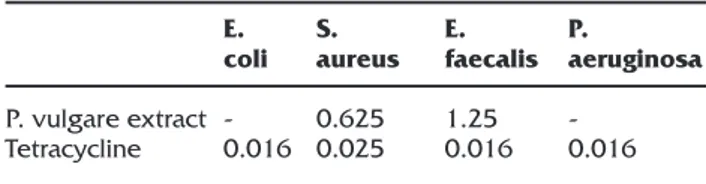
The antimicrobial activity of P. vulgare extracts is showed in Table 2. While the P. vulgare extracts were found to be most sensitive against S. au-reus; P. aeruginosa and E. coli were found to be resistant at tested concentrations (10 mg/mL). MIC value of P. vulgare methanol extract of 0.625 mg/mL against S. aureus was determined.
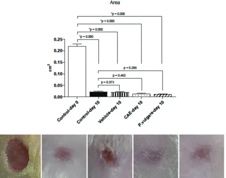
Macroscopic wound healing
Figure 1 exhibited the percentage of macroscopic wound healing rates. On day 10, statistically in-significant difference in wound contraction com-pared to the controls in the P.vulgare group was observed (p<0.2) (Figure 1). No local or system-atic side effects were observed macroscopically during the application of the extract.
Table 1. ABTS and DPPH radical scavenging activities of P. vulgare methanol extract (IC50±SD (mg/mL).
ABTS DPPH Extract IC50 (mg/mL) 3.25±0.03 2.13±0.04 Standards IC50 (mg/mL) 0.034±0.001 (Trolox) 0.009±0.002 (Ascorbic acid) *Statistically significant compared to control, p<0.05.
Table 2. MIC values of P. vulgare MeOH extract on tested concentration (MICs in mg/mL). P. vulgare extract Tetracycline E. coli -0.016 S. aureus 0.625 0.025 E. faecalis 1.25 0.016 P. aeruginosa -0.016
Histology of wound healing
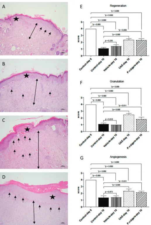
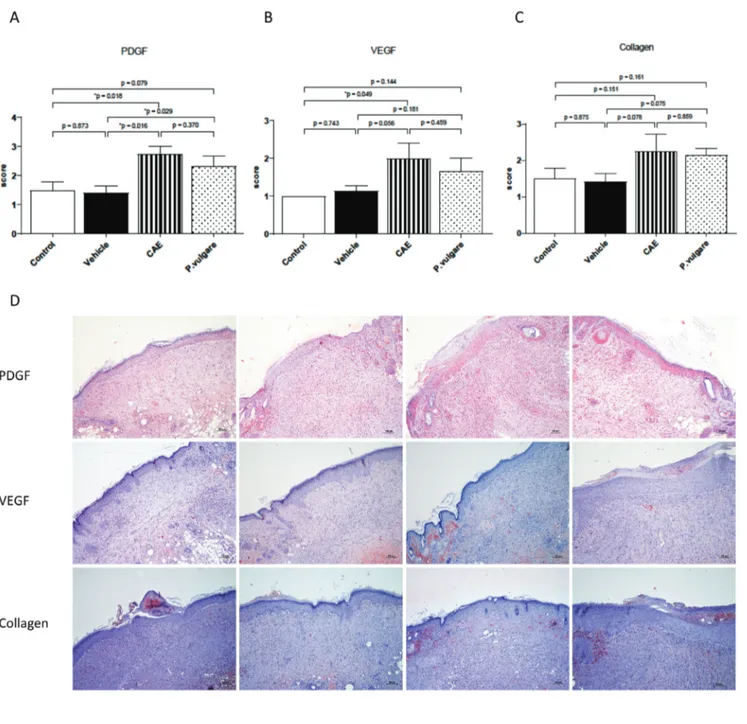
Histological evaluation (H&E evaluation and im-munohistochemical (VEGF, PDGF, and collagen) were appraised separately. Histopathological-ly, H&E (Figure 2 A, B, C, D) and immunohis-tochemical staining were shown in Figure 3 D. CAE (p<0.05) and P. vulgare extract (p<0.05) groups were observed to be more effective than the control and vehicle groups in terms of new vascular organization, epidermal and granulation tissue organization (Figure 2 E, F, G). PDGF, VEGF, and collagen immunohistochemical staining was stronger in the P.vulgare extract and CAE groups
compared to the control and vehicle groups (Fig-ure 3 D). In the P.vulgare and CAE group, PDGF staining intensity was stronger than the control and vehicle groups, although not statistically sig-nificant (Figure 3A), but VEGF and collagen stain-ing in the P.vulgare group were not different from the control group (Figure 3 B,C).
DISCUSSION
Plants rich in antioxidant compounds are used for their wound-healing and anti-aging properties. Also, free oxygen radicals play an important role in apoptosis and cell proliferation mechanisms.
Figure 1. Macroscopic wound healing and healing percentage of wound area in each group (Control, vehicle, CAE, P. vul-gare groups).
Studies have shown that topical application of antioxidant-containing compounds will be ben-eficial for wound healing and protecting tissues
from oxidative damage7,22. In our experiments,
the antioxidant effect of the extract was revealed, and the antioxidant capacity was thought to be
Figure 2. Histopathological view of injured tissues of the control (A), vehicle gel (B), CAE (C) and P.vulgare (D) extract gel on 10th day after wound incision (Original magnification X10). E, F, G; Histological scores of epidermal-dermal regenera-tion, granulation tissue thickness and angiogenesis of control, vehicle gel, CAE, P. vulgare groups. Statistically significant as compared to control; P<0.05. Values are presented as the mean ±SEM. The scale bars represent 100 μm for figure. *: Epidermal regeneration, →: Angiogenesis, blood vessels, ↔: Granulation formation.
effective in wound healing. In our study, the anti-microbial activity of P. vulgare extract was shown. Prevention of microorganisms from multiplying in wound care and the healing process are an im-portant parameters23. Therefore, the antibacterial
effect of the extract against various microorgan-isms found in the human skin flora was investi-gated for this purpose in our study.
The skin is the largest organ in human body, which
Figure 3. A, B, C, Comparison of immunohistochemistry collagen, PDGF and VEGF wound healing scores among groups. Sta-tistically significant as compared to control; P<0.05(*). Values are presented as the mean±SEM. D, Histopathological view of injured tissues of the untreated (control), vehicle, P.vulgare and CAE groups on 10th day after wound incision (Original magnification X10).
acts as the defensive set across physical damage, pathogens, fluid loss, and maintain the body ho-meostasis24. Thus, even a small breakage can
im-pact the health of the individuals. Wound healing is a complex process, although some parts are explained in detail, the rest still need to be ex-plained. The wound healing process is comprised of three different phases. The first is the inflamma-tory stage that leukocytes migrate to the wound area, followed by the proliferation phase, which includes reepithelization, angiogenesis, and gran-ulation tissue formation. The proliferation phase is important and it starts after three days. The liferation phase consists different stages to pro-tect the barrier function of the tissue and provide protection against fluid loss and bacterial entry. The last stage is the restructuring phase when the wound is finally contracted7. Multiple studies
have shown that essential oils of various medici-nal plants increase the wound healing with active ingredients from aromatic plants and fruits7,25. In
this study, we observed that P. vulgare increased wound healing, especially by increasing PDGF. In general, angiogenesis impacts embryonic de-velopment and wound healing. In our study, it was determined that CAE and P. vulgare extracts significantly effect neovascularization, epidermal organization, and granulation tissue formation compared to the control and vehicle groups. Be-sides, neovascularization is essential for wound healing4,7. Epidermal organization, granulation
tissue formation, and revascularization are the critical agents of wound healing. Formation of or-ganized granulation tissue, with the formation of capillary vessels are important criteria in wound healing. During healing process of the wounds, the granulation tissue acts to protect against in-fections and epithelial cells migrate to this area26.
In our study, significant epidermal organization was observed in CAE and P.vulgare groups com-pared to the control and vehicle groups. The TGF-beta, PDGF, fibroblast growth factor (FGF), epider-mal growth factor (EGF), and VEGF decreased in chronic wounds. In addition, grade of tumor ne-crosis factor-alfa (TNF-alfa), interleukins (IL) 1 and
6 decreased in chronic wounds27. it was shown
that the expression of PDGF in CAE and P. vulgare groups was increased relative to that of control and vehicle groups. Angiogenesis improves fi-broblast activity and feeding in the wound area. Fibroblast activity contributes to the formation of granulation tissue. Reactive oxygen specimen (ROS) in wounds decreases fibroblast proliferation and migration; and accordingly, collagen synthe-sis decreases28. In our study collagen staining of P.
vulgare was not different from the control group probably for this reason. Angiogenesis scores of P. vulgare group were not different from the con-trol group.
We observed that P.vulgare enhances skin wound healing. There were marked increases in epider-mal regeneration and granulation tissue in the P.vulgare and CAE groups compared to the con-trol group. In light of all this information, P. vul-gare extract may be a new option in wound heal-ing because of havheal-ing similar properties of CAE. REFERENCES
1. Rieger S, Zhao H, Martin P, Abe K, Lisse TS. The role of nuclear hormone receptors in cutaneous wound repair. Cell Biochem Funct. 2015;33:1-13. [CrossRef]
2. Siafaka PI, Zisi AP, Exindari MK, Karantas ID, Bikiaris DN. Porous dressings of modified chitosan with poly(2-hy-droxyethyl acrylate) for topical wound delivery of levo-floxacin. Carbohydr Polym. 2016;143:90-9. [CrossRef] 3. Ustundag Okur N, Hokenek N, Okur ME, et al. An
alter-native approach to wound healing field; new composite films from natural polymers for mupirocin dermal deliv-ery. Saudi Pharm J. 2019;27:738-52. [CrossRef]
4. Okur ME, Ayla S, Cicek Polat D, Gunal MY, Yoltas A, Biceroglu O. Novel insight into wound healing properties of methanol extract of Capparis ovata Desf. var. palaes-tina Zohary fruits. J Pharm Pharmacol. 2018;70:1401-13. [CrossRef]
5. Shedoeva A, Leavesley D, Upton Z, Fan C. Wound Heal-ing and the Use of Medicinal Plants. Evid Based Comple-ment Alternat Med. 2019;2019:2684108. [CrossRef] 6. Dorai AA. Wound care with traditional,
complemen-tary and alternative medicine. Indian J Plast Surg. 2012;45:418-24. [CrossRef]
7. Ayla S, Okur ME, Gunal MY, et al. Wound healing effects of methanol extract of Laurocerasus officinalis roem. Bio-tech Histochem. 2019;94:180-8. [CrossRef]
8. Kim SH, Dubois GE. Natural high potency sweeteners. 1991:116-85. [CrossRef]
princple of polypodium vulgare. Structure revision. Tet-rahedron Letters. 1992;33:4009-10. [CrossRef]
10. Abbet C, Mayor R, Roguet D, Spichiger R, Hamburger M, Potterat O. Ethnobotanical survey on wild alpine food plants in Lower and Central Valais (Switzerland). J Eth-nopharmacol. 2014;151:624-34. [CrossRef]
11. Delitheos A, Tiligada E, Yannitsaros A, Bazos I. Antiphage activity in extracts of plants growing in Greece. Phyto-medicine. 1997;4:117-24. [CrossRef]
12. Mannan A, Khan RA, Asif M. Pharmacodynamic stud-ies on Polypodium vulgare (Linn.). Indian J Exp Biol. 1989;27:556-60.
13. Saeedi M, Babaie K, Karimpour-Razkenari E, et al. In vitro cholinesterase inhibitory activity of some plants used in Iranian traditional medicine. Nat Prod Res. 2017;31:2690-4. [CrossRef]
14. Glensk M, Tichaczek-Goska D, Sroda-Pomianek K, Wlo-darczyk M, Wesolowski CA, Wojnicz D. Differing antibac-terial and antibiofilm properties of Polypodium vulgare L. Rhizome aqueous extract and one of its purified active ingredients-osladin. J Herb Med. 2019;17-8. [CrossRef] 15. Glensk M, Dudek MK, Ciach M, Wlodarczyk M. Isolation
and structural determination of flavan-3-ol derivatives from the Polypodium vulgare L. rhizomes water extract. Nat Prod Res. 2019:1-10. [CrossRef]
16. Yao CH, Yeh JY, Chen YS, Li MH, Huang CH. Wound-healing effect of electrospun gelatin nanofibres contain-ing Centella asiatica extract in a rat model. J Tissue Eng Regen Med. 2017;11:905-15. [CrossRef]
17. Blois MS. Antioxidant Determinations by the Use of a Sta-ble Free Radical. Nature. 1958;181:1199-200. [CrossRef] 18. Re R, Pellegrini N, Proteggente A, Pannala A, Yang M,
Rice-Evans C. Antioxidant activity applying an improved ABTS radical cation decolorization assay. Free Radical Bi-ology and Medicine. 1999;26:1231-7. [CrossRef] 19. Gunal MY, Okcu Heper A, Zaloglu N. The Effects of
Topi-cal Carvacrol Application on Wound Healing Process in Male Rats. Pharmacognosy Journal 6:10-3. [CrossRef] 20. Galeano M, Altavilla D, Bitto A, et al. Recombinant
hu-man erythropoietin improves angiogenesis and wound healing in experimental burn wounds. Crit Care Med. 2006;34:1139-46. [CrossRef]
21. Tuzun F, Gencpinar P, Ozbal S, et al. Neuroprotective ef-fect of neotrofin in a neonatal rat model of periventricular leukomalacia. Neurosci Lett. 2012;520:6-10. [CrossRef] 22. Kumar B, Vijayakumar M, Govindarajan R, Pushpangadan
P. Ethnopharmacological approaches to wound healing--exploring medicinal plants of India. J Ethnopharmacol. 2007;114:103-13. [CrossRef]
23. Kaplan SL, Hulten KG, Gonzalez BE, et al. Three-year sur-veillance of community-acquired Staphylococcus aureus infections in children. Clin Infect Dis. 2005;40:1785-91. [CrossRef]
24. Cañedo-Dorantes L, Cañedo-Ayala M. Skin Acute Wound Healing: A Comprehensive Review. International Journal of Inflammation. 2019;2019:1-15. [CrossRef]
25. Cavalcanti JM, Leal-Cardoso JH, Diniz LR, et al. The es-sential oil of Croton zehntneri and trans-anethole im-proves cutaneous wound healing. J Ethnopharmacol. 2012;144:240-7. [CrossRef]
26. Somboonwong J, Kankaisre M, Tantisira B, Tantisira MH. Wound healing activities of different extracts of Centella asiatica in incision and burn wound models: an experi-mental animal study. BMC Complement Altern Med. 2012;12:103. [CrossRef]
27. Han G, Ceilley R. Chronic Wound Healing: A Review of Current Management and Treatments. Adv Ther. 2017;34:599-610. [CrossRef]
28. Zeng Z, Zhu BH. Arnebin-1 promotes the angiogenesis of human umbilical vein endothelial cells and accelerates the wound healing process in diabetic rats. J Ethnophar-macol. 2014;154:653-62. [CrossRef]