Effect of Mentofin application on the clearance of Mycoplasma
gallisepticum (MG) from naturally infected layer chickens’ trachea
Serpil KAHYA
1, Kaan ÖNAT
2, Evren ERKÖSE
3, Seran TEMELLİ
3, Aysegul EYİGOR
3,
Kamil Tayfun CARLI
11 Uludag University, Faculty of Veterinary Medicine, Department of Microbiology, Bursa; 2Ministry of Food,Agriculture and Livestock, Balıkesir; Uludag University Faculty of Veterinary Medicine, 3Department of Food Hygiene and Technology, Bursa,
Turkey.
Summary: Aim of this study was to determine if Mentofin would have any effect on Mycoplasma gallisepticum (MG)
clearance from the tracheal epithelium of chickens in commercial layer flocks, which were naturally infected with MG. Results indicated that, compared to the control group, there was a significant and continuous decline in MG infection in chickens of Mentofin group determined by culture and Real-Time Polymerase Chain Reaction (MGrPCR) (P<0,05). Serology results in the control group indicated an increase in MG positivity from 25% to 40% (P>0,05), while there was no change in the Mentofin group (P>0,05). Culture results for MG positivity decreased from 85% to 5% in the Mentofin group, while this decrease was from 80% to 35% in the control group (P<0,05). There was a prominent decrease from 100% to 20% in MGrPCR positives in the Mentofin group (P<0,05) compared to a non-significant change observed from 95% to 80% in the control group (P>0,05). Results of this study indicate that Mentofin clearly had an effect on MG clearance from the tracheal epithelium, supported by detection of decline in MG infection in layers.Keywords: Chicken, Mentofin, Mycoplasma gallisepticum.
Mentofin uygulamasının Mycoplasma gallisepticum (MG) ile doğal infekte yumurtacı tavukların
trakeasından arınması üzerine etkisi
Özet:
Bu çalışmanın amacı Mentofin’in Mycoplasma gallisepticum (MG) ile doğal infekte ticari yumurtacı sürülerin trakeal epitellerinden MG’un arınması üzerine etkisinin belirlenmesidir. Sonuçlar, kontrol grubu ile karşılaştırıldığında Mentofin grubundaki tavuklarda MG infeksiyonunda kültür ve Real-Time Polymerase Chain Reaction (MGrPCR) ile belirlenen belirgin ve sürekli bir düşüş olduğunu göstermiştir (P<0,05). Seroloji sonuçları kontrol grubunda MG pozitiflik %25’den %40’a yükselirken (P>0,05), Mentofin grubunda bir değişiklik olmamıştır (P>0,05). Kültür sonuçlarındaki MG pozitiflik Mentofin grubunda %85’den %5’e düşerken, kontrol grubunda bu düşüş %80’den %35’e olmuştur (P<0,05). Mentofin grubundaki MGrPCR pozitifliğinde belirgin şekilde olan %100’den %20’ye düşüş (P<0,05), kontrol grubunda %95’den %80’e (P>0,05) olan hafif bir düşme olarak gözlenmiştir. Çalışma sonuçları Mentofin’in trakeal epitelden MG arınmasında belirgin bir etkisinin olduğunu yumurtacılarda MG infeksiyonundadüşmenin belirlenmesi ile desteklenen şekilde göstermiştir.
Anahtar sözcükler: Mentofin, Mycoplasma gallisepticum, tavuk.
Introduction
Mycoplasma gallisepticum (MG) causes Chronic
Respiratory Disease (CRD) in chickens and infectious
synovitis in turkeys (11, 19, 28, 30). Main economical
problems of poultry companies in MG infections are loss
in carcass weight, reduction in feed consumption and egg
production, and increase in treatment costs (12).
MG-infected chicken breeder flocks transfer the agent to their
progeny via their eggs leading to airsacculitis in broilers,
respiratory problems and reduction in egg production in
layers (8, 12, 16, 20). Another MG-infection related
problem in poultry production is embryonic deaths in
hatcheries. Since it is almost impossible to eliminate
MG-infection in a poultry flock entirely with antibiotics,
care should be taken to grow MG-free breeders (22).
Additionally, subclinical MG-infections in flocks should
regularly be tested by serological tests (such as Enzyme
Linked Immunosorbent Assay - ELISA), culture (3, 14,
15, 26, 27) and Real-time Polymerase Chain Reaction
(rPCR) (4, 7, 10, 13).
Mentofin, a natural product consisting of some
essential fatty acids and natural herbal essences (10%
eucalyptus oil, 10% menthol, 33% liquid builders, and
47% saponins) has been safely used in broiler and layer
chicken production (5, 6). Previous field trials with
poultry indicated that Mentofin was able to help
preventing respiratory problems, increasing performance
and strengthening the immune system (5, 6, 9).
MG infections in chickens have been an ongoing
problem for many years in Turkey (18). The persistence
of the disease despite many control measures by the
poultry producers made us think of using alternative
approaches for prevention of birds from this infection.
Therefore, we conducted a preliminary study to test
Mentofin by determining its effect on MG clearance from
the tracheal epithelium of MG-infected commercial layer
chickens with serology, culture and rPCR.
Materials and Methods
Samples and sampling plan: Two commercial
Nick-Brown MG-infected layer flocks, diagnosed by serology,
culture, and rPCR prior trial, were selected from
Balikesir, Marmara region/Turkey. Mentofin and control
groups, with flock sizes of 12.690 and 12.105 birds, were
57 and 61 weeks old, respectively. Twenty chickens from
each group were randomly selected, marked, and
sampled for blood (in the 1
st, 3
rdand 8
thweek for the
detection of serum antibodies against MG for Rapid
Slide Agglutination - RSA test and by ELISA) and for
tracheal swabs (in the 1
st, 2
nd, 4
th, 5
th, 6
th, 7
thand 8
thweeks of the trial for culture and rPCR) throughout the 8
week trial period.
Mentofin application: Mentofin (Ewabo Co. Ltd.,
Germany) was applied to the Mentofin group at the 2
ndand 5
thweeks of the trial. Mentofin application was
carried out as spray to flock in the first day, and then
administered by adding it to drinking water in the 2
nd, 3
rdand the 4
thdays of the trial in the dose of 200 ml/1000
liter drinking water.
Serology: RSA (Nobilis
®MG Antigen, Intervet
International Co., Holland, Cat. No: A-650)
and ELISA
(Biocheck, Holland, Cat. No: CK 114) tests were used
for the detection of specific antibodies according to
manufacturers’ instructions.
Culture: A validated MG-culture detection described
in the Manual of Diagnostic Tests and Vaccines for
Terrestrial Animals 2013 of World Organization for
Animal Health (OIE) was adapted (29). Tracheal swabs
were streaked onto Frey’s Agar plates (BBL,
Becton-Dickinson, No. 211456) and incubated in humid and
microaerobic environment (partial 5% CO
2) at 37 °C for
5 days. Each MG-suspect colony observed under
stereomicroscope was transferred into Frey’s Broth
(BBL, Becton-Dickinson, No. 212346) and after 3
consecutive transfers, pure culture of each isolate was
used for identification tests (25) and rPCR.
MGrPCR: A validated MG-specific PCR described
in Manual of Diagnostic Tests and Vaccines for
Terrestrial Animals 2013 of OIE (29) was adapted to
LightCycler 2.0 as MG realtime PCR (MGrPCR) system
(Roche, Germany) and used for the detection of
MG-DNA from tracheal swab samples. After performing the
DNA isolation procedure as addressed in OIE, rPCR was
applied by using the forward and reverse PCR primers
MG1 (GAACGGGGTGCTTGCTTGCACCCA) and
MG2 (TTCAAAGGATACCGTCACAC), which were
selected from a region within the sequence of MG
lipoprotein gene partial codons, with previously
determined sensitivity and specificity for MG and an
expected amplicon size of 400 bp as follows (29): Each
reaction had a volume of 20
µ
l including 18
µ
l of
reaction mixture containing 1 × LC FastStart DNA
SYBR Green I Master Mix (Roche, Germany), MgCl
2(4
mM), and 0.5
µ
M concentration of each primer and 2
µ
l
of template DNA. Cycling parameters used were: Initial
denaturation at 95
C
for 10 min; followed by 40 cycles
of denaturation at 95
C
for 10 sec, annealing at 50
C
for
10 sec, and extension at 72
C
for 20 sec. Melting curve
analysis was automatically performed by LightCycler 2.0
software (Version 3) and the melting peaks were
expected to have melting temperature (T
m) of 82
C.
Statistical analysis: Data were analyzed by
Chi-Square Test. Binomial Test was applied for between
group comparison positive and negative data separately
and exact test was chosen asymptotic only. McNemar
Test was used for inside group comparison. Differences
were considered significant at a probability level of
P<0.05 in all analyses. All statistical analysis was
performed with SPSS software (version 20.0, SPSS Inc,
USA).
Results
Serology: During the study period, there was no
change in the RSA results as numbers of MG-antibody
positive birds of Mentofin group. There was a slight
insignificant increase in the numbers of MG-antibody
positive chickens 4 days after Mentofin application,
where this number decreased to the initial numbers at the
end of the study (P<0,05). In the control group, the
decrease in the number of MG-antibody positive birds by
RSA results, and the slight decrease and then an increase
in the numbers of MG-antibody positive chickens at day
4 and at day 18, respectively was found insignificant
(P>0,01) (Table 1).
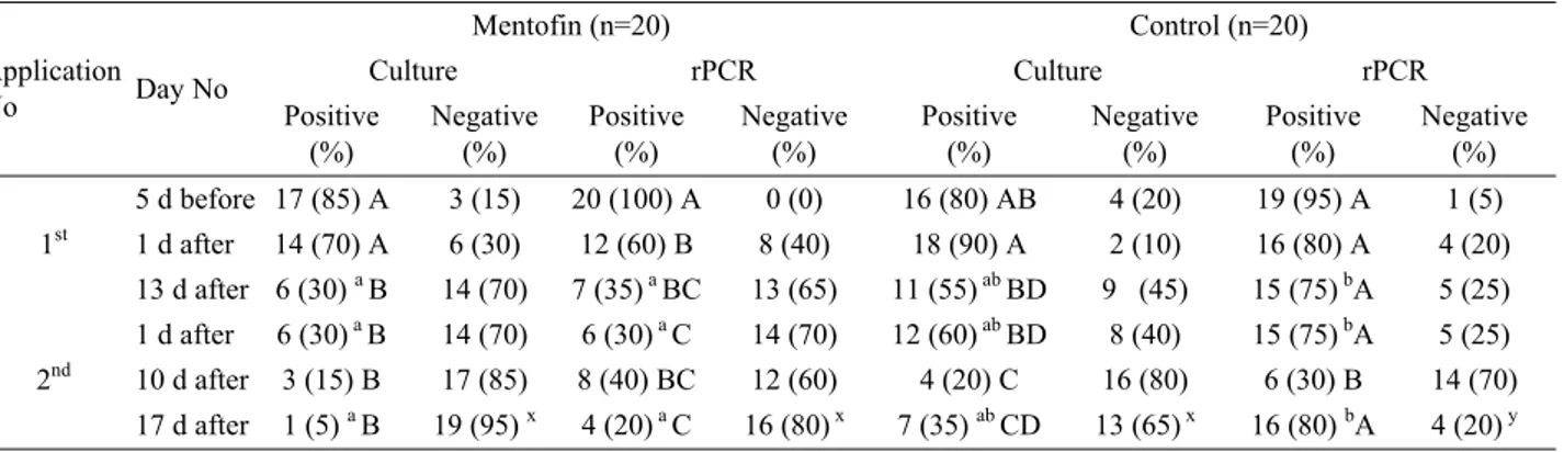
Culture and MGrPCR: There was a significant and
continuous decline in the number of MG-infected birds
in the Mentofin group after Mentofin application
(P<0,05), while a comparably small and a fluctuant
decline in the number of MG-infected birds was
determined in the control group (P>0,05). There was a
significant decline in MG positive birds detected by
rPCR after 1
stand 2
ndmentofin application (P<0,05),
however this decline was found insignificant in both
groups’ culture results (P<0,05) (Table 2).
Discussion and Conclusion
In this study, effect of Mentofin application on
levels of MG-infected birds in naturally infected flocks
was observed. This study was conducted in flocks
selected as typical representatives for Turkish layer
chicken production, which do not have good
management practices. Houses had poor ventilation and
were unclean with non-hygienic cages. Additionally, the
environment around the houses was not properly
managed for cleaning and pest control. There was no
proper structural and organizational biosecurity action
taken in the organization.
There was no substantial change in the number of
MG-antibody positive birds after Mentofin applications
in the Mentofin flock, as expected. This was probably
due to the stable MG-antibody levels produced against
the MG antigen for a long period of time in the serum,
despite the possible elimination of the MG. The
positivity in our ELISA (which detects IgG - the
dominant antibody in chronic infections) test results with
a slight fluctuation in the previously chronically- MG
infected flock, can be related to this. Contrary to the
serology results, there was a significant decline in
MGrPCR results, where MG positive numbers reduced to
less than half of the group. All these findings indicate
that MG-antibody levels were still high due to the
continued persistence of the antibodies in the serum after
chronic infection, but MG was eliminated up to a level,
Table 1. Serological test results for MG-antibody levels in Mentofin and control groups Tablo 1. Mentofin ve kontrol gruplarının MG antikor düzeyini gösteren serolojik test sonuçları
Application
No Day No
Mentofin (n=20) Control (n=20)
RSA ELISA RSA ELISA Positive (%) Negative (%) Positive (%) Negative (%) Positive (%) Negative (%) Positive (%) Negative (%) 1st 5 d before 19 (95) a 1 (5) x 12 (60) ab 8 (40) y 18 (90) a A 2 (10) x 5 (25) b 15 (75) y 1st 4 d after 19 (95) a 1 (5) x 17 (85) a 3 (15) x 19 (95) a A 1 (5) x 3 (15) b 17 (85) y 2nd 18 d 19 (95) a 1 (5) x 12 (60) ab 8 (40) y 12 (60) ab B 8 (40) y 8 (40) b 12 (60) y a, b: Different small letters indicate statistical significance at the same line for positive data (P < 0,05)
x, y: Different small letters indicate statistical significance at the same line for negative data (P < 0,05)
A, B: Different capital letters indicate statistical significance at the same row for inside group comparison positive and negative data together (P < 0,05)
a, b: Farklı küçük harfler aynı sıradaki pozitif verilerin istatistiksel farklılığını belirtir (P < 0,05) x, y: Farklı küçük harfler aynı sıradaki negatif verilerin istatistiksel farklılığını belirtir (P < 0,05)
A, B: Farklı büyük harfler aynı satırdaki pozitif ve negatif verilerin birlikte ve grup içi karşılaştırmasındaki istatistiksel farklılığını belirtir (P < 0,05)
Table 2. Numbers of MG-infected birds detected from Mentofin group by culture and rPCR before and after Mentofin applications in comparison with those of the control group
Tablo 2. Mentofin ve kontrol grubunun Mentofin uygulamasından önce ve sonra MG kültür ve rPCR sonuçlarının karşılaştırılması Application No Day No Mentofin (n=20) Control (n=20) Culture rPCR Culture rPCR Positive (%) Negative (%) Positive (%) Negative (%) Positive (%) Negative (%) Positive (%) Negative (%) 1st 5 d before 17 (85) A 3 (15) 20 (100) A 0 (0) 16 (80) AB 4 (20) 19 (95) A 1 (5) 1 d after 14 (70) A 6 (30) 12 (60) B 8 (40) 18 (90) A 2 (10) 16 (80) A 4 (20) 13 d after 6 (30) a B 14 (70) 7 (35)a BC 13 (65) 11 (55)ab BD 9 (45) 15 (75) bA 5 (25) 2nd 1 d after 6 (30) a B 14 (70) 6 (30)a C 14 (70) 12 (60)ab BD 8 (40) 15 (75) bA 5 (25) 10 d after 3 (15) B 17 (85) 8 (40) BC 12 (60) 4 (20) C 16 (80) 6 (30) B 14 (70) 17 d after 1 (5) a B 19 (95) x 4 (20)a C 16 (80)x 7 (35) ab CD 13 (65) x 16 (80) bA 4 (20)y a, b: Different small letters indicate statistical significance at the same line for positive data (P < 0,05)
x, y: Different small letters indicate statistical significance at the same line for negative data (P < 0,05)
A-D: Different capital letters indicate statistical significance at the same row for inside group comparison, positive and negative data together (P < 0,05)
a, b: Farklı küçük harfler aynı sıradaki pozitif verilerin istatistiksel farklılığını belirtir (P < 0,05) x, y: Farklı küçük harfler aynı sıradaki negatif verilerin istatistiksel farklılığını belirtir (P < 0,05)
A-D: Farklı büyük harfler aynı satırdaki pozitif ve negatif verilerin birlikte ve grup içi karşılaştırmasındaki istatistiksel farklılığını belirtir (P < 0,05)
as shown by reduction in positive birds tested by
MGrPCR.
Antibacterial properties of eucalyptus derivates
have been previously reported in studies by Babayi et al.
(1), Barbour et al. (2), Jain et al. (17), Mohamed and
Ibrahim (21), Nair et al. (23), and Navarro et al. (24). In
this study we used culture and rPCR methods to detect
MG-infected birds and found that MG-infected bird
numbers had significantly and continuously decreased in
the Mentofin
group, despite no considerable change in
the control group. This dramatic decrease was found
significant only in MGrPCR results, indicating its
superiority over culture. Therefore, we recommend the
use of rPCR in MG detection in the flocks, since it is not
uncommon to experience difficulties in MG isolation,
leading to false negative results in culture compared to
PCR.
In conclusion, results of this study indicate that
Mentofin clearly had an effect on MG clearance from the
tracheal epithelium, supported by detection of decline in
MG infection in layers. The actual action mechanism of
Mentofin on MG clearance from naturally infected
chicken trachea is still unknown, and requires further
detailed investigations.
References
1. Babayi H, Kolo I, Okogun JI, Ijah UJJ (2004): The antimicrobial activities of methanolic extracts of Eucalyptus camaldulensis and Terminalia catappa against some pathogenic microorganisms. Biokemistri, 16, 106-111. 2. Barbour EK, Yaghi RH, Shaib HA, Tayeb IT, Sleiman
FT (2008): Evaluation of an essential oil in treatment of immunosuppressed-coinfected broilers. Am Eurasian J Sustain Agric, 2, 212-218.
3. Barua SR, Prodhan AM, Islam S, Chowdhury S (2006): Study on Mycoplasma gallisepticum in chickens in selected areas of Bangladesh. J Vet Med Sci, 4, 141-142.
4. Blanchard B, Chevallier B, Vermissey Y, Charbonnier A, Guennec JLE (2001): Detection by PCR of avian Mycoplasmas from environmental swabs of poultry houses. Br Poult Sci, 42, 59-63.
5. Bragg B (2004): Evaluation of the safety of the continual use of the product, Mentofin® for spraying of layer
chickens. Evaluation of the safety of the continual use of the product, Mentofin® for spraying of broiler chickens.
Available at: http://www.ewabo.de/sites/default/files/ downloads/16_08_mentofin_gb_folder.pdf
6. Bragg RR (2006): Determination of the minimum inhibitory concentrations (MIC) of Mentofin® against Newcastle Disease virus with a 15 min contact period. Available at: http://www.ewabo.de/sites/default/files/downloads/16_08_ mentofin_gb_folder.pdf
7. Carli KT, Eyigor A (2003): Real-time polymerase chain reaction for detection of Mycoplasma gallisepticum in chicken trachea. Avian Dis, 47, 712-717.
8. Carli KT, Kahya S (2011): Kanatlı Hayvanların İnfeksiyöz Hastalıkları. Uludağ Üniversitesi Veteriner Fakültesi Yayınları, Bursa.
9. Carli KT, Onat K, Gunaydin E (2008): Application of Mentofin® in broilers with clinical Infectious Bursal
Disease to reduce Escherichia coli related problems after vaccination against Newcastle Disease. Turk J Vet Anim Sci, 32, 73-79.
10. Dovc P, Bencina D, Antes R, Mann W (1994): Recombinant DNA probes and polymerase chain reaction for detection of Mycoplasma gallisepticum strains. FEMS Microbiol Lett, 122, 79-84.
11. Esendal OM (2002): Mikoplazma infeksiyonları. 79-95. In: Kanatlı Hayvan Hastalıkları. Medisan Yayın, Ankara. 12. Evans JD, Leigh SA, Branton SL, Collier SD, Pharr
GT, Bearson SMD (2005): Mycoplasma gallisepticum: Current and developing means to control the avian pathogen. J Appl Poult Res, 14, 757-763.
13. Fan HH, Kleven H, Jackwood MW, Johansson KE, Petterson B, Levisohn S (1995): Species identification of avian Mycoplasmas by polymerase chain reaction and restriction fragment length polymorphism analysis. Avian Dis, 39, 398-407.
14. Haaheim H, Vorland L, Gutteberg TJ (2001): Laboratory diagnosis of respiratory diseases: PCR versus serology. Nucleosides, Nucleotides, Nucleic Acids, 20, 1255-1258. 15. Hossain KMM, Ali MY, Hauque MI (2006):
Seroprevalence of Mycoplasma gallisepticum infection in chicken in the greater rajshahı strict of Bangladesh. J Vet Med Sci, 5, 9-14.
16. İzgür M (2006): Mycoplasma, Ureaplasma, Acheloplasma, Spiroplasma ve Erysipelothrix infeksiyonları. 293-304. In: Veteriner Mikrobiyoloji (bakteriyel hastalıklar). İlke Emek Matbaacılık ve Yayıncılık, Ankara.
17. Jain P, Nimbrana S, Kaila G (2010): Antimicrobial activity and phytochemical analysis of Eucalyptus tereticornis bark and leaf methanolic extracts. Int J Pharm Sci Rev Res, 4, 126-128.
18. Kahya S, Temelli S, Eyigor A, Çarlı KT (2010): Real-Time PCR, bacteriology and serology for the diagnosis of Mycoplasma gallisepticum in chicken breeder flocks. Vet Microbiol, 144, 319-324.
19. Kleven SH, Yoder HW (1989): Mycoplasmosis: A
Laboratory Manual for the Isolation and Identification of Avian Pathogens. Pages 57–62 in: American Association of Avian Pathologists. 3rd ed. H. G. Purchase, L. H. Arp, C. H. Domermuth, and J. E. Pearson, ed. Kennett Square, PA, USA.
20. Ley DH (2003): Mycoplasma gallisepticum infection. 722-743. In: Diseases of Poultry, Iowa State University Press, IA, USA.
21. Mohamed GA, Ibrahim SRM (2007): Eucalyptone G, A new phloroglucinol derivative and other costituents from Eucalyptus globulus Labill. Arkıvoc, XV, 281-291. 22. Mohammed HO, Carpenter TE, Yamamoto R (1987):
Economic impact of Mycoplasma gallisepticum and Mycoplasma synoviae in commercial layer flocks. Avian Dis, 31, 477-82.
23. Nair R, Vaghasiya Y, Chanda S (2008): Antibacterial activity of Eucalpytus citriodora Hk. oil on few clinically important bacteria. Afr J Biotechnol, 7, 25-26.
24. Navarro V, Villarreal ML, Rojas G, Lozoya X (1996): Antimicrobial evaluation of some plants used in Mexican traditional medicine for the treatment of infectious diseases. J Ethnopharmacol, 53, 143-147.
25. Poveda J.B (1998): Biochemical Charactristics in Mycoplasma Identification. 69-78. In: Methods in Molecular Biology. Mycoplasma Protocols. Humana Press Inc, Totowa, NJ, USA.
26. Sarkar SK, Rahman M, Amin KMR, Khan MFR, Rahman MM (2005): Seroprevalence of Mycoplasma gallisepticum infection of chickens in model breeder poultry farms of Bangladesh. Int J Poult Sci, 4, 32-35. 27. Sikder AJ, Islam MA, Rahman MM, Rahman MB
(2005): Seroprevalence of Salmonella and Mycoplasma gallisepticum infection in the six model breeder poultry farms at Patuakhali district in Bangladesh. Int J Poult Sci, 4, 905-910.
28. United States Department of Agriculture (2011): Standart test procedures for Mycoplasma. Subpart A, Section 147-7. National Poultry Improvement Plan and Auxiliary Provisions, USDA, Animal and Plant Health Inspection Service.
29. World Organization for Animal Health (2013): Avian Mycoplasmosis (Mycoplasma gallisepticum, M. synoviae). Chapter 2.3.5. Manual of Diagnostic Tests and Vaccines for Terrestrial Animals.
30. Yoder HW (1984): Mycoplasma gallisepticum infection. 190-212. In: Diseases of Poultry. Iowa State University Press, Iowa, IA, USA.
Geliş tarihi: 27.01.2014 / Kabul tarihi: 02.05.2014 Address for correspondence:
Prof. Dr. Ayşegül Eyigör Uludag University,
Faculty of Veterinary Medicine,
Department of Food Hygiene and Technology, 16059, Görükle Campus, Bursa, Turkey e-mail: [email protected]