Journal Of Ankara University Faculty of Medicine 2011, 64(2)
Cengiz Kara, Berkan Reșorlu, Ural Oğuz, Ali Ünsal 97
Endoscopic Treatment of Simultaneously Detected
Fibroepithelial Polyps of Ureter and Bladder
Mesane ve Üreterde Eș Zamanlı Olarak Tespit Edilen Fibroepitelyal Poliplerin Endoskopik Tedavisi
Cengiz Kara, Berkan Reșorlu, Ural Oğuz, Ali Ünsal
Ankara Keçiören Eğitim ve Araștırma Hastanesi Üroloji Anabilim Dalı Üriner sistemin fibroepitelyal polipleri ender görülen ve günümüzde endoskopik olarak tedavi
edilen iyi huylu tümörlerdir. Endoskopik tedavi küçük boyutlu polipler için altın standart olarak kabul edilmekle birlikte, büyük boyutlu poliplerin tedavisi hakkında kesin bir görüș birliği yok-tur. Burada sistoskopi esnasında mesane ve üreterde eșzamanlı olarak tespit edilen fibropitelyal polipleri olan vakamızı ve üreterdeki 7 cm uzunluğundaki polipin holmium:YAG laser ile endos-kopik olarak çıkartılmasını tarifliyoruz.
Anahtar Sözcükler: Fibroepitelyal polip, Bladder, Üreter, Endoskopik Tedavi
Fibroepithelial polyps of the urinary tract are rare benign tumors that usually treated with endoscopically today. Endoscopic treatment have been regarded as the gold standart for small lesions, while the management modality for long lesion is stil controversial. We report a case of simultaneous fibroepithelial polyps of bladder and ureter which is incidentally detected on cystoscopy. We review our experience with ureteroscopic management of long (7 cm) ureteral fibroepithlial polyp by holmium: yttrium- aluminum- garnet (YAG) laser resection.
Key Words: Fibroepithelial polyp, Bladder, Ureter, Endoscopic treatment
Fibroepithelial polyps (FEPs) are the most common benign lesions of the urinary tract (1). Although the exact etiology of them is uncertain, some have suggested both congenital factors in children and chronic infection in adults (2). FEPs are predominantly located in the upper third of the ureter and the ureteropelvic junction (3). The most common symptoms are hematuria and flank pain, which may be attributable to either torsion or intussusception of the polyp or an obstruction. The endoscopic approach is applied for diagnostic and therapeutic evaluation. Relatively few reports have been published of excision of FEPs with use of flexible ureteroscopy in conjuction with the Holmium:YAG laser (4, 5). To our knowledge, there is no report have been published about coexistence of FEPs in ureter and bladder. We report our experience on endoscopic holmium:YAG laser treatment of this large FEP.
Case Report
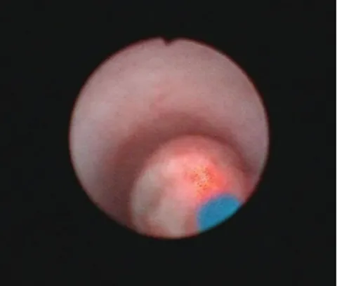
A 43-year-old woman was referred to us with intermittent right flank pain and hematuria. She had undergone left adrenelectomy 7 years ago. Physical examination and routine blood tests were normal. Urine analysis showed mild microscopic hematuria with a sterile urine culture. Abdominal ultrasonography showed a solid mass in the urinary bladder. The rest of abdomen was normal. No hydronephrosis was seen in the right kidney. The patient subsequently underwent diagnostic cystoscopy. Cystoscopy revealed a right ureteral polyp and a polypoid masses (2x2 cm size) around the right ureteral orifice (Figure 1). A urine sample was taken for cytology. The polypoid masses in the bladder were transurethral resected by electroresection and couterized. Ureteroscopic evaluation of the right ureter showed the 7 cm lenght polyp (Figure 2). The polyp in the distal ureter was grasped with Ankara Üniversitesi Tıp Fakültesi Mecmuası 2011, 64 (2)
DOI: 10.1501/Tıpfak_000000791 CERRAHİ BİLİMLER/SURGICAL SCIENCES
Case Report / Olgu Sunumu
Received: 12.08.2009 • Accepted: 28.10.2010 Corresponding Author
Dr. Cengiz KARA
Ankara Keçiören Eğitim ve Araștırma Hastanesi Üroloji Anabilim Dalı
Fax : +90 312 356 90 02 GSM : 0505 255 38 90 E-Posta : [email protected]
Ankara Üniversitesi Tıp Fakültesi Mecmuası 2011, 64(2)
Mesane ve Üreterde Eș Zamanlı Olarak Tespit Edilen Fibroepitelyal Poliplerin Endoskopik Tedavisi 98
biopsy forceps and punch biopsies were done with ureteroscope. The histological examination revealed a fibroepithelial polyp of the ureter and bladder. The result of urine cytology
was negative. The patient underwent a routine intravenous urography (IVU) and computerized tomography (CT) urography evaluation and elongated filling defect involving the
distal part of the right ureter (Figure 3). Three week later second look ureteroscopy was performed with an 8.4/5.3F flexible ureteroscope (Olympus) for removal of ureteral FEP. Before ureteroscopy a 0.038 hydrophilic guidewire was advenced through the orifice and the ureteroscope was advenced via the hydrophilic guidewire. A holmium: Yag laser was operated at a wavelenght of 2100 nm, and the frequency was usually set between 5 to 10 Hz. The base of polyp was ablated with a 365 nm holmium laser fibre operating at 10W to 15W. The polyp was then removed from the ureteral wall with 3F grasping forceps (Figure 4). Hemostasis was achieved with laser coagulation. A 4.8F ureteral stent was placed at the completion of the procedure and removed 6-week follow-up visit. Histologic analysis demonstrated FEP with no evidence of malignancy. The operative time was 55 minutes. No complication was observed during the procedure. Subsequent intravenous urography (IVU) at 12 months was normal. No recurrence or ureteral stricture was detected.
Discussion
Fibroepithelial polyps are rare, benign, mesodermal tumors of the urinary tract that are histologically composed of fibroous stroma covered with a transitional epithelium (3). Other rare urinary tract tumors, which are even less common than FEPs, are leiomyomas, lymphangiomas, fibromas, endometriomas, and neurofibromas (6). FEPs typically appear as smooth, mobile, pedinculated mass. In some cases it is difficult to differentiate them from transitionel cell carcinoma, based only on imaging findings and their potential for malignant transformation is extremly rare (7). Based on current literature, FEPs can occure in newborns and adults older than 70 years but commonly present in adults in the third through the fifth decades, with a male/female ratio of 3 to 2. Most FEPs occur in the Figure 1. Transurethral resection of bladder
polyp Figure 2. Ureteral resection of fibroepithelial polyp with laser
Figure 3. IVU and CT urography reveals a right distal ureteral filling defect (arrows)
Journal Of Ankara University Faculty of Medicine 2011, 64(2)
Cengiz Kara, Berkan Reșorlu, Ural Oğuz, Ali Ünsal 99
ureter, 15% occur in the renal pelvis, and small number develop in the posterior urethra or bladder (3, 4). The usual location of a FEP of the ureter is in the proximal part, with the left side being more commonly involved than the right side. They are almost always unilateral and solitary, however, cases of bilateral or multipl polyps in a ureter have been described (4, 8).FEPs are tought to be either congenital slow-growing lesions or lesions that develop as a result of chronic urethelial irritants, such as infection, inflammation, or obstruction (2). However, the definitive etiologic factors of these tumors are still unknown. The most common symptoms are hematuria and flank pain, which may be attributable to either torsion or intussusception of the polyp or an obstruction. Urinary frequency, dysuria, and pyuria are less common findings (4).
Preoperative radiological diagnosis of FEPs is difficult. IVU shows a filling defect within the renal pelvis or ureter. Ultrasonography and CT can be used to rule out radiolucent calculi, sloughed papillae, fungi, or blood clots and allows the extent and morphological features of these tumors to be determined (9). The role of CT urography in the diagnosis of FEPs is determinitive in the preoperative assesment, but other authors doubt
its ability to provide information complementary toIVU (10).It is important to distinguish fibroepithelial polyps from upper urinary tract carcinoma, because the management and prognosis can be significantly different. Cystoureteroscopy, which enables a biopsy, is very important in the study of filling defects located in the excretory system (6). Cystoscopy is essential following an episode of hematuria for the evaluation of urothelial tumors in the bladder. All patients should have biopsy proven histologic confirmation before beginning definitive therapy (4).
Treatment of FEPs includes local coagulation by laser, polypectomy by ureteroscopy, segmental resection with ureteroureterostomy or nephroureterectomy. In the past management of FEP was excision of the polyp and reanastomosis with an open procedure. Recently, with the advent of ureteroscopes, endoscopic biopsy and resection of these tumors has become more popular.The holmium:YAG laser is another modality for endoscopic resection. Carey et al successfully ablated multiple polyps in one ureter by using the holmium laser and removed each polyp from the ureteral wall with grasping forceps (5). Percutaneous antegrade excision should be available for treating polyps in the renal pelvis and
the upper ureter (4). The holmium laser is well suited to the treatment of these entities. The depth of penetration of the holmium:YAG laser is desirable to avoid inadvertent thermal injury to the ureteral wall during coagulation of resection site. Ureteroscopic resection can be difficult in patients with a long, thick polypoid lesion similar in our case. This difficulty is often caused by inability to access the base of the stalk because of hindrance from the pedunculated body of the lesion (11). Furthermore visualization may also be
poor, and little working space is available, making it difficult to differentiate the ureteral wall from the polyp itself. Therefore, before the procedure a hydrophilic guide wire was placed past the polyp base in the patient. It is important that to find the base of the polyp is essential for the succesfull endoscopic treatment. In this study we report a case of a simultaneous bladder and ureteral FEP which is incidentally demonstrated on cystoscopy. We have also demonstrated that with the proper equipment and endoscopic experience highly complex cases of FEP in ureter may be treated with by minimally invasive means with the preservation of the renal unit.
KAYNAKLAR
1. Bolton D, Stoller ML, Irby P: Fibroepithelial ureteral polyps and urolithiasis. Urology 1994; 44: 582-7. 2. Wang ZJ, Meng MV: Ureteral
fibroepithelial polyp. J Ultrasound Med 2008; 27: 1647-9.
3. Williams TR, Wagner BJ, Corse WR: Fibroepithelial polyps of the urinary tract. Abdom Imag 2002; 27: 217-21.
4. Lam JS, Bingham JB, Gupta M: Endoscopic treatment of fibroepithelial polyps of the renal pelvis and ureter. Urology 2003; 62: 810-3.
5. Carey RI, Bird VG: Endoscopic management of 10 separate fibroepithelial polyps arising in a single ureter. Urology 2006; 67: 413-5.
6. Kiel H, Ullrich T, Roessler W, et al: Benign ureteral tumors: four case reports and review of the literature. Urol Int 1999; 63: 201-5.
7. Brady JD, Korman HJ, Civantos F, et al: Fibroepithelial polyp of the renal pelvis: nephron sparing surgery after false-positive biopsy for transitionel carcinoma. Urology 1997;49:201-5
8. Tekdogan UY, Canaklı F, Aslan Y, et al: Bilateral ureteral fibroepithelial polyps and review of the literature. Int J Urol 2005; 12: 98-100.
9. Kumar A, Das SK, Trivedi S, et al: Genito-urinary polyps: summary of the 10 -years experiences of a single institute. Int Urol Nephrol 2008; 40: 901-7. 10. Yeh CC, Lin HC, Chen CC, et al: Ureteral
fibroepithelial polyp prolapsed into the bladder cavity suspending a bladder stone. J Urol 2001; 165: 1627-8.
11. Sun Y, Xu C, Wen X, et al: Is endoscopic management suitable for long ureteral fibroepitelial polyps? J Endourol 2008; 22: 1459-62.